


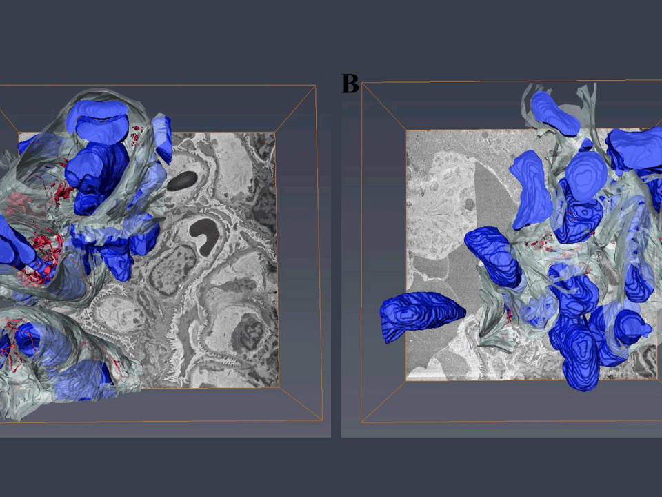


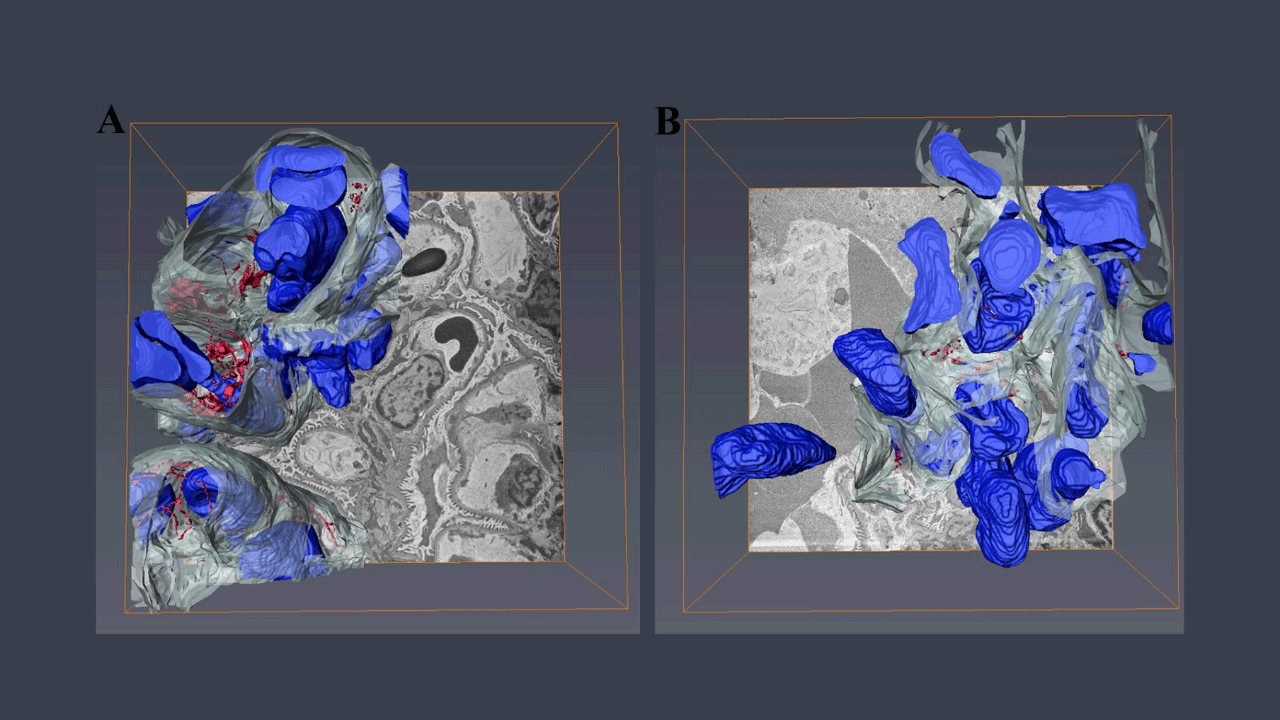
 shown in red, capillary in white, and cells in blue. Superior plane.")
 shown in red, capillary in white, and cells in blue. Superior plane.")
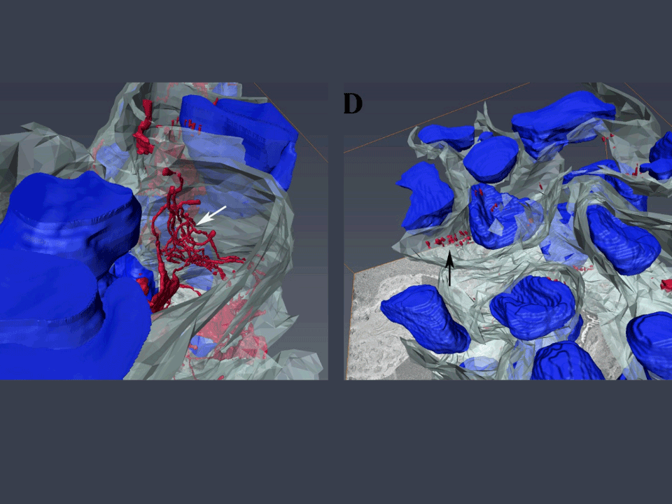
 shown in red, capillary in white, and cells in blue. Superior plane in higher magnification. Wild type (left) presents FEFS arranged in a tubular-shaped network (white arrow) within the capillary. MFS (right) presents fractured FEFS (black arrow), and loss of capillary structure.")
 shown in red, capillary in white, and cells in blue. Superior plane in higher magnification. Wild type (left) presents FEFS arranged in a tubular-shaped network (white arrow) within the capillary. MFS (right) presents fractured FEFS (black arrow), and loss of capillary structure.")
 shown in red, capillary in white, and cells in blue. Superior plane in higher magnification. Wild type (left) presents FEFS arranged in a tubular-shaped network (white arrow) within the capillary. MFS (right) presents fractured FEFS (black arrow), and loss of capillary structure.")
 shown in red, capillary in white, and cells in blue. Superior plane in higher magnification. Wild type (left) presents FEFS arranged in a tubular-shaped network (white arrow) within the capillary. MFS (right) presents fractured FEFS (black arrow), and loss of capillary structure.")
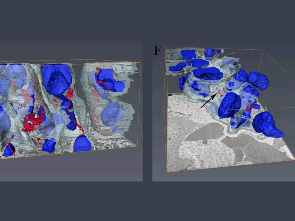
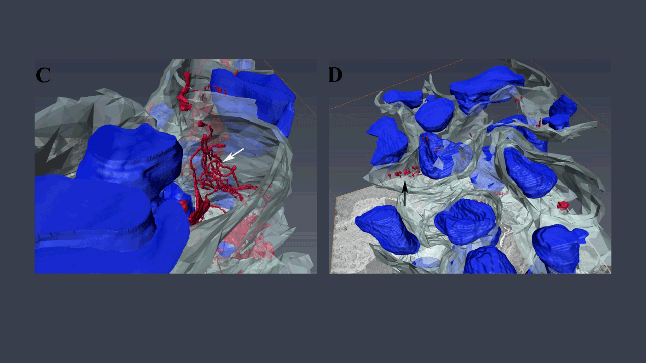
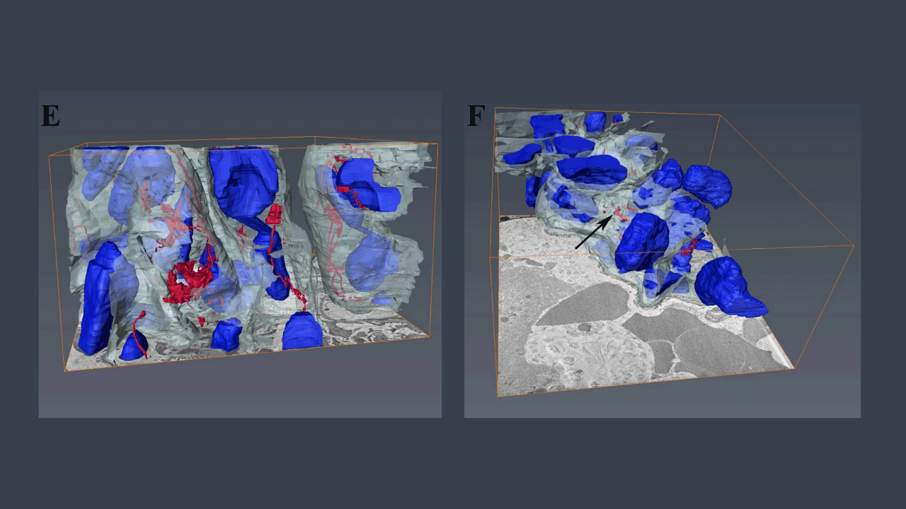
 shown in red, capillary in white, and cells in blue. Lateral plane. Wild type (left) presents FEFS arranged in a tubular-shaped network (white arrow) within the capillary. MFS (right) presents fractured FEFS (black arrow), and loss of capillary structure.")
 shown in red, capillary in white, and cells in blue. Lateral plane. Wild type (left) presents FEFS arranged in a tubular-shaped network (white arrow) within the capillary. MFS (right) presents fractured FEFS (black arrow), and loss of capillary structure.")


Potential New Clinical Aspects
Professor Barbosa and colleagues propose that alterations in kidney vessels and tissues resulting from Marfan Syndrome could be important to understanding patients with this genetic condition and may be something to consider in the clinical management of the disease.
Read the full article in PLOS One and see their additional work using histology, transmission electron microscopy (TEM), fluorescence microscopy, and macro and micro blood flow measurements in the kidney.

