Section Heading
Text that elaborates on the main headline above
Request a trial
Please fill this form to request a 30-day ZEISS arivis free trial.3D imaging and analysis solutions for organoid screening
Courtesy of Daniel Reumann and Jürgen Knoblich
ZEISS Lightsheet 7
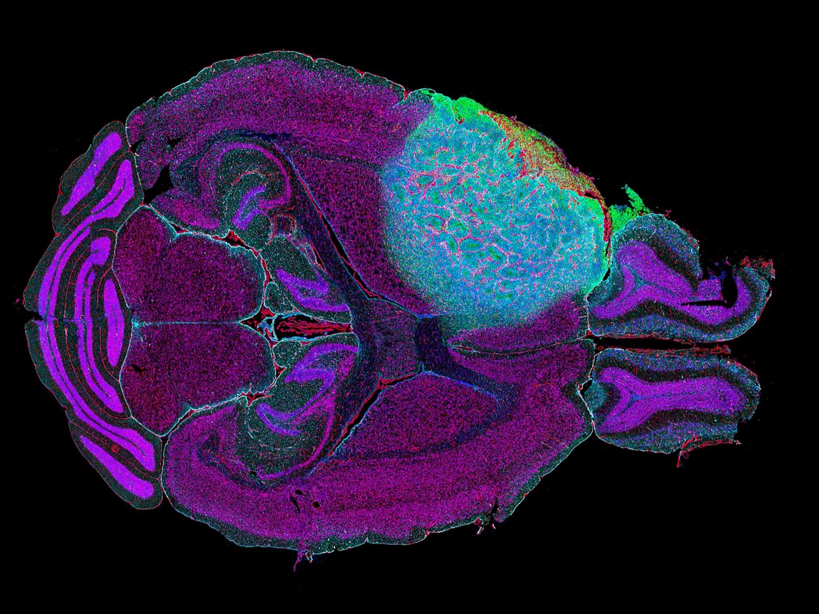
Explore the complexities of neuronal morphology in 3D
Courtesy of C. Kuo, Stanford University
ZEISS LSM 990
Acquire multiple images simultaneously, reducing acquisition time and preserving sample integrity
Section Heading
Text that elaborates on the main headline above


Inflammation and autoimmunity


Uncovering pathways and cell-types in inflammatory processes
Immuno-Oncology

Evaluating immunotherapy effectiveness and tumor immune interactions
Infectious Disease and vaccines


Mapping immune responses to pathogens and vaccine efficacy
Other applications

● Cardiovascular diseases
● Transplant, GVHD
● Regenerative medicine
● Gastrointestinal Diseases
Metabolic diseases

Investigating tissue-specific metabolic pathways and immune cell infiltration in conditions like diabetes and obesity
Neurology

Exploring immune-neural interactions and their role in neurological disorders
What You Can Do
Product comparison
Find you which product is right for you


Product comparison
Find you which product is right for youCancer Research


Workflow demos for biotech & pharma
Select the demo that suits your needs- Virtual demo: Personalized insights into ZEISS technology
- Demo at ZMCC: Visit ZEISS Microscopy Customer Center (ZMCC)
- Select your experience: Choose the demo that fits your schedule
Workflow demos for biotech & pharma
Select the demo that suits your needs- Virtual demo: Personalized insights into ZEISS technology
- Demo at ZMCC: Visit ZEISS Microscopy Customer Center (ZMCC)
- Select your experience: Choose the demo that fits your schedule
Microscopy demos for biotech & pharma
Select the demo that suits your needs- Virtual Demo: Personalized insights into ZEISS technology
- Demo at ZMCC: Visit ZEISS Microscopy Customer Center (ZMCC)
- Select your experience: Choose the demo that fits your schedule
Enabling Efficient, Reproducible Multiplex Spatial Profiling at Scale
Are you a translational, immuno-oncology researcher or service laboratory looking for a comprehensive solution that enables you to work at high throughput while ensuring accurate and reproducible data? The ZEISS tissue multiplexing workflow for biopharma, Academic Medical Centers and CROs enables routine histopathology laboratories with no prior experience of spatial biology methods to deliver rich biomarker data with high consistency across large cohorts.
Benefit from the efficiency of this tissue multiplexing workflow in your large cohorts, even under tight deadlines. Standardized methods allow you to conduct multi-site clinical trials with consistency over time, ensuring reproducible and consistent results. In addition, our solution streamlines data analysis and reporting, making it easier to evaluate multiple parameters and gain actionable insights.

Cell Biology: Quantify with Precision
From single cells to comprehensive populations, extract meaningful data automatically
Transform your cell biology research with ZEISS arivis's powerful quantification capabilities for complex cellular phenotypes. Easily segment, track, and analyze heterogeneous cell populations in complex microenvironments that better mimic in vivo conditions. Obtain reproducible measurements that capture subtle phenotypic changes without manual intervention.
- Easily automated cell and nuclei counting with AI-based segmentation
- Cell or nuclei tracking across time series to analyze division and migration
- Confluency measurement for growth monitoring
- Phenotypic classification based on multiple parameters using AI or custom feature patterns
- Quantification of cell-cell relationships, such as distance measuerments and colocalization
- Batch processing for batch and high-content experiments
Related Applications
Workflow demos for biotech & pharma
Select the demo that suits your needs- Virtual demo: Personalized insights into ZEISS technology
- Demo at ZMCC: Visit ZEISS Microscopy Customer Center (ZMCC)
- Select your experience: Choose the demo that fits your schedule
Leasing & Financing Plans for Biopharma


Microscopy Application Services


Demo Offerings


Demos & Leases
Microscopy Demos for Biotech & Pharma
Virtual, Hands-On & Personalized Education & TrainingZEISS provides flexible, tailored demo experiences to showcase its cutting-edge microscopy solutions for biotech and pharmaceutical research. Whether you're remote or onsite, our experts bring the technology—and the insights—directly to you:
- Virtual Demos offer curated, guided sessions led by ZEISS application specialists. Experience live walkthroughs of systems and software, tailored proof-of-concept experiments using sample data, and application training—all without leaving your lab.
Zeiss - In-Person Demos at the ZEISS Microscopy Customer Center (ZMCC) allow hands-on interaction with the full range of ZEISS imaging platforms. Benefit from personalized consultations, workflow testing with your own or provided samples, and one-on-one support from our specialists.
Scroll animation items
Revolutionizing fertility
Breakthroughs in IVF research transforming family dreamsVisualize critical structures like the zona pellucida, polar body, and meiotic spindle with advanced contrast techniques
including circular polarization, PlasDIC and iHMC, while maintaining temperature stability and ergonomic control throughout ICSI, IMSI, and embryo assessment.

Leverage ZEISS developed iHMC for clear visualization of fine cellular details
Especially the nucleus, nucleoli, and nuclear shape within embryosEnsure precise ICSI timing and optimal oocyte selection
With non-invasive spindle imagingStreamline semen analysis
With high-resolution imaging, smart documentation, and versatile contrast modes so you can work faster, with greater confidence.Organoid Research Use Cases
Resources, Articles & More

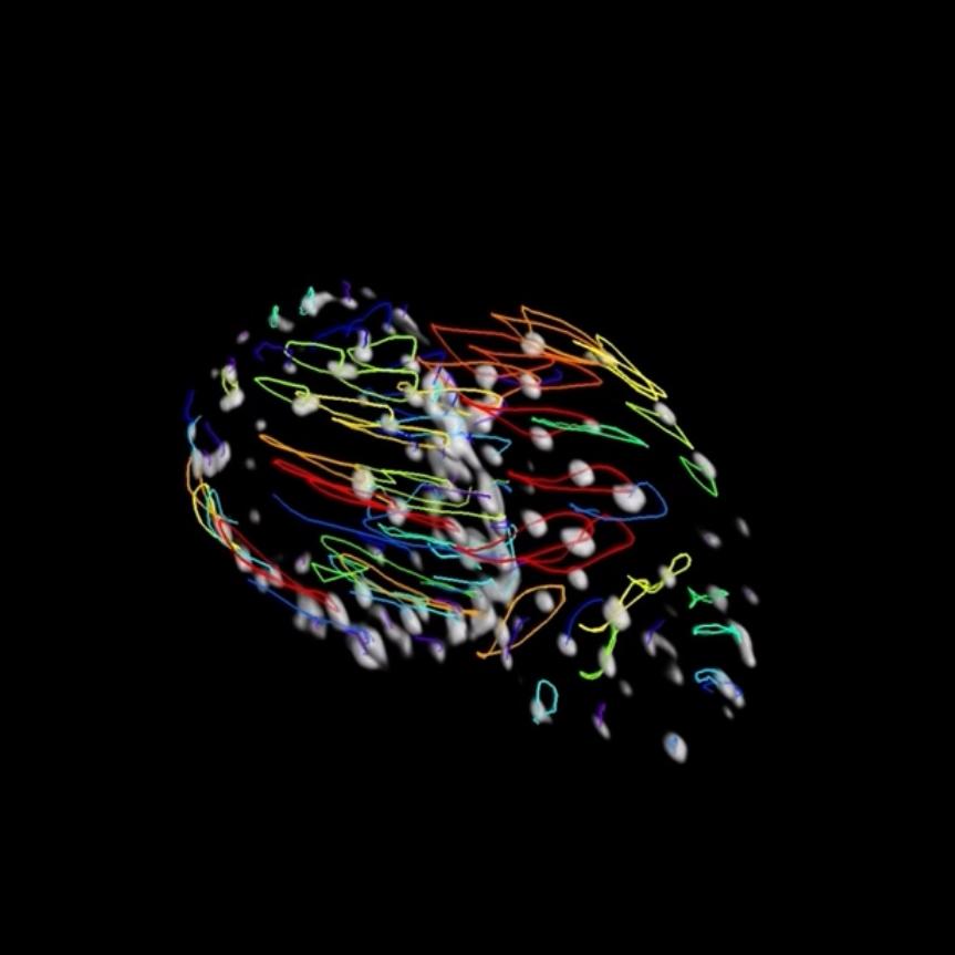
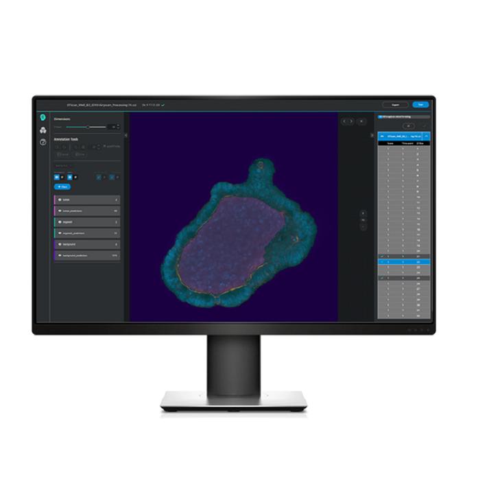
3D preview of analysis results in which individual cells in the outer layer of an organoid are visualized in different colors
This solution can be used to segment organoids, their lumen, and nuclei within the organoids using a combination of AI tools and conventional image analysis steps in ZEISS arivis Pro. Using the nuclei masks, the cell bodies of the individual cells within the outer layer of the organoids are segmented.

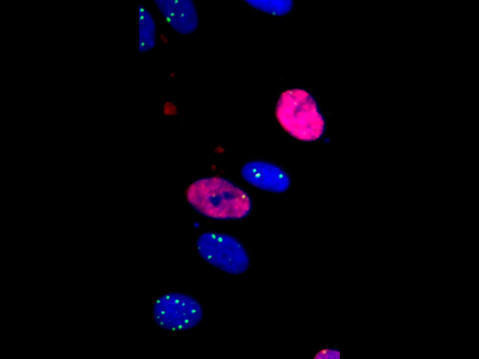

High Content Imaging for Genotoxicity
In this series "From Image to Results", explore various case studies explaining how to reach results from your demanding samples and acquired images in an efficient way. For each case study, we highlight different samples, imaging systems, and research questions. In this second episode, we set up an application example to perform DNA-damage analysis by employing ZEISS Axio Observer and the ZEISS arivis Pro image analysis software.

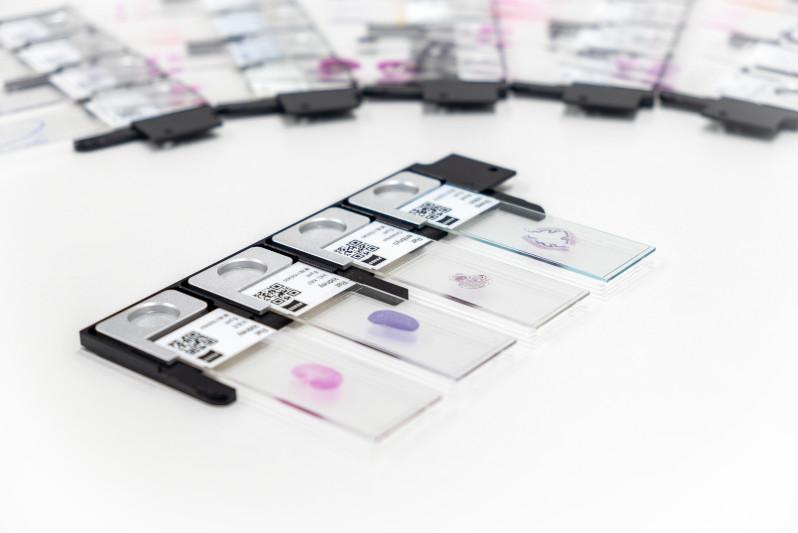
ZEISS Axioscan 7
Discover high-performance digital slide scanning tailored to your application needs. Whether your focus is spatial biology at scale, life science research, clinical applications or geology, the Axioscan 7 slide scanning microscope brings you advanced automation capabilities and exceptional image quality.

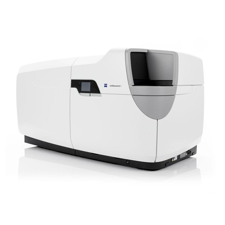
ZEISS Celldiscoverer 7
If your research requires explorative high-content imaging, you are often faced with a trade-off between the desired image quality and the need to capture large amounts of data efficiently. ZEISS Celldiscoverer 7 is your research companion for collecting statistically relevant data, giving you easy access to high-quality imaging, adaptability to demanding experiments, and stable long-term operation.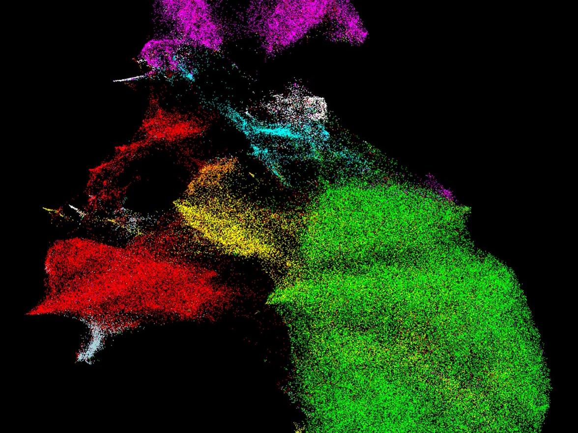

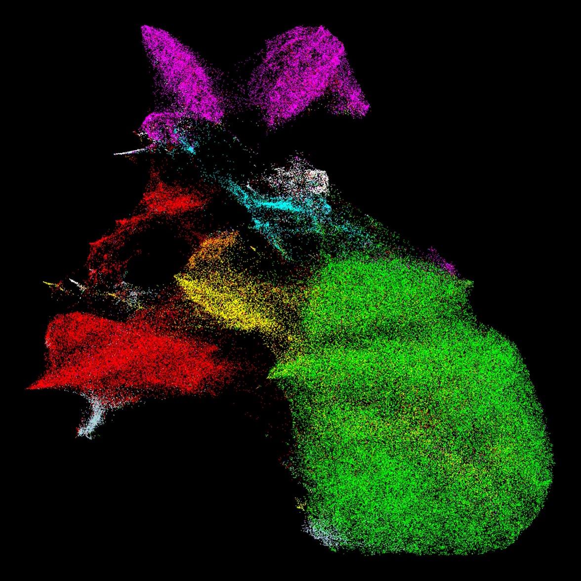
Brain Tumor Microenvironment Spatial Biology with High-Throughput Hyperplexed Imaging
Dr. Spencer S. Watson, a Research Fellow in Professor Joyce’s lab, published an article investigating how the brain tumor microenvironment responds to radiotherapy, not just in terms of changes in different cell populations, but also regarding how the cells spatially organize themselves. In order to do this, the team worked to create a new multiplex workflow, called Hyperplexed Immunofluorescence Imaging (HIFI), to study the spatial biology of tumors before and after treatment.

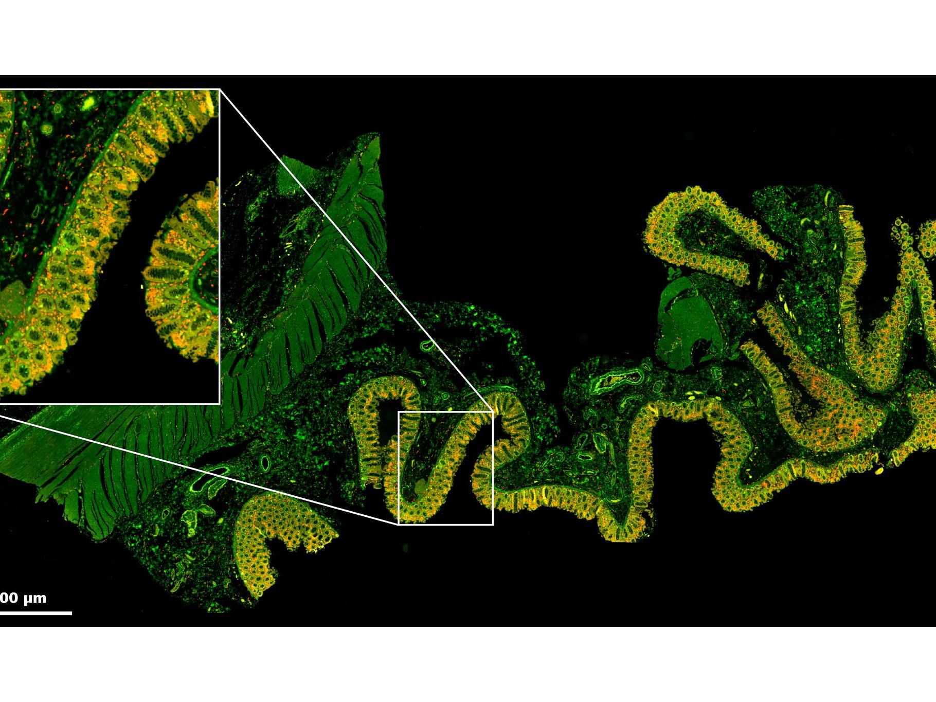
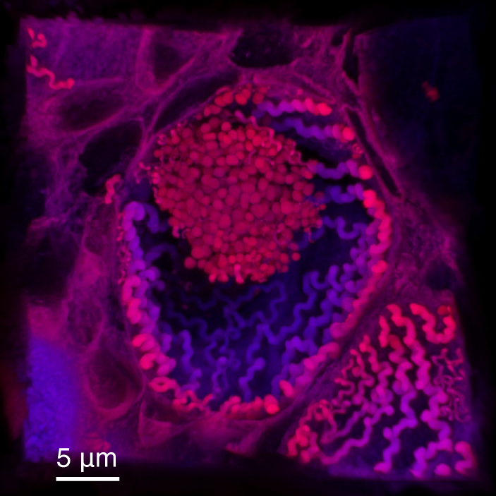
7 Color Multiplex Immunofluorescence Protocol for Spatial Biology with a Digital Slide Scanner
Dr. Caroline Bouzin is the facility manager of the 2IP Imaging Platform at the Institute of Experimental and Clinical Research at the UCLouvain, Brussels, Belgium. She has worked with oncologists and pulmonologists to fine-tune a protocol for customized mIF stainings on formalin-fixed paraffin-embedded (FFPE) tissue samples followed by data collection with the ZEISS Axioscan digital slide scanner. A combination of signal amplification (TSA) with carefully selected filtersets matched to a panel of seven fluorophores results in the fast generation of ready-to-analyze digital images without the need for image post-processing, such as spectral unmixing or tissue alignment. This workflow is equipping researchers in her facility for high-throughput, spatial biology analyses, such as the immune microenvironment of tumors.


From Image to Results | Organoid Analysis
In this new series "From Image to Results", explore various case studies explaining how to reach results from your demanding samples and acquired images in an efficient way. For each case study, we highlight different samples, imaging systems, and research questions.

Advanced Microscopy for Biotech & Pharma
From 3D-cellular imaging to tissue analysis for pre-clinical studies, our devices deliver the precision, scalability, and automation needed to drive regulatory-compliant drug discovery workflows.Brightfield Workflow

Fluorescence Workflow
In-Booth Presentations
Attend educational presentations in the ZEISS booth.-
Super-resolution imaging of the membrane-associated periodic skeleton in neurons uncovers constitutive dynamics
Presented by Evan Heller, Research Associate in Chemistry and Chemical Biology, Harvard University
Description....
Evan Heller, PhD.
Add bio
-
TITLE
Presented by Dr, Rose Creed, University of California San Francisco


Rose Creed, PhD
Rose B. Creed is a postdoctoral fellow at the University of California, San Francisco, in the laboratory of Alexandra Nelson, M.D., Ph.D. There, she takes a systems neuroscience approach to examine how impairments in the basal ganglia circuit underlie different types of movement disorders. Rose obtained her Bachelor of Science degree in Biology at Stetson university in 2014 and received her Ph.D. from the University of Alabama at Birmingham (UAB) in summer 2020. While at UAB she worked in the labs of Drs. Matthew Goldberg and Lori McMahon, investigating how loss of function of the mitochondrial targeted kinase PINK1 impacts the striatal circuit and its implication for Parkinson’s disease.

Take the Next Step to Starting Your Own Digital Classroom
Fill out the form below to get started on setting up your own digital classroom. A ZEISS representative will be in contact to answer any questions you have or to help get you started!