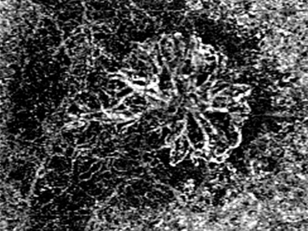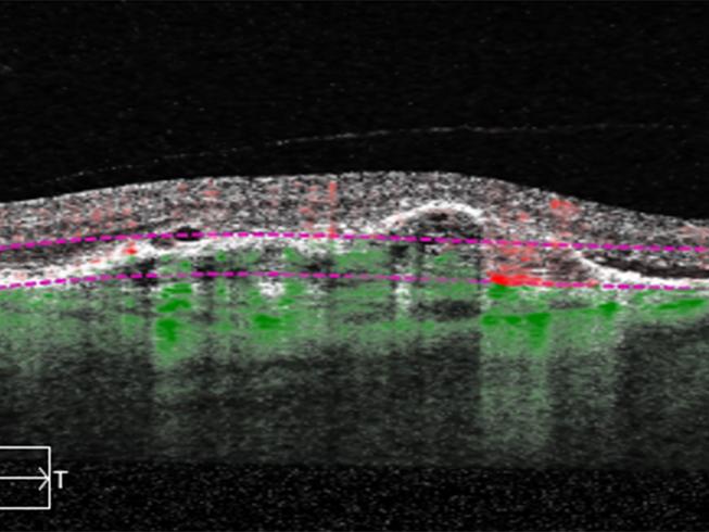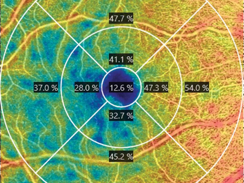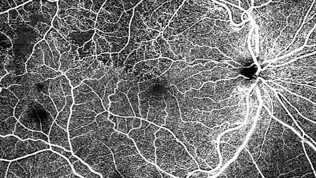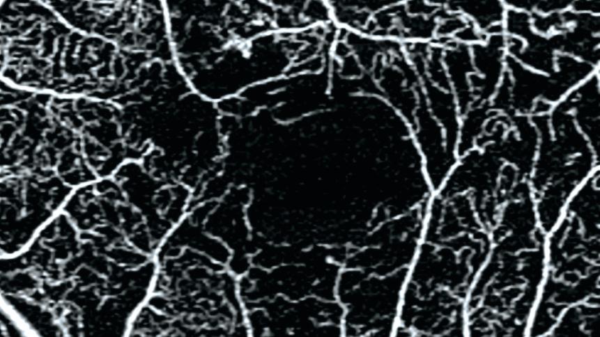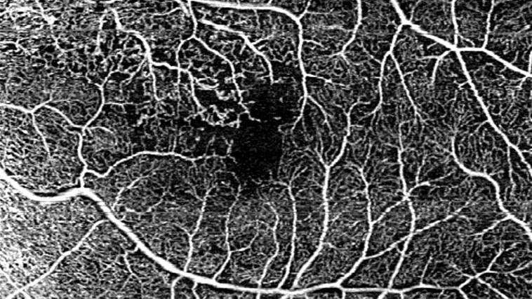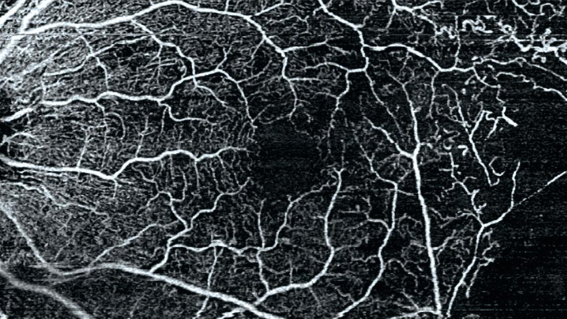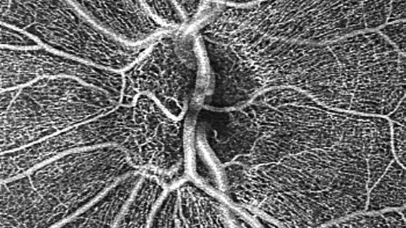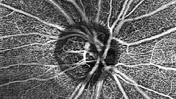

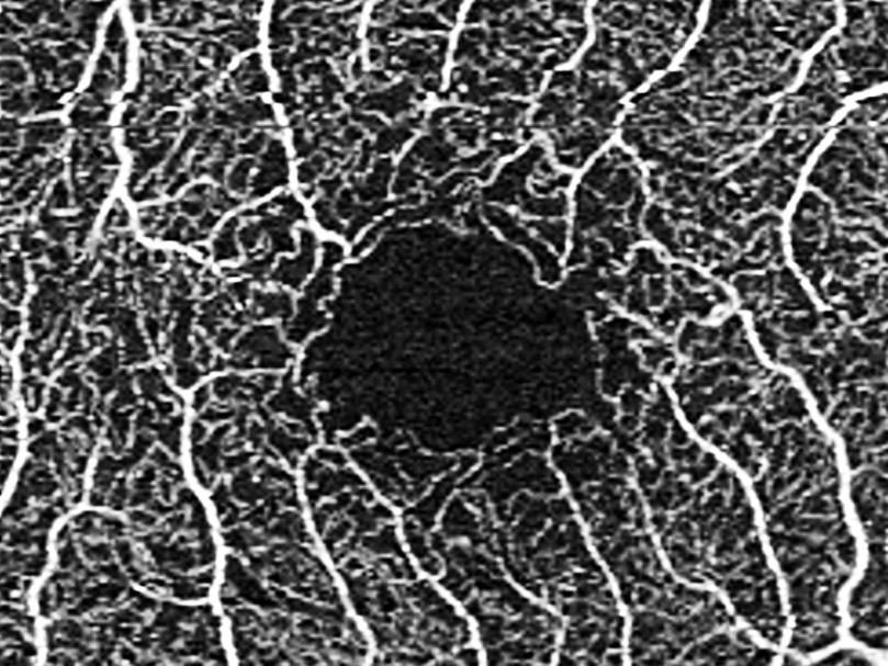


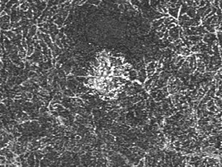

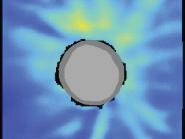


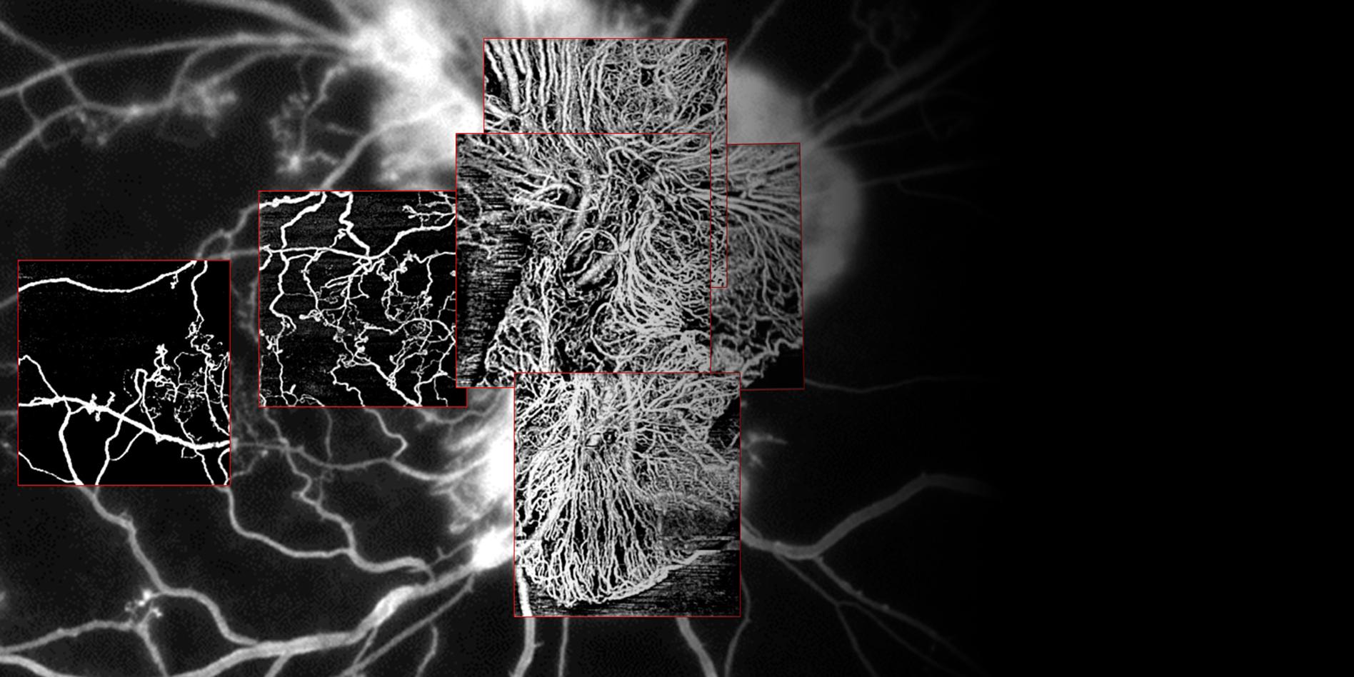

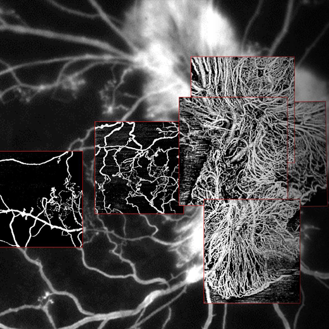

Visualize vascular abnormalities
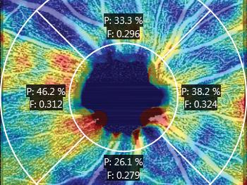
AngioPlex OCT Angiography - powered by ZEISS CIRRUS - enables you to do more: gain workflow efficiency, deliver care with ease, quantify vascular change and manage more with confidence.
Skip to main content
You are on our international English website. This site features our entire product portfolio worldwide. The products featured may not be available in the US. If you are a citizen from the US, please visit your country website for local information and contacts.
You are on our international English website. This site features our entire product portfolio worldwide. The products featured may not be available in the US. If you are a citizen from the US, please visit your country website for local information and contacts.
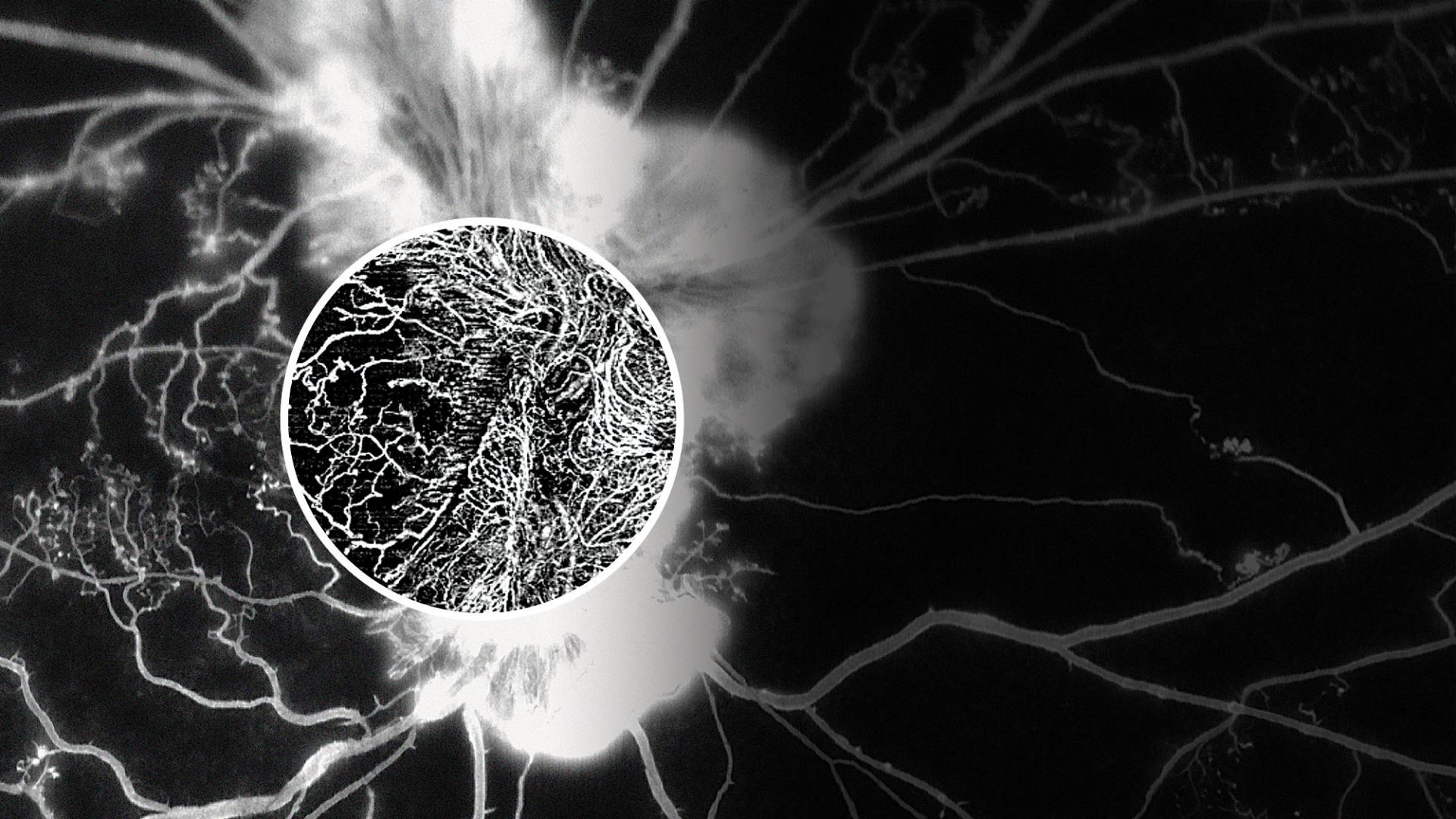
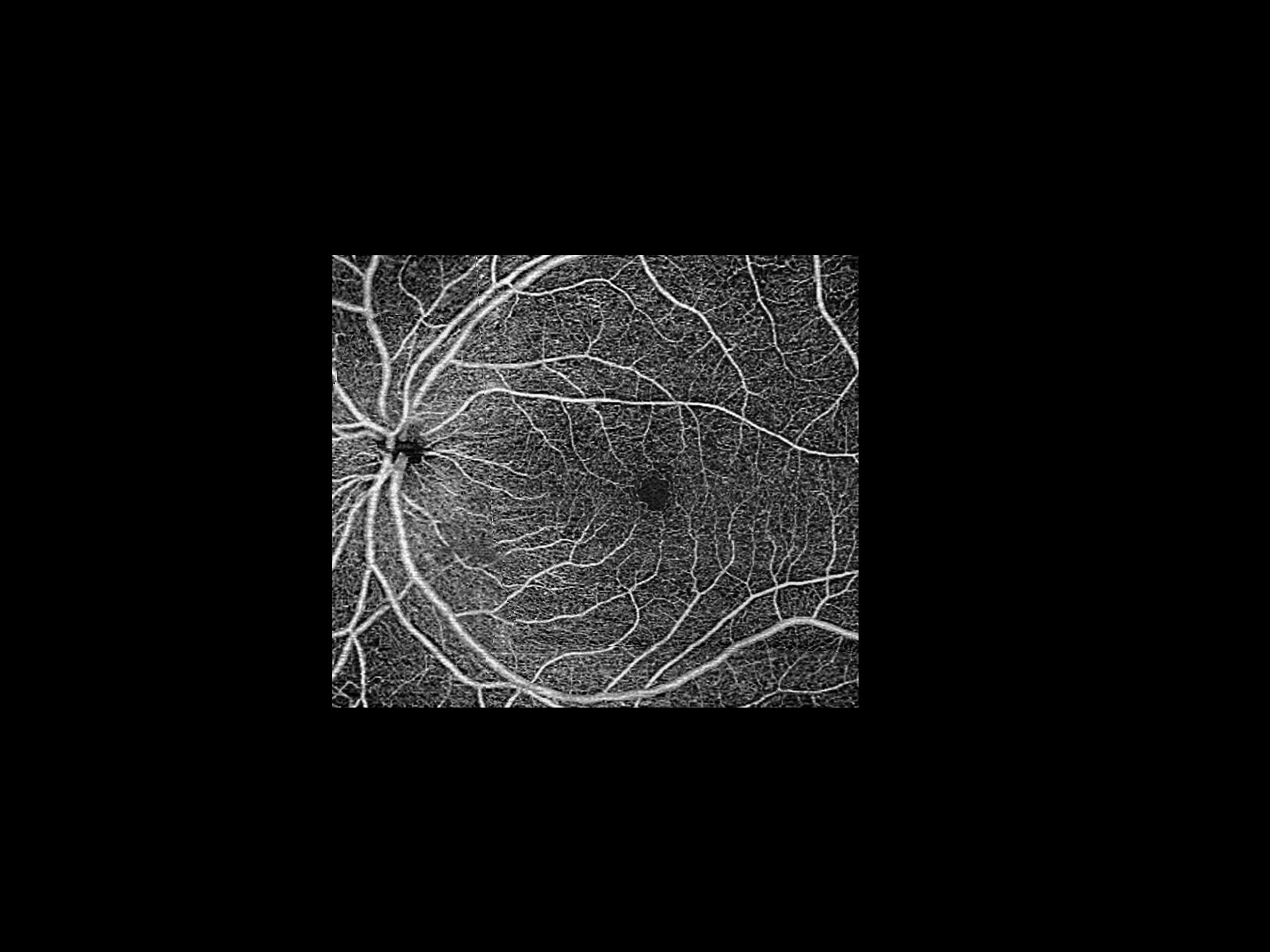
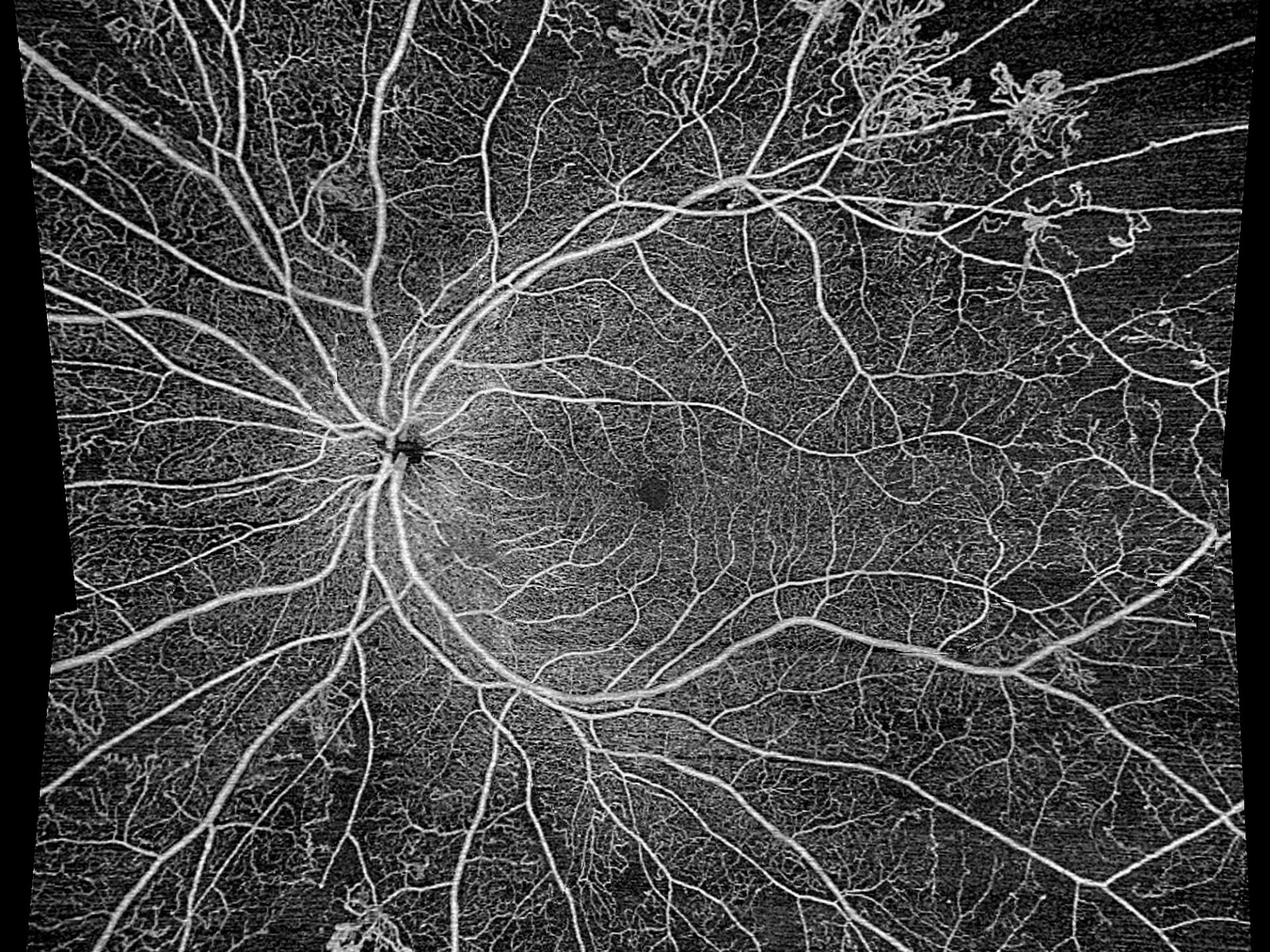
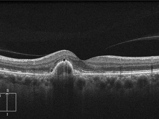
AngioPlex® OCT Angiography from ZEISS ushers in a new era of eye care with non-invasive imaging of retinal microvasculature - taking glaucoma and retinal disease management and treatment planning to the next level.


Integrating OCT Angiography into your practice provides advanced care for your patients and the advanced technology you need to support the demands of today's aging population.
AngioPlex image capture with Single Scan Simplicity and FastTrac™ lets you scan at the highest resolution, minimizing artifacts from blinks and other eye movements, and without sacrificing patient throughput. Watch the video to understand how it works.
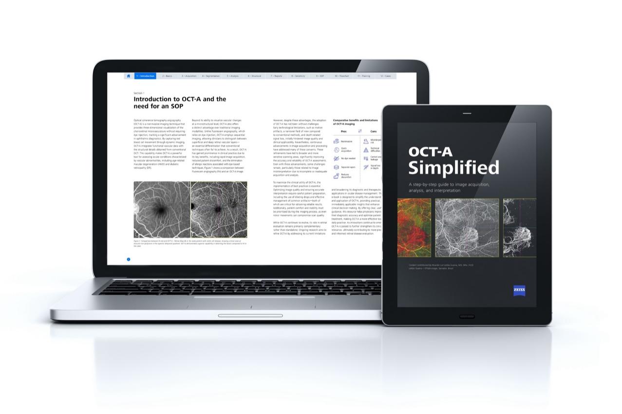

Authored by Ricardo Luz Leitão Guerra, MD, MSc, FICO, in collaboration with ZEISS, the OCT-A Simplified e-book is a practical resource for clinicians seeking to adopt OCT-A technology with greater confidence and clarity.