

Make every second count with high-performance OCT
ZEISS CIRRUS 6000
Performance OCT
100,000 scans per second to power your practiceCIRRUS® 6000 is the next-generation OCT from ZEISS, delivering high-speed image capture with HD imaging detail and a wider field of view so you can make more informed decisions and spend more time with the patients who need it.
Learn how you can maximize patient throughput and practice efficiency with ZEISS CIRRUS 6000.


")
")


")
")


Making the revolutionary, routine
ZEISS AngioPlex OCT AngiographyAngioPlex® OCT Angiography from ZEISS ushers in a new era of eye care with non-invasive imaging of retinal microvasculature - taking glaucoma and retinal disease management and treatment planning to the next level. By offering the industry’s most comprehensive tools for assessing and analyzing a range of pathologies, ZEISS provides a complete OCT Angiography (OCTA) solution.


Proven analytics
CIRRUS-powered treatment decisionsAs the pioneering OCT technology, the CIRRUS platform offers clinicians extensive, clinically-validated applications—for retina, glaucoma and anterior segment—that allow for precise analysis, faster throughput, and smarter decision-making across a range of clinical conditions and patient types.


Multi-layer segmentation
- Inner Limiting Membrane
- Retina Nerve Fiber Layer
- Inner Plexiform Layer
- Inner Nuclear Layer
- Outer Plexiform Layer
- IS/OS Junction
- Retinal Pigment Epithelium

")
")


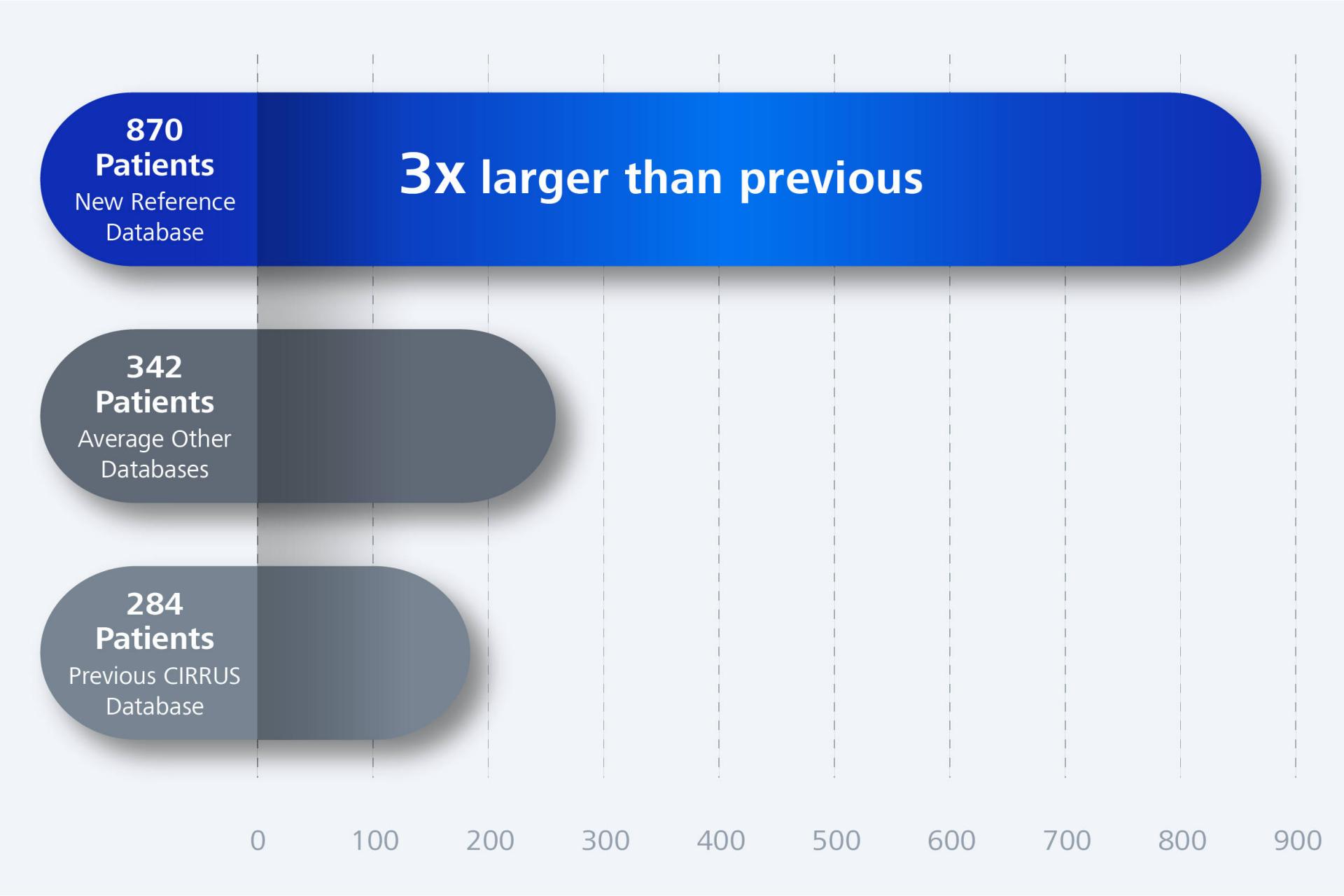
ZEISS CIRRUS expanded reference database - supporting your practice with more data. Average values calculated from publicly available statistics as of April 2024.
Expanded reference database
The continually advancing ZEISS CIRRUS Reference Database now includes 870 patients, more than triple that of previous versions, and with greater diversity, taking into account different optic disc sizes in addition to age. Comparing macular thickness, ganglion cell thickness, optic disc and RNFL measurements to a reference range for healthy eyes 18 to 88+ years, interpolated from quantile regressions using additional statistical models.


Patient-first design
Unique patient-centric platform designed for the futureWith ZEISS CIRRUS, your patient data is never left behind. CIRRUS is the platform that allows seamless transfer of raw, dynamic patient data from previous generations, allowing clinicians to manage their patients with utmost care, across generations.


ZEISS CIRRUS – offering next-level data protection
ZEISS CIRRUS software 11.7: enhanced cybersecurity
New enhanced cybersecurity features are designed to meet ever-evolving compliance and security needs. For the large institution IT requirements of today and tomorrow, ZEISS CIRRUS offers features such as enhanced password security, enterprise-scale security requirements and more.
- Whether at rest or in transit, your CIRRUS data is secure with BitLocker encryption and DICOM Transport Layer Secure (TLS) protocol.
- New InterBase ultra-fast embeddable database, offering top-of-the-line data security and instant disaster recovery. Supports Windows 10 configuration to run in Federal Information Processing Standards (FIPS) mode.
- Share DICOM OP and OPT compressed data with ZEISS FORUM and electronic medical records (EMRs) using JPEG2000(J2K) or JPEG Baseline methods.
- CIRRUS Review Station supports installation on Windows 10, Windows 11, and Windows Server 2012R2, 2016R2, and 2019 operating systems.
 with cystoid macular edema post-cataract surgery")
 with cystoid macular edema post-cataract surgery")
")
")


Discover the advanced imaging capabilities of ZEISS CIRRUS / CLARUS / products through our curated clinical cases. To improve your experience, utilize the filters on the ZEISS multi-modal clinical case library to search by ZEISS product, imaging modality, or condition (available in English only).
Ready to unlock the full potential of your clinic?
ZEISS Retina & Glaucoma WorkflowThe ZEISS CIRRUS 6000 could be just the beginning: Take your workflows to a new level and learn how our products and applications add value beyond the mere sum of their parts.
Downloads


Download the CIRRUS Essentials for Glaucoma
The concise and complete guide for every glaucoma practice

This guide applies across multiple CIRRUS OCT models and generations.


