You are on our international English website. This site features our entire product portfolio worldwide. The products featured may not be available in the US. If you are a citizen from the US, please visit your country website for local information and contacts.
You are on our international English website. This site features our entire product portfolio worldwide. The products featured may not be available in the US. If you are a citizen from the US, please visit your country website for local information and contacts.
The video player is blocked due to your cookie preferences. To change the settings and play the video, please click the button below and consent to use of "Functional" tracking technologies.
Bring your patient’s story to light
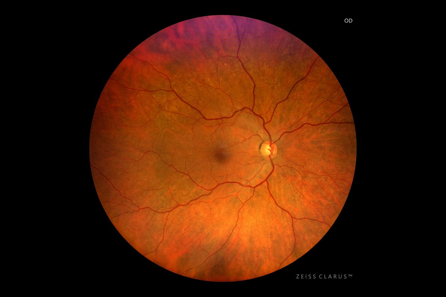
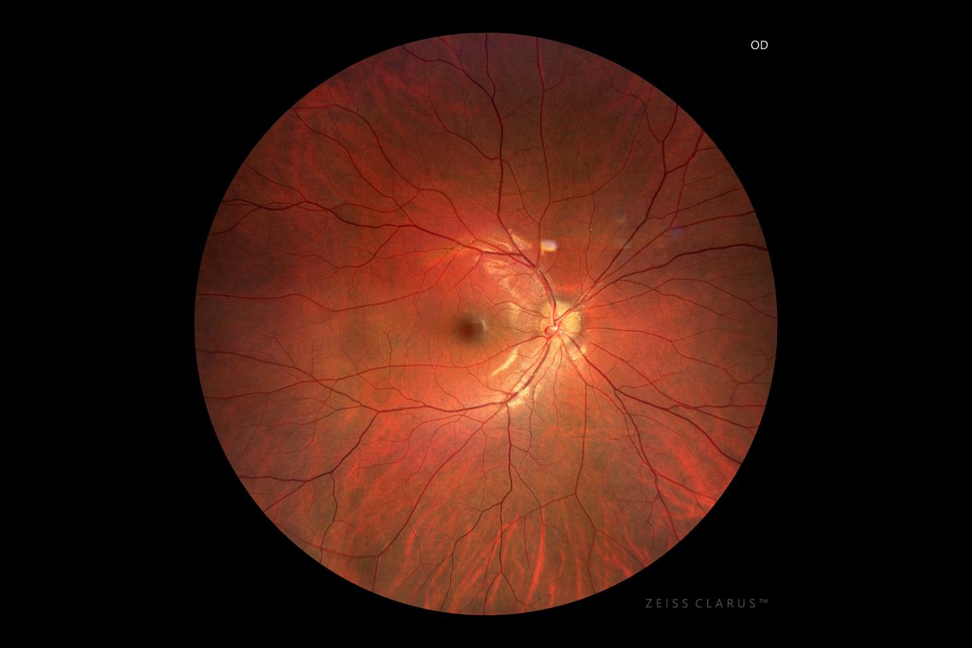
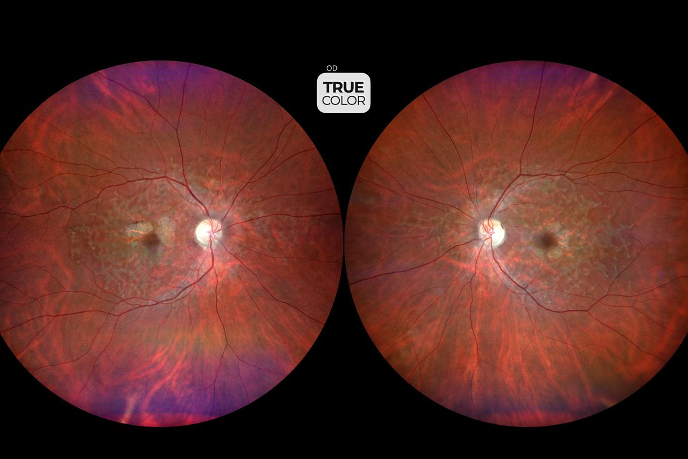
Learn about the case of a 76-year-old, asymptomatic male who presented for a routine eye examination. His medical and ocular histories were unremarkable except for an oral statin for hypercholesteremia.
After being screened with CLARUS, fundus photography revealed a Hollenhorst plaque OD. An urgent carotid duplex showed carotid arteriolar stenosis of right 50-69%, left 15% due to arteriosclerosis. In this instance, a quickly taken true color, widefield image of the retina uncovered a subtle, incidental finding, giving practitioners the opportunity to prevent a potentially life threatening event.
The video player is blocked due to your cookie preferences. To change the settings and play the video, please click the button below and consent to use of "Functional" tracking technologies.
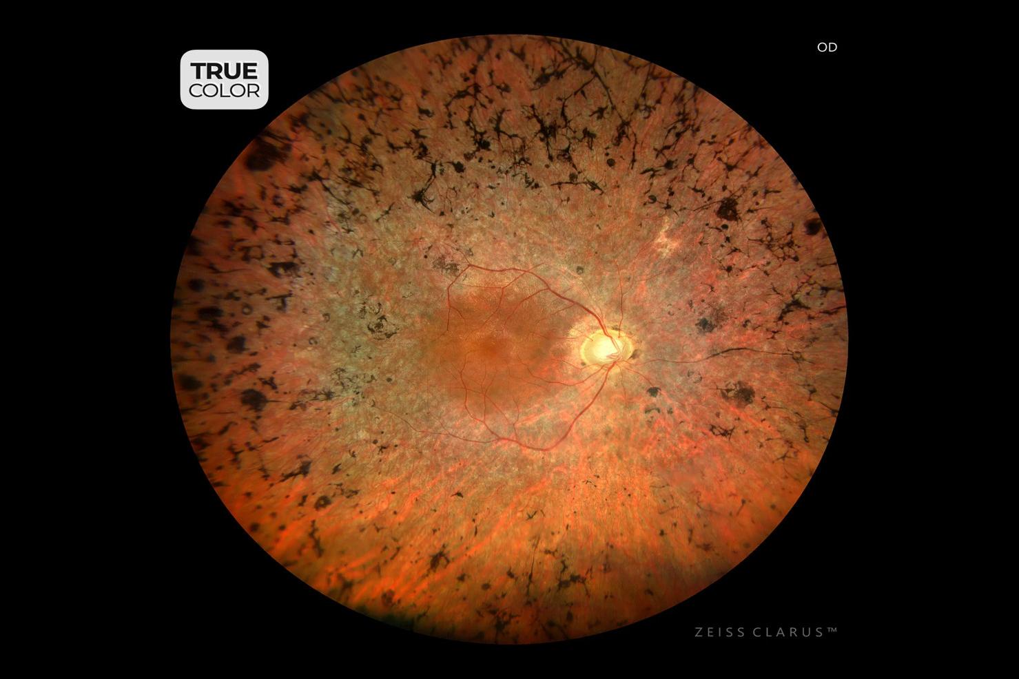
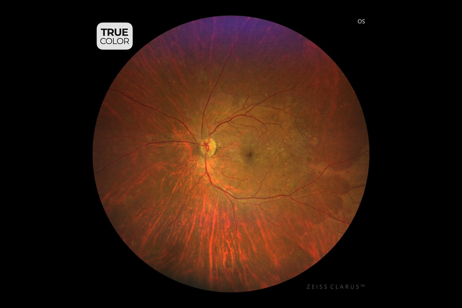
Retinitis Pigmentosa (RP)
Multi-modal retinal analysis (true color, FAF-blue and FAF green) of a 67-year-old male diagnosed with Retinitis pigmentosa 30 years ago.
The widefield, True color fundus image allows the identification of peripheral retinal pigmentary changes such as areas of hypopigmentation and bone-spicule pigments, disuse arteriolar atrophy and waxy disc pallor.
Fundus autofluorescence (both green and blue) are helpful in determining the extension and pattern of RPE atrophy as well as the increased ring of hyper-autofluorescence at the macula.
However, FAF-green excitation wavelength is less absorbed by luteal pigments and provides more details regarding the RPE function at the macular area. Notice that, even presenting posterior capsule opacities in the left eye, the image quality does not compromise the analysis."
Image courtesy of Ricardo Luz Leitão Guerra, MD, Clínica de Olhos Leitão Guerra, Brazil
The video player is blocked due to your cookie preferences. To change the settings and play the video, please click the button below and consent to use of "Functional" tracking technologies.
Branch Retinal Artery Occlusion (BRAO)
Multi-modal retinal analysis (true color, FAF green and Fluorescein angiography) of a 71-year-old male diagnosed with branch retinal artery occlusion.
FIGURE 1) True color fundus image allow the identification of a yellowish cholesterol emboli (Hollenhorst plaque) obstructing the inferno-temporal artery branch and the retinal whitening of the affected area.
FIGURE 2) Fundus autofluorescence presented a marked hyperautofluorescence of the cholesterol emboli.
FIGURE 3, 4 and 5) Fluorescein angiography is extremely helpful determining the extension of the ischemia, as well as the presence of retrograde filling, which is a sign of a better prognosis.
Image courtesy of Ricardo Luz Leitão Guerra, MD, Clínica de Olhos Leitão Guerra, Brazil
The video player is blocked due to your cookie preferences. To change the settings and play the video, please click the button below and consent to use of "Functional" tracking technologies.
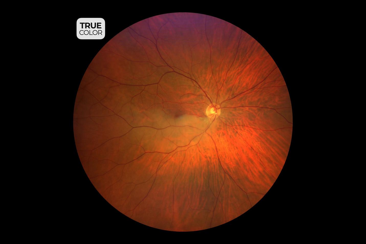
Open-Angle Glaucoma
True color image of a 83-year-old male diagnosed with primary open angle glaucoma. Optic disc analysis shows several signs of glaucoma, such as cup-to-disc ratio, Susanna Streaks (striate pattern of lamina cribrosa openings), nasal displacement of central retinal vessels, notch of the neuroretinal rim and Hoyt sign (localized RNFL loss).
Image courtesy of Ricardo Luz Leitão Guerra, MD, Clínica de Olhos Leitão Guerra, Brazil
The video player is blocked due to your cookie preferences. To change the settings and play the video, please click the button below and consent to use of "Functional" tracking technologies.
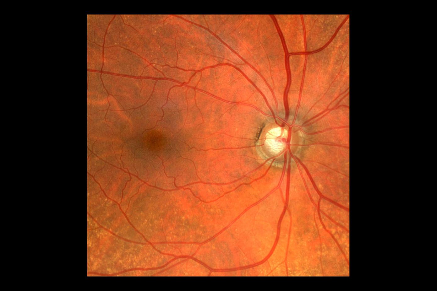
Hypertensive Retinopathy
Fundus imaging of a 61-year-old female diagnosed with arterial hypertension with irregular control. True color image allow the identification of hypertensive retinopathy signs, such as:
engorged venules
copper-wire arterioles and
AV nicking
Image courtesy of Ricardo Luz Leitão Guerra, MD, Clínica de Olhos Leitão Guerra, Brazil
The video player is blocked due to your cookie preferences. To change the settings and play the video, please click the button below and consent to use of "Functional" tracking technologies.
Optic Disc Drusen
Multi-modal retinal analysis (true color, FAF-blue and FAF green) of a 23-year-old male referred for a retina specialist evaluation due to an optic disc abnormality.
In this case, fundus autofluorescence both green and blue were able to identify the presence of an optic disc drusen.
Image courtesy of Ricardo Luz Leitão Guerra, MD, Clínica de Olhos Leitão Guerra, Brazil
The video player is blocked due to your cookie preferences. To change the settings and play the video, please click the button below and consent to use of "Functional" tracking technologies.
Stargardt Disease
Multi-modal retinal analysis (true color and FAF green) of a 51-year-old female diagnosed with Stargadt disease (genetic test positive for ABCA4).
True color images shows areas of RPE atrophy at the macula and multiple flecks at the posterior pole and mid periphery.
Fundus autofluorescence is an extremely helpful tool in this case, enhancing the identification of the RPE atrophy and flecks.
An interesting thing about FAF in Stargardt disease is the preserved RPE area surrounding the optic disc.
Image courtesy of Ricardo Luz Leitão Guerra, MD, Clínica de Olhos Leitão Guerra, Brazil
The video player is blocked due to your cookie preferences. To change the settings and play the video, please click the button below and consent to use of "Functional" tracking technologies.
PDR with IRMA and PRP Treatment
Multi-modal retinal analysis (true color and fluorescein angiography) of a 49-year-old male diagnosed with Proliferative diabetic retinopathy.
True color image shows several microvascular abnormalities, exudates and neovascularization.
Fluorescein angiography enhance the identification of the microvascular abnormalities, such as microaneurysms, IRMA’s, ischemic areas and several neovascularizations.
Treatment was performed by panretinal photocoagulation (PRP) in one session using Visulas Green VITE (multi spot laser).
Image courtesy of Ricardo Luz Leitão Guerra, MD, Clínica de Olhos Leitão Guerra, Brazil
The video player is blocked due to your cookie preferences. To change the settings and play the video, please click the button below and consent to use of "Functional" tracking technologies.
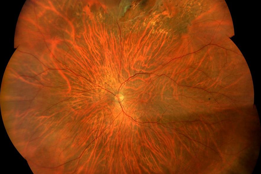
Ultra-widefield (UWF) of Retinal Tear
Ultra wide field (UWF) image (acquired by a montage of six wide field images) presenting a retinal tear partially blocked by laser scars.
This UWF high resolution image allows the identification of the untreated area and is a useful tool for treatment planning and follow up.
Image courtesy of Ricardo Luz Leitão Guerra, MD, Clínica de Olhos Leitão Guerra, Brazil
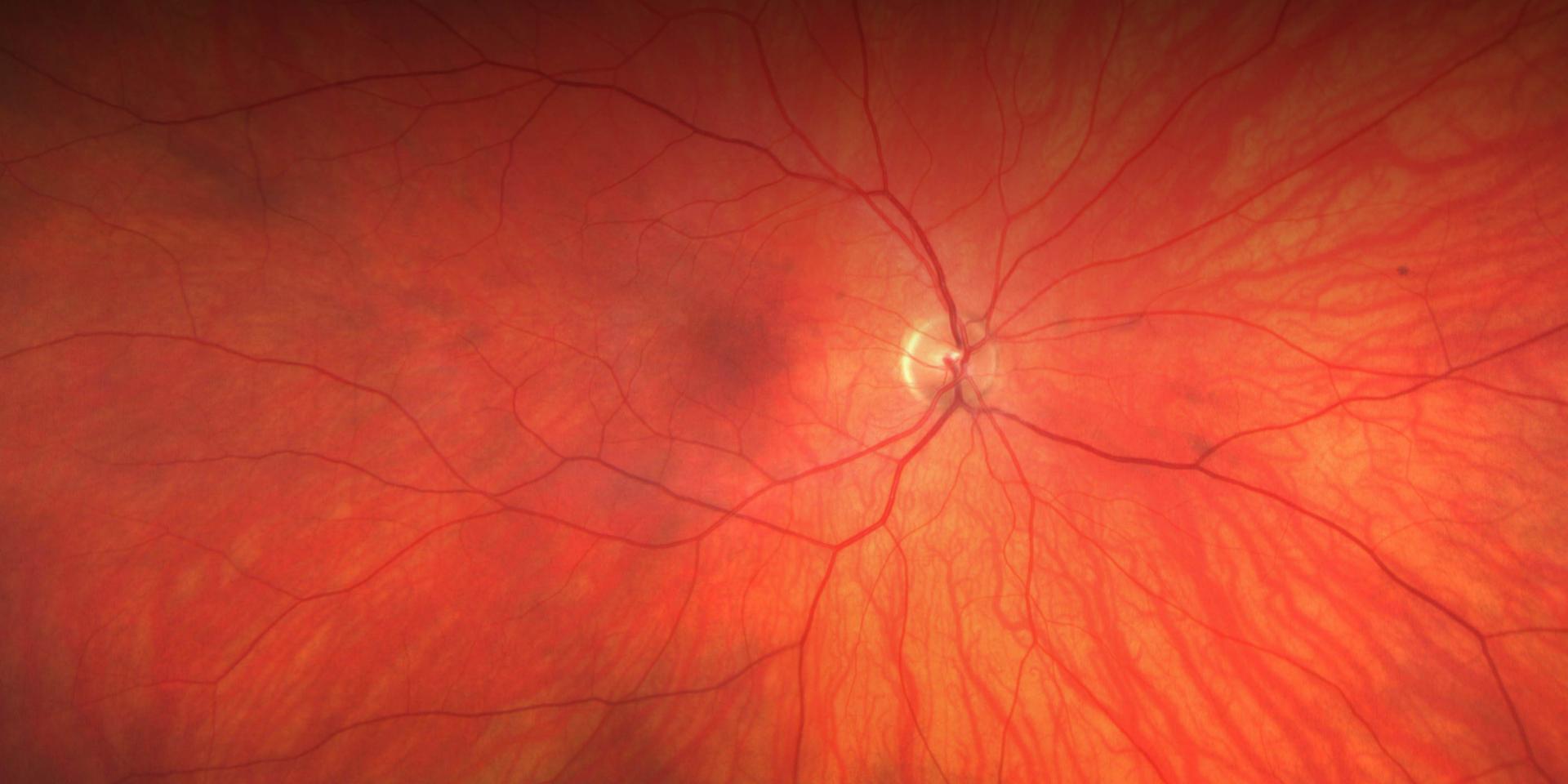
Each high-resolution capture reveals a new chapter of your individual patient’s story. ZEISS CLARUS utilizes a technique called Broad Line Fundus Imaging (BLFI) that is capable of capturing a broader range of autofluorescence generated at the fundus because it illuminates/excites at two wavelength ranges. ZEISS CLARUS illuminates in two wavelength ranges, FAF-Blue (435-500 nm) and FAF-Green (500-585 nm). All fluorophores that absorb light within those ranges will be detected if the fluorophores emit light within the band pass filter range, which is 532-650 nm for FAF-Blue and 630-750 nm for FAF-Green. Consequently, the ZEISS CLARUS image may look different from an image obtained by cSLO, which merely detects light emitted from a smaller range of fluorophores.
See the bigger picture while facilitating the deconstruction of RGB channels for a detailed analysis of each layer of the retina.
BLFI enables the combination of an ultrawidefield
view and a full range of retinal
imaging modes—capturing images that
resemble the coloration of the fundus as
seen during clinical examination.
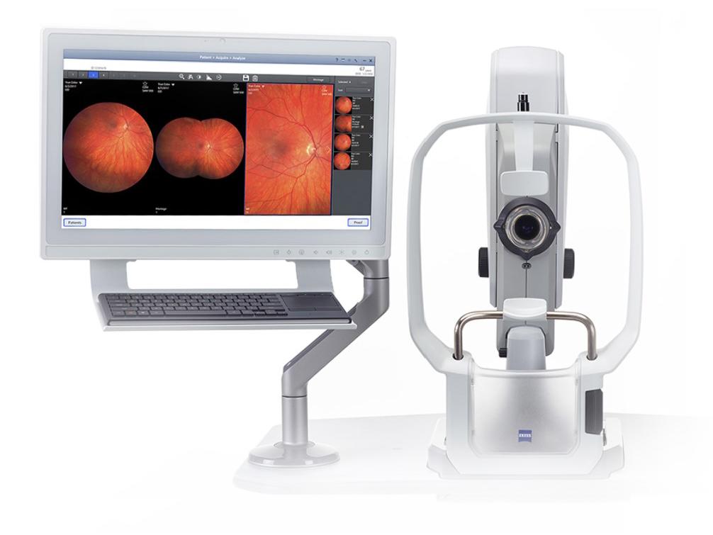
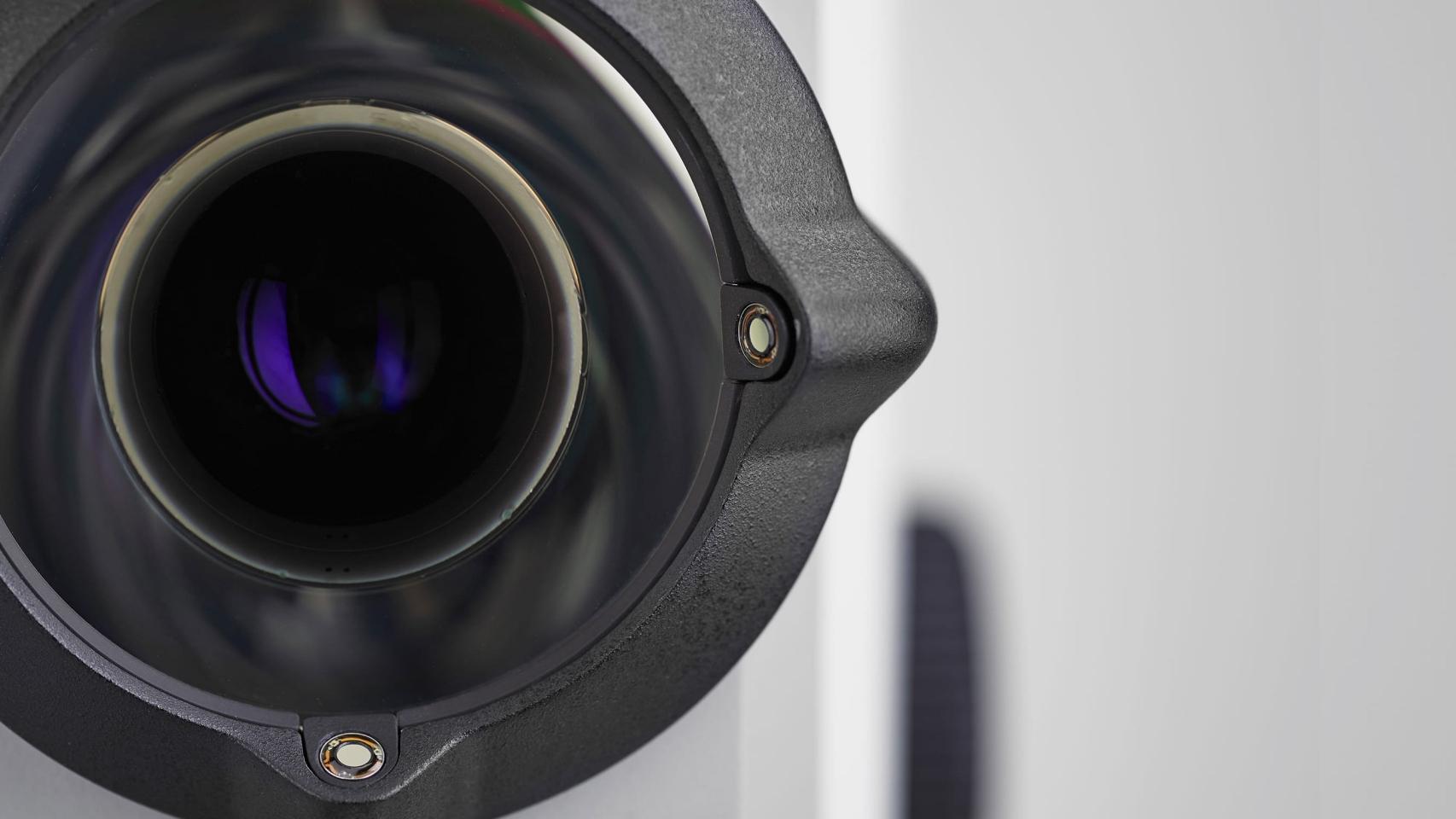
CLARUS 500
Color. Clarity. Comfort.
The fundus imaging system that provides true color and high-resolution across an entire ultra-widefield image.
ZEISS CLARUS 500 at a Glance
True color images aid in diagnosis and documentation
Image in high resolution anywhere in the retina
Create a comfortable, satisfying patient experience that provides images
free of obstructions
The video player is blocked due to your cookie preferences. To change the settings and play the video, please click the button below and consent to use of "Functional" tracking technologies.
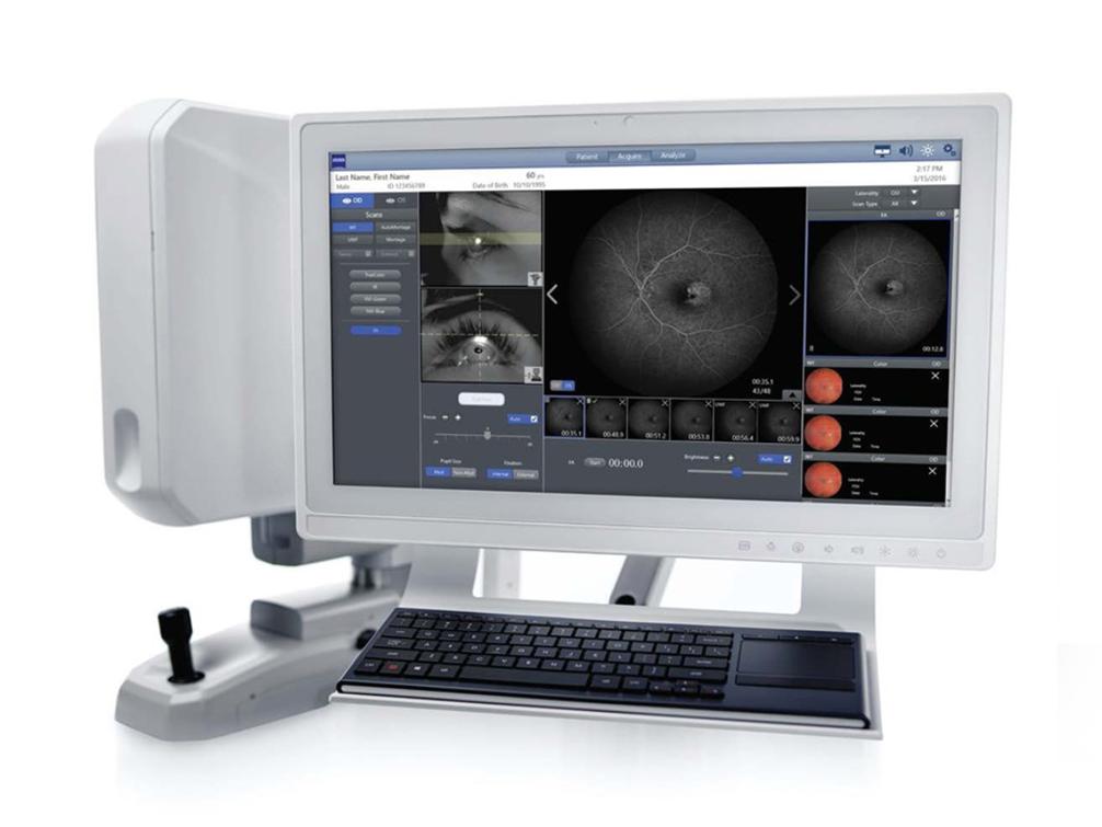
Stronger together with workflows
CLARUS is powerful on its own but unstoppable together with ZEISS Retina and Glaucoma solutions. Connecting ZEISS devices, data and applications enables a seamless integration from office to the OR and back.
Uncover your own patient’s stories with CLARUS
Our commitment to helping elevate the experience of your patients in practice extends beyond fundus imaging.
Schedule a demo
Discover how CLARUS enables you to communicate your patients' ocular health