

ZEISS CLARUS 500
Imaging ultra-wide without compromise.The introduction of widefield retinal imaging has shown us that indications of disease are often located in the far periphery of the retina. CLARUS™ 500 from ZEISS, is an advanced fundus imaging system from ZEISS that provides True Color and high-resolution across an entire ultra-widefield image.
Field of View Comparisons


Advanced Imaging Features
ZEISS CLARUS 500 has several features that allow you to focus on what matters most – your patient. Through patented technology, these features using algorithms will maximize your workflow efficiency and decrease chair time. ETDRS Grid, an overlay of grids used to support clinical studies and patient education.


True color
ZEISS CLARUS 500, the ultra-widefield retinal camera from ZEISS, allows clinicians to use color to their diagnostic advantage. ZEISS CLARUS 500 generates images that closely resemble the coloration of the fundus as seen during clinical examination. Color fundus imaging can aid in the diagnosis and documentation of ocular disease, ensuring confidence when evaluating the optic disc, nevi and lesions where color is important.


True Color
The ZEISS CLARUS ophthalmic camera captures true color images that represent the retina as seen through a direct ophthalmoscope. Utilizing Broad Line Fundus Imaging, the ZEISS CLARUS 500 employs sequential illumination by broad spectrum red, green, and blue LDs, allowing practitioners to achieve a familiar view similar to what they would experience during a direct examination. This advanced technology ensures that the images reflect the true color of the fundus and support differential diagnosis.
Comprehensive imaging modalities for differential diagnosis
ZEISS CLARUS 500 allows clinicians to track subtle changes in pathology over time. In addition to True Color imaging, it also captures high resolution fundus autofluorescence (FAF) images–FAF-Blue and FAF-Green–and Infrared (IR) and external eye images. Along with an intuitive review software, the ultra-high-resolution of this next-generation retinal camera from ZEISS allows clinicians to manage change with confidence.


True color
True color
True Color images can be separated into red, green and blue channel images, enhancing the visual contrast of details in certain layers of the retina. Color accuracy aids in the documentation and diagnosis of ocular disease.


Red channel
Red channel
Red channel images reveal the choroid in more detail. This can be helpful in visualizing choroidal lesions such as nevi or tumors.


Green channel
Green channel
Green channel images provide excellent contrast of the retina, especially of vasculature and hemorrhages.


Blue channel
Blue channel
Blue channel images increase visibility of the anterior retinal layers, allowing easier visualization of the retinal nerve fiber layer and epiretinal membranes.


Powered by Broad Line Technology: Download a free white paper
This white paper explains Broad Line Fundus Imaging – the technology that powers ZEISS CLARUS 500 & 700– and its benefits for capturing fundus autofluorescence (FAF) images on the device.


Exceptional clarity
ZEISS CLARUS 500 captures clear and accurate images from the macula to the far periphery. Early indications of disease can often be subtle and difficult to see through direct observation or low resolution fundus imaging. Leveraging ZEISS optics, ZEISS CLARUS 500 is an ultra-widefield retinal camera that captures a high-resolution image down to 7 microns.
From the macula to the periphery with ONE system
Legacy ultra-widefield imaging systems require doctors to maintain a traditional high-resolution fundus camera for optic nerve and macular disease diagnosis and management. ZEISS CLARUS 500 was the first fundus imaging system to provide True Color and clarity within an ultra-wide field of view, enabling clinicians to capture high-resolution fundus images from macula to the far periphery.
Watch Jean-François Korobelnik, MD, University of Bordeaux, France shares his experience using the ZEISS CLARUS 500.


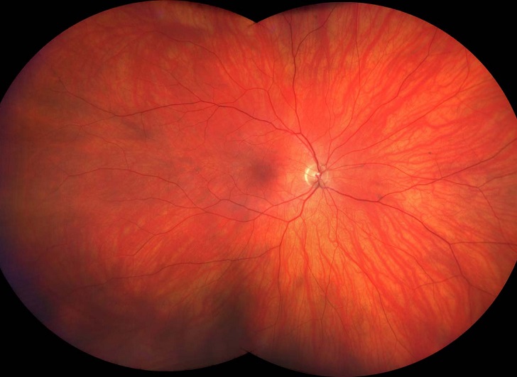

Ultra-widefield image of a healthy eye
Ultra-widefield image of a healthy eye


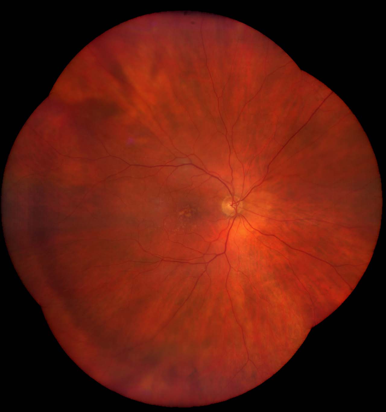

Dry age-related macular degeneration
Dry age-related macular degeneration


FAF-Blue image of geographic atrophy
FAF-Blue image of geographic atrophy


FAF-Green image of dry age-related macular degeneration


Comfort
Simple, stable and intuitive, ZEISS CLARUS 500 is an ultra-widefield retinal camera that has been purposefully designed to optimize each patient's experience.
A satisfying patient experience
By bringing the optics to the patient, ZEISS CLARUS 500 helps create a comfortable, satisfying patient experience that provides images free of obstructions, such as lids and lashes, and requires fewer recaptures.

How the ZEISS CLARUS 500 supports the Glaucoma Workflow
See how Dr. Pilar Casas de Llera from Fernández Casas Oftalmólogos uses the ZEISS CLARUS optic nerve retinography to evaluate patients for Glaucoma. Discover how ZEISS CLARUS is enhancing patient care and transforming ophthalmic practices.


Discover the advanced imaging capabilities of ZEISS CIRRUS / CLARUS / products through our curated clinical cases. To improve your experience, utilize the filters on the ZEISS multi-modal clinical case library to search by ZEISS product, imaging modality, or condition (available in English only).
Discover insights from your peers through testimonials and engaging videos


Clinical Spotlights & Reference Materials


Specifications
ZEISS CLARUS 500Ready to unlock the full potential of your clinic?
ZEISS Retina & Glaucoma WorkflowThe ZEISS CLARUS 500 could be just the beginning: Take your workflows to a new level and learn how our products and applications add value beyond the mere sum of their parts.


