

Expanding insights with ultra-wide imaging
Color. Clarity. Complete.A complete fundus system
Comprehensive in every way to maximize workflow efficiency.CLARUS® 700 from ZEISS provides the ability to identify disease using the most comprehensive suite of fundus imaging modalities to increase efficiency in your workflow. With ZEISS CLARUS, you can capture clear and accurate images from the macula to the far periphery, all with a single instrument.
A comprehensive imaging system
ZEISS CLARUS 700 combines ultra-widefield imaging with true color, excellent clarity and a complete suite of imaging modalities. Now you can manage all fundus imaging modalities without compromising image quality.

True Color with RGB Channel Separation


Red channel: reveals the choroid in more detail. This may be helpful in visualizing choroidal lesions such as nevi or tumors.


Green channel: provides excellent contrast of the retina, especially of vasculature and hemorrhages.


Blue channel: increases visibility of anterior retinal layers, allowing easier visualization of retinal nerve fiber layers defects and epiretinal membranes.


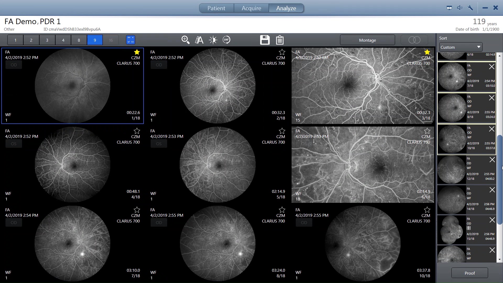
FA of proliferative diabeticretinopathy
")
")
ICGA of Central Serous Chorioretionapathy (CSCR)


FAF-Green image of dry age-related macular degeneration


FAF-Blue image of geographic atrophy


Stereo image pairs can be captured for stereoscopic evaluation of the fundus.


External eye
Introduction to ZEISS CLARUS 700
Andrea Colombo shares the latest innovations for fundus imaging.Unsurpassed color and clarity
Powered by Broadline Fundus Imaging (BLFI)A broadline of illumination from the ZEISS CLARUS 700 scans across the retina. Red, green and blue Light Emitting Diodes (LEDs) sequentially illuminate to generate true color images. The broad bandwidth of LEDs gives a true color rendering. Blue and green LED illumination enables Fundus Autofluorescence (FAF) imaging.
By illuminating only, a narrow strip of the retina at a time, the illumination stays out of the viewing path, keeping haze and fluorescence of the anterior segment out of the retinal image and allowing a clear view of much more of the retina than the annular-ring illumination used in traditional fundus cameras.
Advanced imaging features
ZEISS CLARUS 700 has introduced several new features that allow you to focus on what matters most – your patient. Through patented technology, these features using algorithms will maximize your workflow efficiency and decrease chair time.


Bring your patient’s story to light
There’s a story behind every capture. Each unique to your patient. Explore with us the stories we’ve uncovered with ZEISS retina imaging solutions.
Discover the advanced imaging capabilities of ZEISS products through our curated clinical cases. The ZEISS multi-modal clinical case library showcases the extensive visualization capabilities of our retina imaging solutions. Each case presents a unique pathology illustrating how your peers utilize ZEISS technologies for effective diagnosis, monitoring and treatment of patients.


See what your peers are saying: Watch these testimonials


Clinical and reference materials


Ready to unlock the full potential of your clinic?
ZEISS Retina & Glaucoma WorkflowThe ZEISS CLARUS 700 could be just the beginning: Take your workflows to a new level and learn how our products and applications add value beyond the mere sum of their parts.


