You are on our international English website. This site features our entire product portfolio worldwide. The products featured may not be available in the US. If you are a citizen from the US, please visit your country website for local information and contacts.
You are on our international English website. This site features our entire product portfolio worldwide. The products featured may not be available in the US. If you are a citizen from the US, please visit your country website for local information and contacts.
Root canal location: Pain points and main challenge
The root canal anatomy of teeth can be very variable and missed canals are a major cause of failure of root canal treatment. Commonly missed canals are the MB2 canal in maxillary molars and to a lesser degree, the mid-mesial canal in mandibular molars, buccal canals of lower incisors and second and third canals in premolars.1
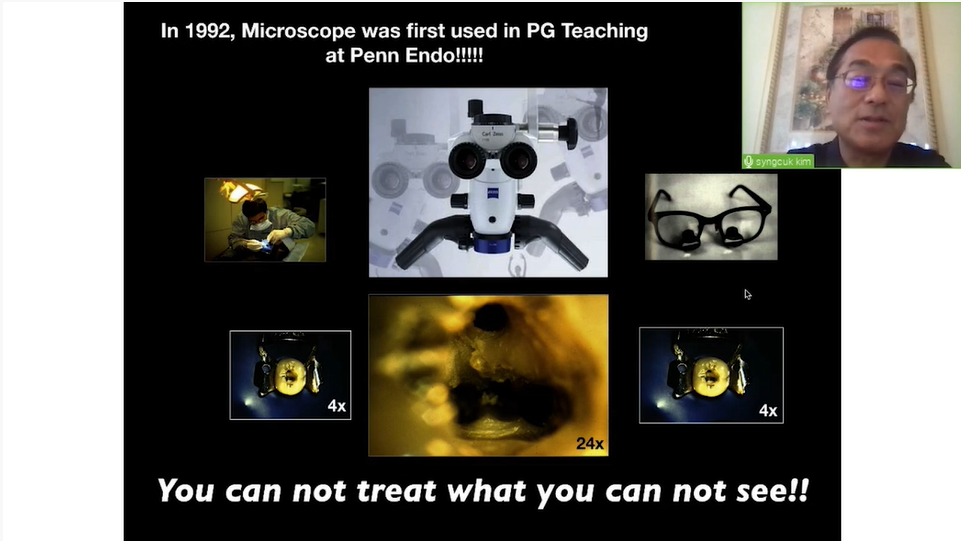
One of the greatest challenges in endodontics is locating canals, especially calcified canals. Canals sclerose from coronal to apical and several millimetres of sclerotic dentine may have to be removed before the canal is found.2
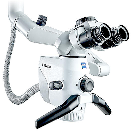
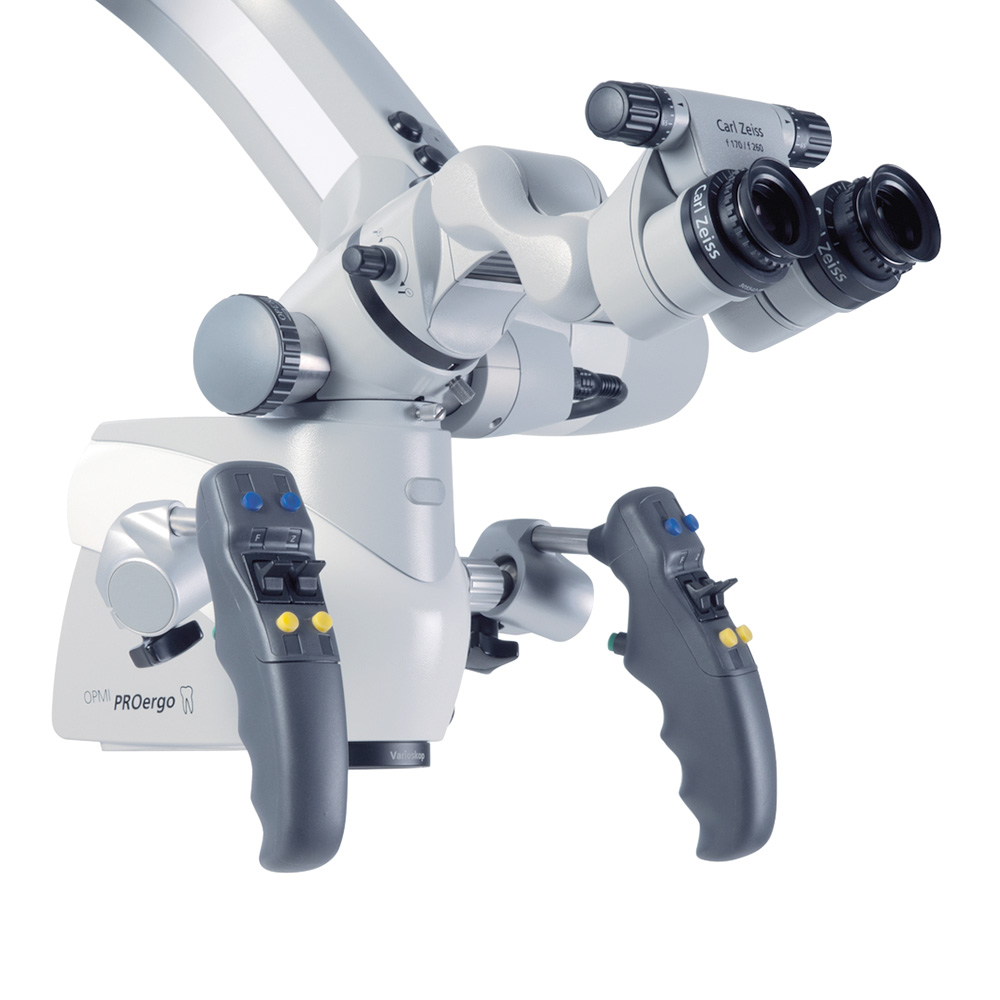
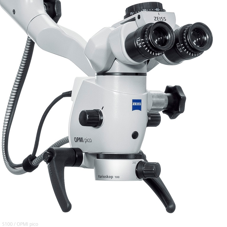
Endodontic procedures are often done with limited visibility that require precision and great attention to detail. Fractions of millimeters may influence the outcome of treatment. […] Use of magnification and illumination is not a fantasy anymore rather it has become a necessity to improve the vision of the operational field.
The OPMI plays a vital role in helping to identify accessory canals at whatever level they may be3
Small canals can be located using the OPMI at higher magnification and with medium to high illumination.
MB2 canals are often sclerosed in the pulp chamber and require small diameter, surgical-length, slow-speed and round burs or ultrasonic tips.
For improved precision, the tip of the bur or ultrasonic instrument should be visible at all times during canal location.
Flexible ultrasonic tips which have been pre-curved are particularly useful in these situations.
Once the canals are located, very small hand files should be used to negotiate canals.