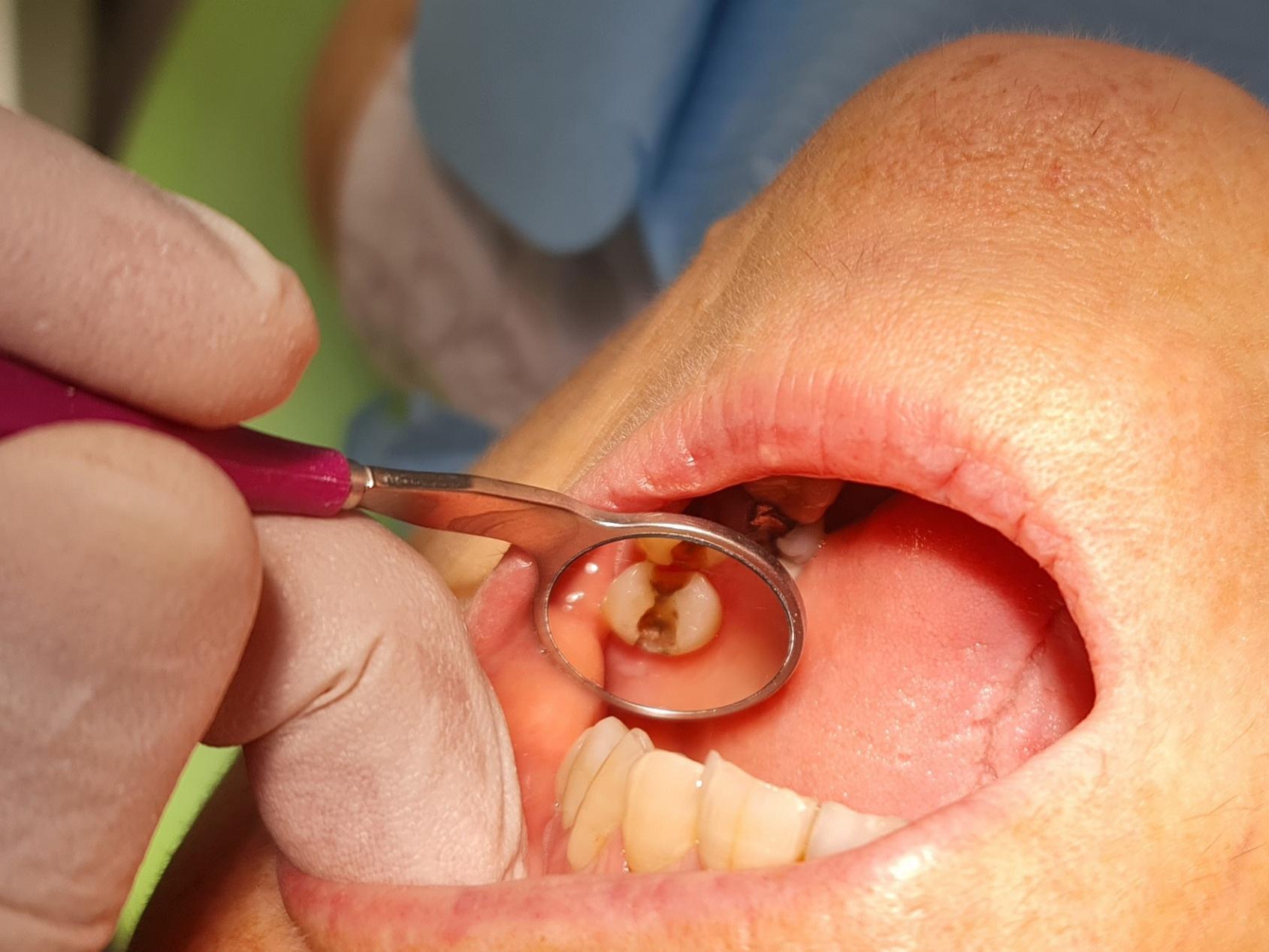
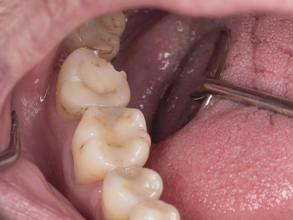
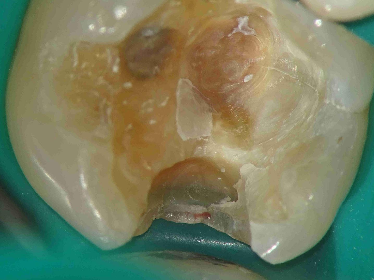
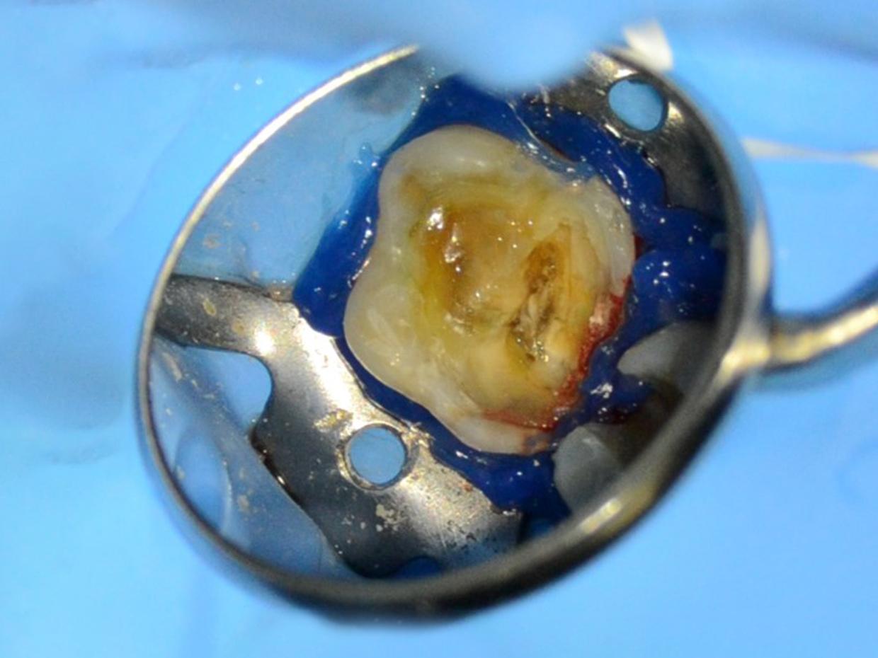
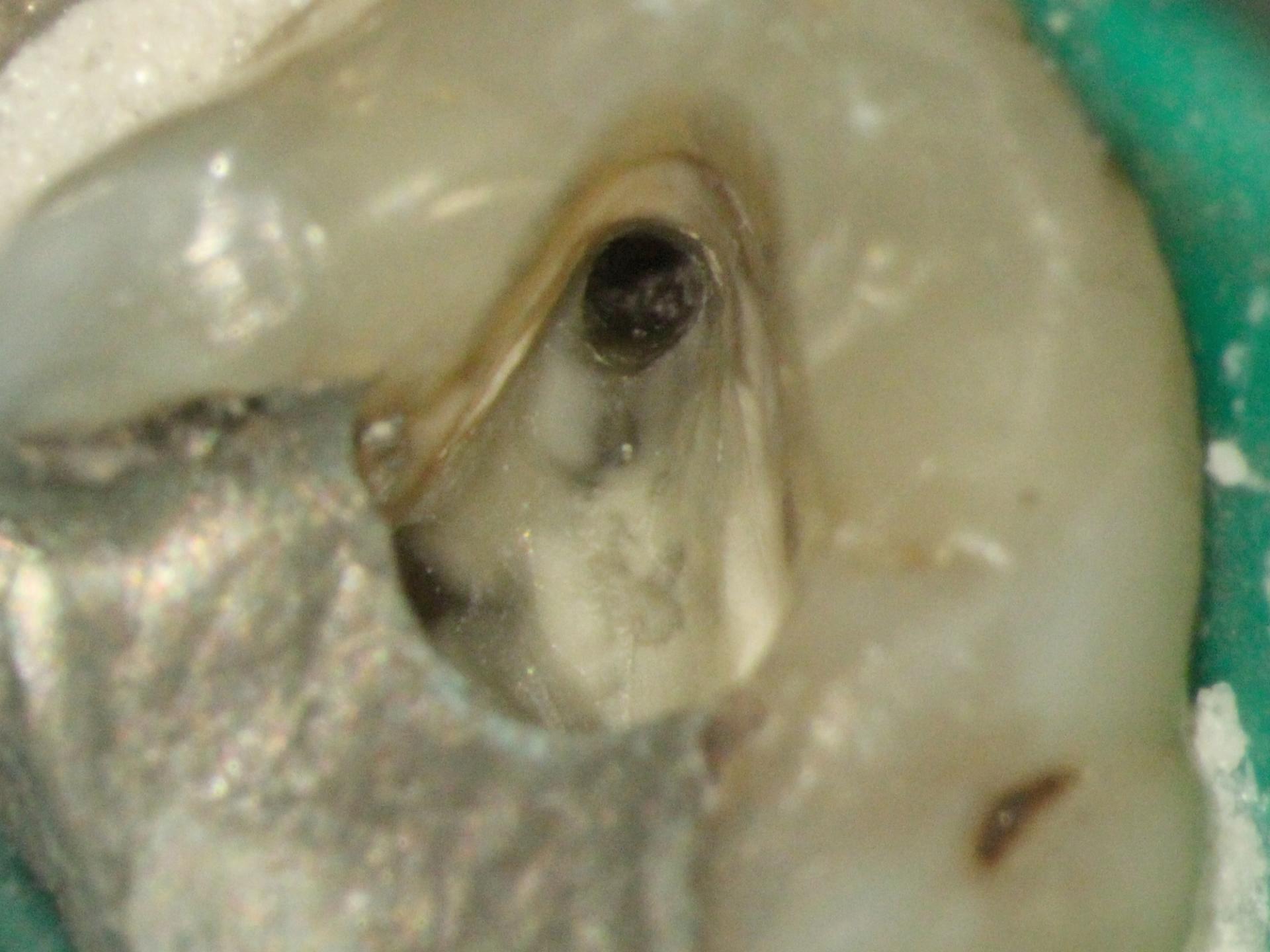
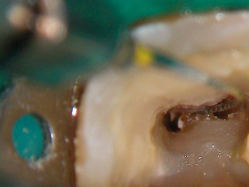
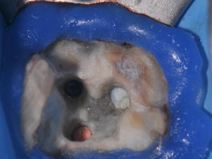
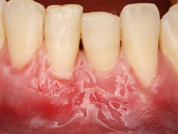
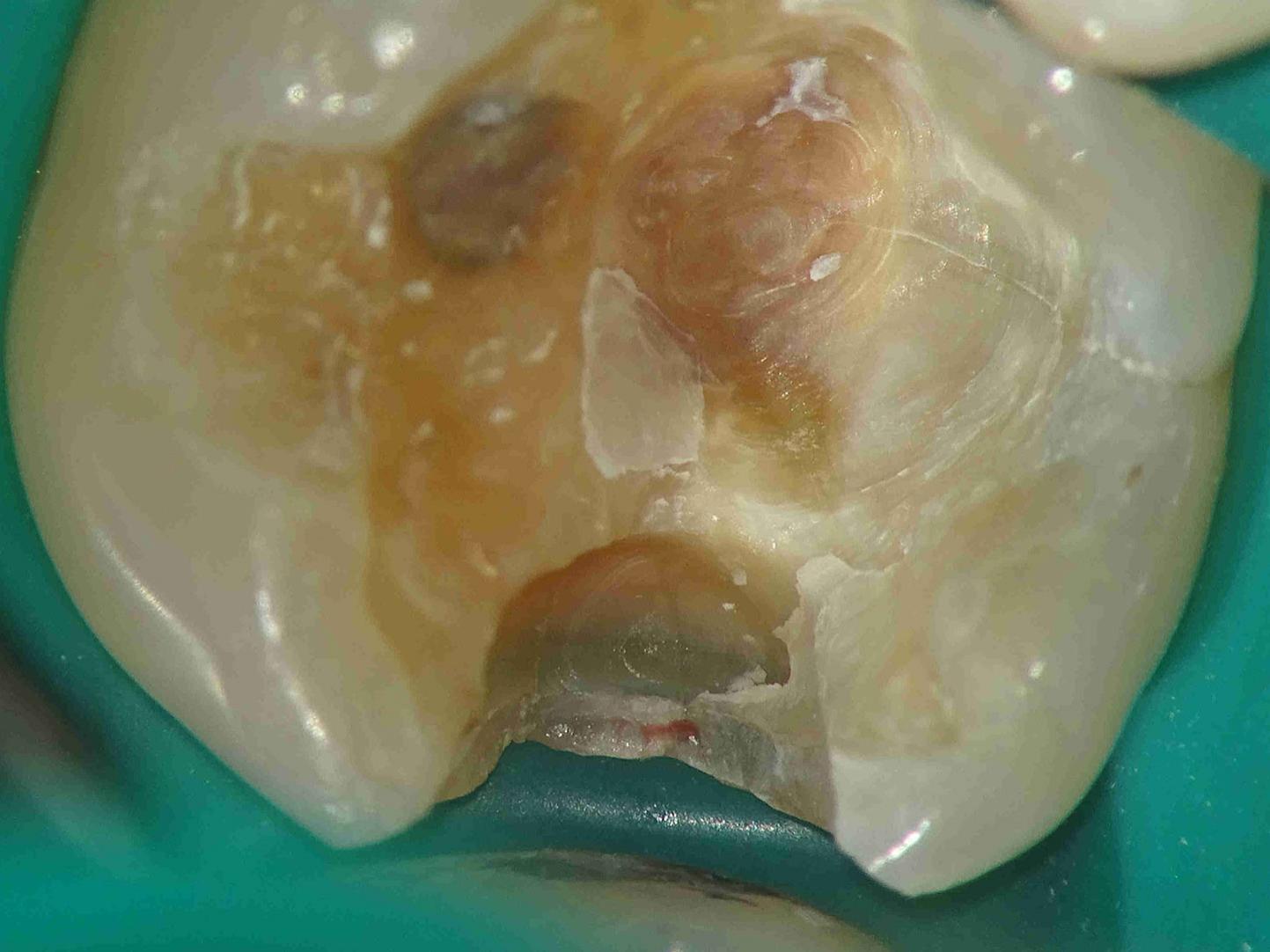
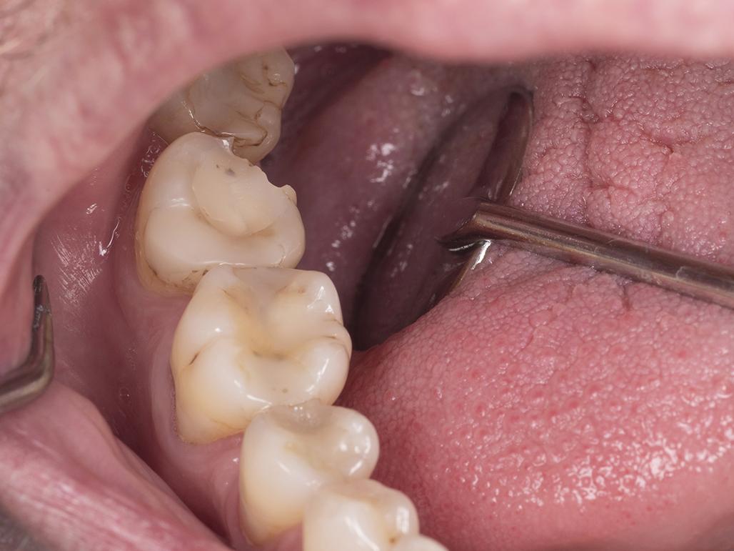
A medical professional wearing a mask is using a surgical microscope to examine a patient seated in an examination chair.
dental visualization

Optimize performance and your workflow

Clinical challenges in dentistry

A healthcare professional using a dental instrument to examine a patient's mouth.

Better diagnosis of fissures and cracks

Visual diagnosis has its limitations because of the resolution of the human eye. This invisible eye gap is a handicap when it comes to visual fissure and crack detection.

An intraoral view showing dental instruments being used inside a patient's mouth, highlighting teeth and soft tissue.

Avoiding over-reduction or under-reduction

Eye limitation has a direct impact on the functional durability of indirect dental restoration. Using optics improves the diagnosis, preparation, and cementation protocol.

Close-up view of a discolored dental crown showcasing signs of wear and possible decay, set against a green background.

Removal of old defective restoration

High magnification through the operative microscope, is essential for enhanced longevity, aesthetics, and functionality in everyday restorative practice. Every removal of dental materials from the tooth diminishes dental structure.
Close-up of a surgical instrument holding a specimen within a sterile blue environment, featured prominently for clinical use by ZEISS Meditec.

Dealing with calcified chambers

Calcified chambers can be difficult to visualize due to their narrow and intricate nature. This can pose a challenge during treatment.

Close-up view of a dental observation featuring a tooth structure and surrounding material.

Locating root canals

The root canal anatomy of teeth can be very variable and missed canals are a major cause of failure of root canal treatment.  

Close-up view of a cross-section of anatomical tissue surrounded by surgical tools on a green drape, likely used for medical procedures associated with ZEISS Meditec.

Non-surgical re-treatment

Endodontic re-treatment is considered to be one of the most challenging procedures in endodontics. In these situations, the OPMI is essential.
Close-up image of a dental impression with a blue material surrounding a detailed structure.

Endodontic instrument removal

Broken endodontic instrument removal includes limited visibility, accessibility within narrow and curved root canals. These challenges are critical.
Close-up image of a human gum line showing signs of inflammation and receding gums around the teeth.

Mucogingival surgery

Executing mucogingival surgery using the naked eye can be challenging. The absence of magnification potentially causing collateral tissue trauma. Magnification tools can overcome these challenges, leading to successful outcomes.

Close-up view of a discolored dental crown showcasing signs of wear and possible decay, set against a green background.

Removal of old defective restoration

High magnification through the operative microscope, is essential for enhanced longevity, aesthetics, and functionality in everyday restorative practice. Every removal of dental materials from the tooth diminishes dental structure.

An intraoral view showing dental instruments being used inside a patient's mouth, highlighting teeth and soft tissue.

Avoiding over-reduction or under-reduction

Visual diagnosis has its limitations because of the resolution of the human eye. This invisible eye gap is a handicap when it comes to visual fissure and crack detection.

A medical professional wearing a mask to examine a patient seated in an examination chair.

Reducing neck and back pain

Over 70% of dentists suffer musculoskeletal disorders due to awkward positioning and poor posture.1

A healthcare professional is applying a treatment to a patient’s arm in a modern clinical setting.

Convincing patients of treatment

Explaining treatments and desired results to patients to ensure a thorough understanding of the benefits of the procedure is sometimes challenging.

Discover the portfolio for dentistry from ZEISS

A medical microscope on a wheeled stand, set against a blue background, showcasing the advanced design of ZEISS Meditec equipment.

ZEISS EXTARO 300

Perfecting your art

EXTARO® 300 from ZEISS provides breakthrough visualization modes that introduce new applications to microdentistry. From more efficient caries detection to a similar tooth restoration workflow, ZEISS EXTARO 300 is poised to revolutionize and differentiate your practice.

A dental professional wearing magnifying glasses and gloves is examining a patient's mouth in a modern clinical setting.

ZEISS EyeMag Medical Loupes

Treating with precision

Over 100 years ago, ZEISS invented the medical loupe. Based on this vast experience, the portfolio has been constantly refined to offer excellent optical performance for first-time and experienced users in dentistry. Today, you can find the right loupe that suits your individual needs in our portfolio of medical loupes.

Two medical professionals in white uniforms and face masks are performing a procedure on a patient under a ZEISS surgical microscope in a bright clinical setting.

ZEISS OPMI pico

The most frequently sold ZEISS surgical microscope worldwide, is the trusted choice among doctors. It makes details and fine structures clearly visible. It enables you to better visualize regions of interest and consistently provide your patients with high-quality examinations and treatments.

A healthcare professional wearing a lab coat is using a ZEISS microscope, focusing intently through the eyepieces.

ZEISS OPMI PROergo

OPMI® PROergo from ZEISS is excellence at your fingertips. It offers a variety of convenient, motorized functions that support effortless handling and mechanical stability. The virtually free-floating system allows smooth and precise movement in an appealing, sleek and compact design.

Curious to learn more about the ergonomic benefits of dental microscopes?

Depth+ Infographic

We created a guidebook on how to position ergonomically with a dental microscope during endodontic and restorative treatments.

Register now and download the guidebook.

Form is loading...

If you want to have more information on data processing at ZEISS please refer to our data privacy notice.


  • 1

    Gopinadh A, et al. Ergonomics and musculoskeletal disorder: as an occupational hazard in dentistry. J Contemp Dent Pract 2013;14:299-303. [Abstract]