You are on our international English website. This site features our entire product portfolio worldwide. The products featured may not be available in the US. If you are a citizen from the US, please visit your country website for local information and contacts.
You are on our international English website. This site features our entire product portfolio worldwide. The products featured may not be available in the US. If you are a citizen from the US, please visit your country website for local information and contacts.
One of the common errors of tooth preparation is over-reduction of tooth structure. This will lead to gingival retraction in the future and visible margins between the tooth and prosthodontic replacement, or it can cause irritation, leading to the gingival inflammation.1 Tooth preparation needs to have specific characteristics to assure retention and resistance to vertical and lateral forces. Overprepping the axial walls and the occlusal surface can jeopardize tooth vitality and lead to the decementation of crowns, bridges, etc.2 The most challenging aspects of tooth preparation include marginal preparation as well as the removal of all of the undermined parts and the roughness of the tooth surface to facilitate better impressions or scans.
The human eye, without the help of magnification, has the ability to distinguish two lines as single ones if they are separated by at least 0.2 mm. Therefore, magnification improves the eye’s ability to notice more minor defects, tooth cracks, and greater details.3
When it comes to achieving functional and aesthetic indirect restoration, there are many factors influencing the final design. Tooth preparation is the hardest and most technically demanding part of fixed prosthodontic therapy nowadays. Marginal preparation placement and type determination are best assessed using microscope magnification.3,4
In conclusion, eye limitation has a direct impact on the functional durability of indirect dental restoration. Using optics improves the diagnosis, preparation, and cementation protocol.
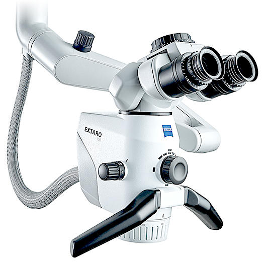
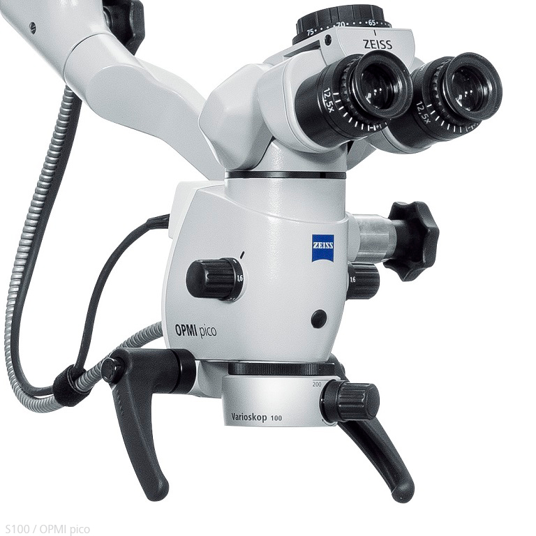
The dental microscope makes all the difference in the preparation and cementation of prosthetic work. By combining the digital protocol and the microscope in minimally invasive dentistry, we achieve wonderful results in terms of precision and speed, without any disadvantages in terms of functionality and aesthetics.
Easier diagnosis and more precise tooth preparation
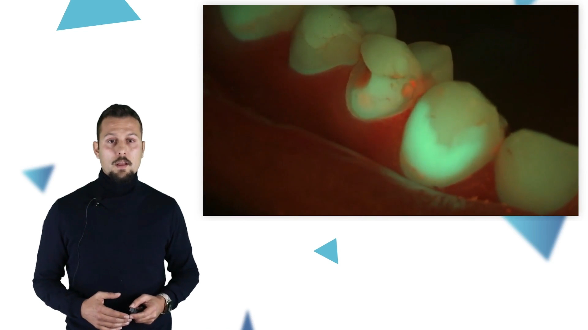
Microscopes offer a wide range of augmented visualization modes nowadays that are useful in restorative and fixed prosthodontic dental therapy, such as fluorescent technologies that show the difference between natural tooth structure, plaque, caries, and restorations, allowing the dentist to make a diagnosis and execute treatment plans more easily.5
Microscopes play a big part in indirect restoration. By relying on magnification with optical devices, we can see the marginal edges and fit of the restoration, assuring good placement. During the cementation phase, a therapist using a microscope can see excess resin cement residue, especially translucent ones, which needs to be removed in order to ensure healthy gums and avoid inflammation.3
By using a dental microscope, a therapist is working in an operating field that is magnified by 20 to 40 times, which allows them to move precisely while using aggressive burs at high speeds, usually in the presence of cooling water and tooth debris, preventing damage to the tooth structure or vitality. Adjacent teeth are also preserved during proximal or crown preparation.4
Improved diagnosis, preparation, and cementation protocol
Image courtesy: Dr. Marko Jakovac, Zagreb, Croatia
Diagnosis without magnification (left) and with high magnification (right)
Image courtesy: Dr. Marko Jakovac, Zagreb, Croatia
Diagnosis without magnification (left) and with high magnification (right)
Image courtesy: Dr. Marko Jakovac, Zagreb, Croatia
Tooth preparation without magnification (left) and with high magnification (right)
Image courtesy: Dr. Marko Jakovac, Zagreb, Croatia
Tooth preparation without magnification (left) and with high magnification (right)
Image courtesy: Dr. Marko Jakovac, Zagreb, Croatia
Try-in and cementation without magnification (left) and with high magnification (right)
Image courtesy: Dr. Marko Jakovac, Zagreb, Croatia
Try-in and cementation without magnification (left) and with high magnification (right)
Deepak, Anupama, Dhanraj Ganapathy, and M. Jeevitha. Prevalence of errors in tooth preparation in patients visiting a university dental hospital – a retrospective study. European Journal of Molecular & Clinical Medicine. 2020; 7(01).
2
Rosella, Daniele, et al. A tooth preparation technique in fixed prosthodontics for students and neophyte dentists. Annali di stomatologia. 2015; 6(3-4):104.
3
Van As, Glenn A. The use of extreme magnification in fixed prosthodontics. Dentistry Today. 2003; 22(6):93-99.
4
Bud, Marius, et al. The advantages of the dental operative microscope in restorative dentistry. Medicine and Pharmacy Reports 2021; 94(1):22.
5
Jakovac M., editors. Protokol. Zagreb: Stega tisak; 2022. 437-446.


 and with high magnification (right)")
 and with high magnification (right)")
 and with high magnification (right)")
 and with high magnification (right)")
 and with high magnification (right)")
 and with high magnification (right)")
 and with high magnification (right)")
 and with high magnification (right)")
 and with high magnification (right)")
 and with high magnification (right)")
 and with high magnification (right)")
 and with high magnification (right)")
 and with high magnification (right)")
 and with high magnification (right)")