You are on our international English website. This site features our entire product portfolio worldwide. The products featured may not be available in the US. If you are a citizen from the US, please visit your country website for local information and contacts.
You are on our international English website. This site features our entire product portfolio worldwide. The products featured may not be available in the US. If you are a citizen from the US, please visit your country website for local information and contacts.
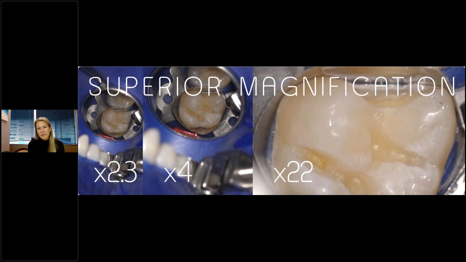
Achieve exceptional dental work with high magnification
When replacing an old dental restoration, paying attention to small details becomes crucial for achieving optimal results1. For example, even if one believes they have effectively removed all previous restorative material and caries, a thorough examination with the naked eye (Fig. 1) might not uncover everything2. However, a more in-depth assessment using the OPMI reveals the need for further work. Furthermore, when employing the selective etch bonding technique, using self-etching adhesives like FL bond II from Shofu, it is vital to exclusively condition the enamel with phosphoric acid, avoiding the dentin3. This becomes notably challenging without proper magnification, particularly along the gingival wall (Fig. 2). Additionally, determining whether a proximal matrix has been correctly placed often requires high magnification4,5, Sources: (Fig. 4, Fig.5). These examples highlight the significance of minor details in achieving exceptional dental work with the OPMI.
For 24 years, I've used a microscope in my dental practice, enhancing precision, predictability, and ergonomics in all treatments. Increasingly, I'm certain that high magnification can benefit all dental fields. Just integrate it into your practice persistently, and you'll be amazed by the results.
Precision beyond the scope of the bare eye
Achieving optimal results with modern restorative techniques requires precision beyond the naked eye's capabilities6 High magnification, particularly through the operative microscope, is essential for enhanced longevity, aesthetics, and functionality in everyday restorative practice. This is especially important during old restoration replacements7. Every removal of dental materials from the tooth diminishes dental structure, underscoring the need for precision to avert premature re-replacement2. High magnification improves care quality, enabling amplification, versatile image resolution, optimal illumination, and the best ergonomic posture8,9. Microscopes crucially enhance visibility for accurate diagnosis, material and caries removal, tooth preparation, matrix and composite placement, and finishing of restorative procedures, always preserving most of the sound tooth structure8,9,10
Restoration with magnification
Image courtesy: José Roberto Moura Junior, Taubate, Brazil
Fig. 0 - Image of the old restoration on the first left lower molar to be replaced through the naked eye.
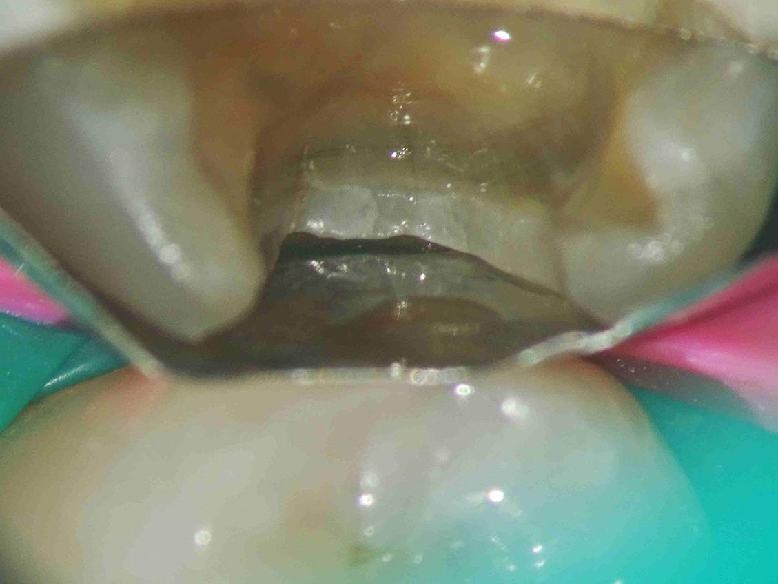
Fig. 1 - Image of the cavity after first attempt of removing old restoration and caries with the naked eye.
Fig. 2 - Image of the cavity after first attempt of removing old restoration and caries with 10X magnification. We can still see old restorative material, caries and a crack.
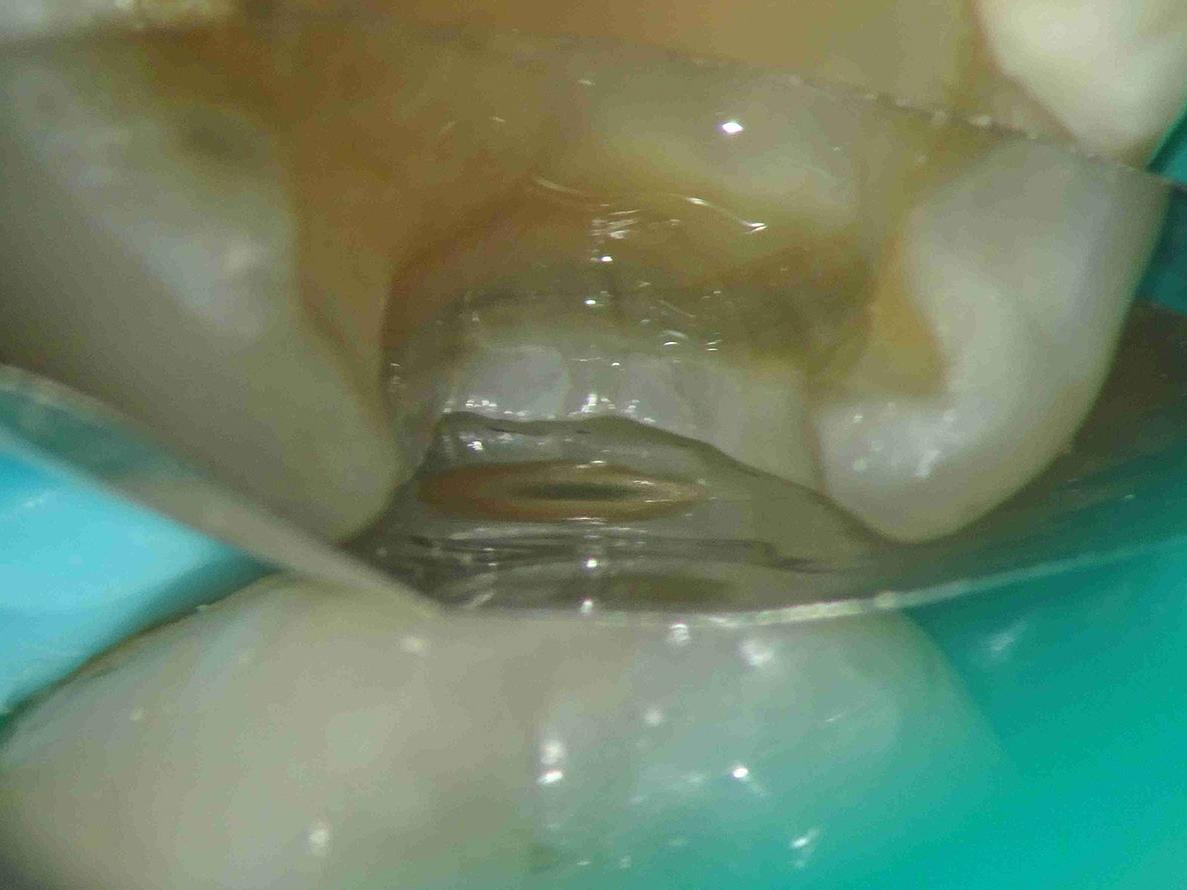
Fig. 3 - Enamel conditioning with phosphoric acid in the proximal box without touching dentin with 10X magnification.
Fig. 4 - View of a sectional matrix not properly placed at the gingival margin. 12X magnification.
Fig. 5 - View of a sectional matrix correctly adapted at the gingival margin. 12X magnification.