You are on our international English website. This site features our entire product portfolio worldwide. The products featured may not be available in the US. If you are a citizen from the US, please visit your country website for local information and contacts.
You are on our international English website. This site features our entire product portfolio worldwide. The products featured may not be available in the US. If you are a citizen from the US, please visit your country website for local information and contacts.
Executing mucogingival surgery, which corrects soft tissue defects around teeth, using the naked eye can be challenging. This approach lacks precision and control, vital for aesthetically and functionally successful outcomes. Without magnifying tools, visualization is limited, making it difficult to distinguish minor tissue variations, crucial for graft placement and suturing1.
Moreover, the absence of magnification negatively affects trimming, shaping, and suturing accuracy, potentially causing collateral tissue trauma. Precise measurements of defect dimensions are essential for successful surgery. The naked eye, however, often fails to provide these accurate measurements, compromising graft sizing, positioning, aesthetics, and function2.
Another concern is hemostasis control — inadequate visualization could lead to postoperative bleeding or jeopardize graft blood supply. Aesthetic results demand a nuanced understanding of gingival architecture, precise incisions, and gentle tissue handling. The naked eye, however, may overlook these subtleties, resulting in aesthetic failures like color mismatch, scarring, or uneven gingival margins3.
Furthermore, insufficient visualization could lengthen surgical procedures due to time spent on adjustments and corrections, increasing patient discomfort and affecting the success rate. Inaccuracies and imprecisions could also hamper the biological healing process, leading to slower healing, infection risks, or graft failure. Hence, incorporating appropriate magnification tools can overcome these challenges, leading to successful outcomes 4.
As a periodontist, the ZEISS microscope has revolutionized my practice. Its enhanced visualization and precision have significantly improved my surgical outcomes, especially in complex mucogingival procedures. It's a game-changer, enabling better patient care and more predictable esthetic results. I couldn't imagine performing periodontal treatment without it.
Revolution of mucogingival surgery
Dental microscopes have revolutionized mucogingival surgery5, a delicate dental procedure:
Enhanced vision: Dental microscopes magnify and illuminate the area, offering a detailed view. This is vital in detailed tasks such as the tunneling technique, where precise flap elevation is crucial to avoid tissue damage.
Microsurgical tools: Tailored for magnified work, these tools combined with microscopes allow for precise tissue handling. The result is reduced trauma and faster, more predictable healing6.
Graft preparation: Preserving Shape & Volume: Magnification allows for cautious shaping of grafts, maintaining their vital components, critical for aesthetic and functional success. Refined tissue selection: Microscopes enable the selective removal of unnecessary epithelium and submucosa, optimizing graft adaptability.
Graft positioning: Enhanced visualization ensures grafts bond closely with recipient sites, fostering better blood flow and healing.
Reduced post-op issues: The precision provided by the microscope and tools minimizes tissue trauma, reducing pain and post-op complications7,8.
In summary, dental microscopes amplify clinicians' abilities in mucogingival surgeries. The fusion of detailed vision with specialized tools offers unmatched precision, optimizing outcomes and patient experiences.
Mucogingival surgery – step by step
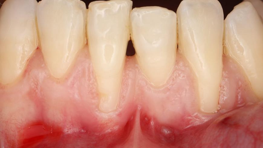
Delicate flap elevation (tunneling)
Delicate flap elevation (tunneling)
Delicate flap elevation (tunneling)
Before meticulous graft preparation
Before meticulous graft preparation
Before meticulous graft preparation
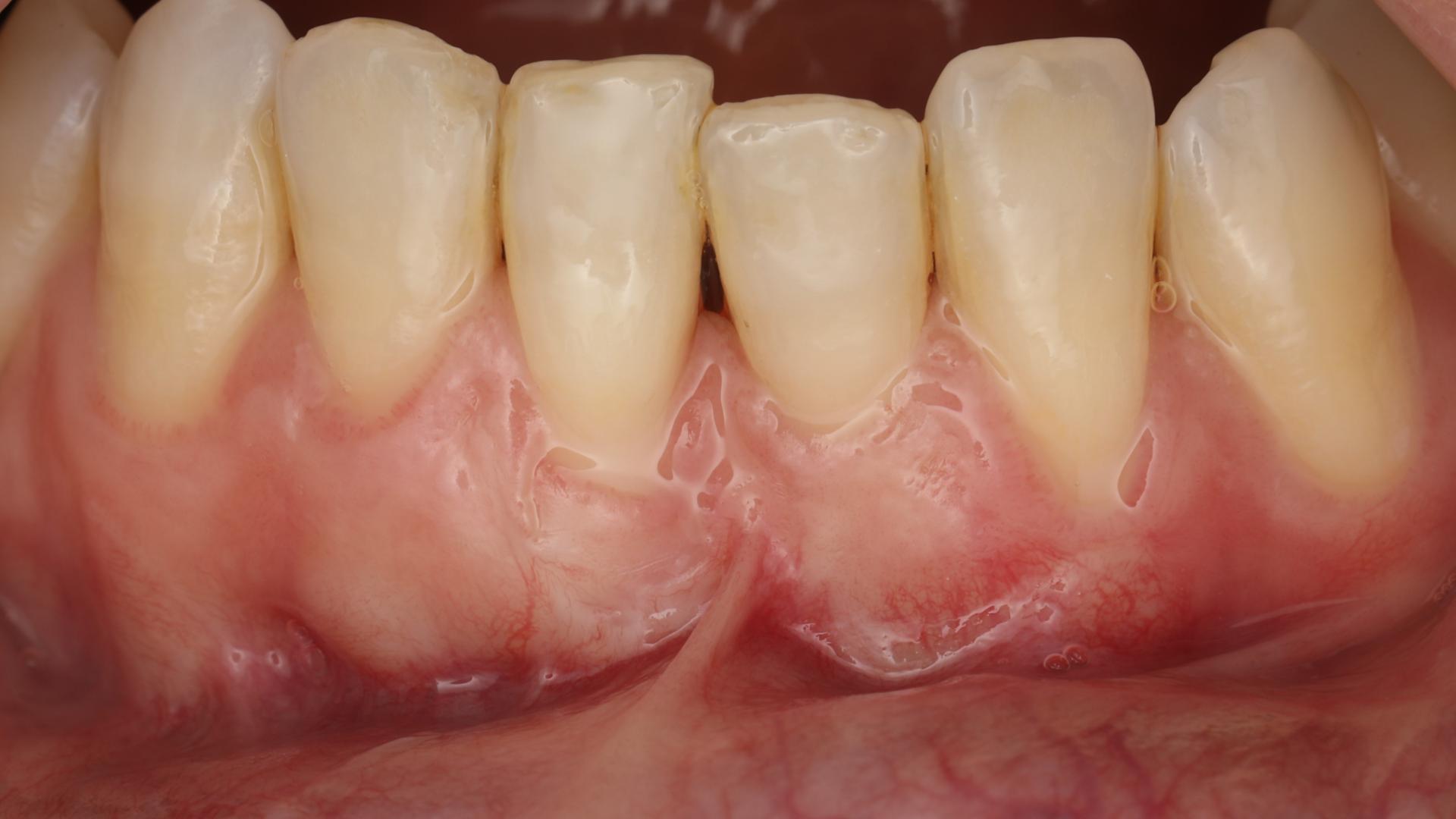
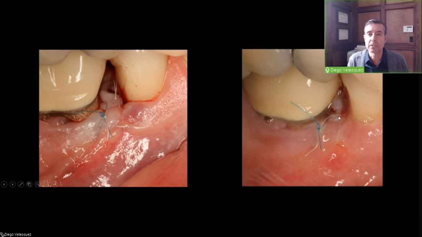
After meticulous graft preparation
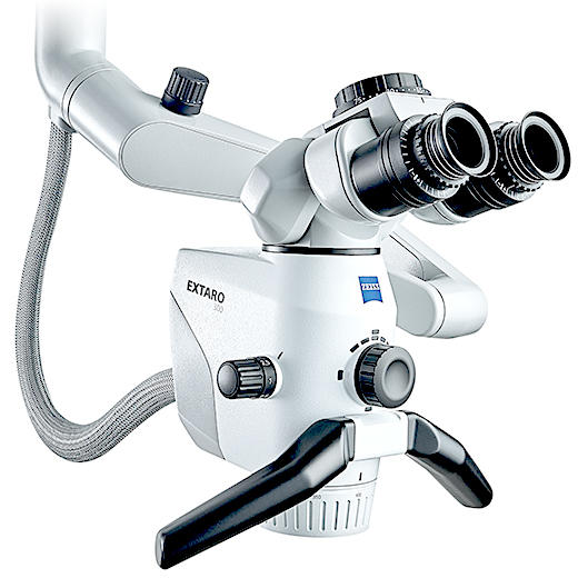
After meticulous graft preparation, photo captured with x10 magnification of ZEISS EXTARO 300
After meticulous graft preparation, photo captured with x10 magnification of ZEISS EXTARO 300
Ma L, Fei B. Comprehensive review of surgical microscopes: technology development and medical applications. J Biomed Opt. 2021 Jan;26(1):010901.
2
Cairo, F., Rotundo, R., Miller Jr, P. D., & Pini Prato, G. P. (2009). Root coverage esthetic score: a system to evaluate the esthetic outcome of the treatment of gingival recession through evaluation of clinical cases. Journal of periodontology, 80(4), 705-710.
3
Griffin, T. J., Cheung, W. S., Zavras, A. I., & Damoulis, P. D. (2006). Postoperative complications following gingival augmentation procedures. Journal of periodontology, 77(12), 2070–2079.
4
Sultan, N., Jafri, Z., Sawai, M., & Bhardwaj, A. (2020). Minimally invasive periodontal therapy. Journal of oral biology and craniofacial research, 10(2), 161–165.
5
Belcher J. M. (2001). A perspective on periodontal microsurgery. The International journal of periodontics & restorative dentistry, 21(2), 191–196.
6
Jain, R., Kudva, P., & Kumar, R. (2014). Periodontal microsurgery-magnifying facts, maximizing results. J Adv Med Dent Sci Res, 2, 24-34.
7
Prato, G. P., Clauser, C., & Cortellini, P. (1995). Periodontal plastic and mucogingival surgery. Periodontology 2000, 9(1), 90-105.
8
Zuhr, O., & Hürzeler, M. (2015). Plastic-Esthetic Periodontal and Implant Surgery: A Microsurgical Approach. Quintessence Publishing.


")
")


")
")
")
")