You are on our international English website. This site features our entire product portfolio worldwide. The products featured may not be available in the US. If you are a citizen from the US, please visit your country website for local information and contacts.
You are on our international English website. This site features our entire product portfolio worldwide. The products featured may not be available in the US. If you are a citizen from the US, please visit your country website for local information and contacts.
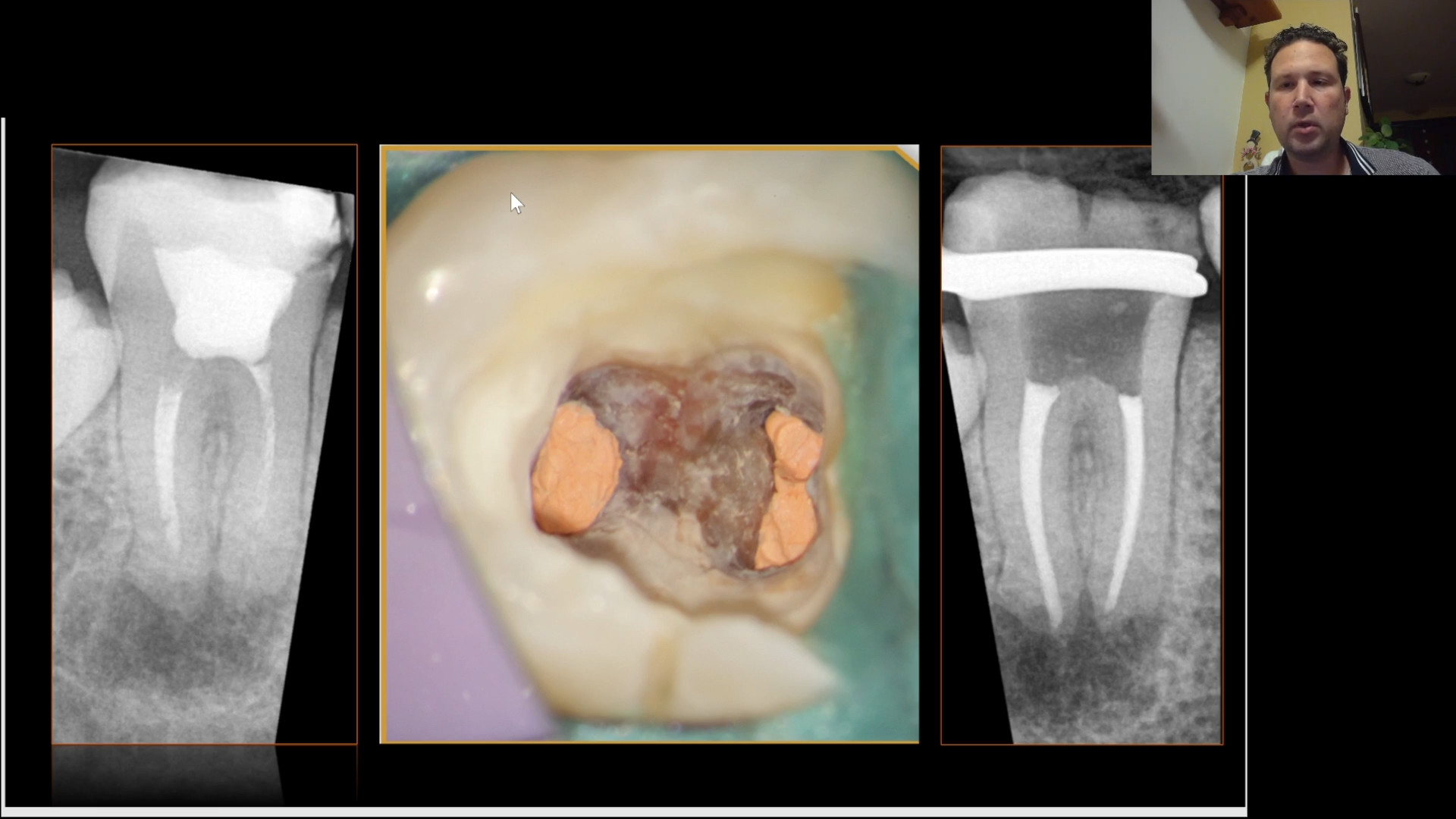
Demonstration of access without the use of magnification where we can observe how difficult it is to see the pulp chamber and the pulp stone found there.
Calcified chambers: Pain points and main challenges
In endodontics, calcified chambers can pose a challenge during treatment.1,2
Calcified chambers can be difficult to visualize due to their narrow and intricate nature. The limited visibility makes it challenging to accurately locate and navigate the canals within the chamber, increasing the risk of missed canals or incomplete treatment.2
This type of chamber may have constricted or tortuous canals, making it difficult to gain access and navigate the root canal system. The narrow spaces and calcified obstructions make it challenging to effectively clean, shape, and disinfect the canals.1,3
Dealing with calcified chambers can prolong treatment time and increase the complexity of the procedure. Dentists may need to employ various techniques and instruments to overcome the challenges posed by calcifications, which can require additional expertise and skill.2
To address these challenges, dentists may utilize advanced tools and techniques such as microscopes, ultrasonic instruments, rotary files, and magnification aids to enhance visualization, improve access, and increase precision. Additionally, continuing education and keeping up to date with the latest advancements in endodontics can help dentists effectively manage calcified chambers and achieve favorable treatment outcomes.1,2,4
The use of the microscope in my professional life has been of great help because I have been able to solve complex cases that on many occasions, without its help, could have taken me more than 3 appointments. In addition to this, magnification also allows me to perform my work in my endodontic practice without developing musculoskeletal problems. I opted to start using magnification in my endodontic practice in 2009, and it has been a great support in my day-to-day work ever since.
When dealing with calcified chambers in endodontics microscopes provide several clinical solutions
Improved chamber localization: The microscope’s high magnification and illumination allow the calcified chamber to be precisely located. The enhanced visualization helps identify subtle anatomical details, making it easier to pinpoint the exact location of the chamber within the tooth.5,7 Accurate canal negotiation: The microscope's magnification enables improved visualization of narrow and tortuous canals within the calcified chamber. This allows the endodontist to negotiate the canals more accurately, reducing the risk of missed canals or procedural errors.6,7 Enhanced instrumentation: The precise focus and fine adjustments of the microscope enable endodontists to perform intricate instrumentation within the calcified chamber. This includes accessing and shaping canals, removing calcifications, and preparing the root canal system for disinfection and obturation.5,7,8 Thorough cleaning and disinfection: The magnification provided by the microscope permits a detailed view of the root canal walls and intricate anatomical complexities within the calcified chamber. This ensures thorough cleaning and disinfection, as the endodontist can effectively remove debris, infected tissue, and bacteria from the entire canal system.7,8 Reduced risk of perforation: The microscope's high magnification helps the endodontist accurately assess the thickness of dentinal walls within the calcified chamber. This reduces the risk of perforation during instrumentation, as the operator can precisely navigate the canals without damaging the surrounding tooth structure.6,7,8 Precise obturation: The microscope enables precise evaluation and placement of the obturation material within the calcified chamber. This ensures optimal sealing of the root canal system, reducing the chances of post-treatment complications and promoting long-term success.7,8
With the help of the ultrasonic tips, we begin to eliminate the pulp stone starting from the entrance of the previously identified canals using 12% Ethylenediaminetetraacetic acid (EDTA) to remove the inorganic tissue and to be able to adequately observe the color changes of the pulp calculus and the dentin of the pulp chamber.
Dr. Cynthia Mercado Velazquez, Mexico City
Opening access to identify the calcified chamber.
Dr. Cynthia Mercado Velazquez, Mexico City
Identifying the entrance of the canals using ultrasonic tips.
The entrance of the mesiobuccal (MB) and distobuccal (DB) ducts is observed at the different magnifications 0.4x and 1.0x.
Dr. Cynthia Mercado Velazquez, Mexico City
Field of view under lower magnification (0.4x).
Dr. Cynthia Mercado Velazquez, Mexico City
Field of view under higher magnification (1.0x).
The pulp stones are observed with a grayish color compared to the yellowish walls of the pulp chamber. In this way, by distinguishing changes in color in the chamber, we can learn to safely eliminate pulp stones adhering to the chamber. Without the help of magnification, it would be very difficult to discern these changes, and EDTA would have to be used.
Dr. Cynthia Mercado Velazquez, Mexico City
Identifying the MB and DB canals through the calcified chamber under 1.6x magnification.
Subsequently, after eliminating part of the pulp stone located at the entrance of the MB canal, with the help of the ultrasonic tip we can observe the floor of the pulp chamber with the presence of pulp tissue. If observed at 0.4x and 1.0x magnification, we can see that the different structures located in this area are adequately discernible at 1.0x magnification.
Dr. Cynthia Mercado Velazquez, Mexico City
Identifying the MB and DB canals through the calcified chamber under 0.4x magnification.
Dr. Cynthia Mercado Velazquez, Mexico City
Identifying the MB and DB canals through the calcified chamber under 1.0x magnification.
Comparison of two types of 1.6x and 1.0x magnification respectively, where more details of the floor can be observed at 1.6x magnification than the 1.0x magnification.
Dr. Cynthia Mercado Velazquez, Mexico City
Identifying the MB 1 under 1.6x magnification.
Dr. Cynthia Mercado Velazquez, Mexico City
Indentifying the MB 1 under 1.0x magnification.
Following the path where the floor of the pulp chamber was already located, the elimination of the pulp stone continues little by little until all the molar canals are located.
Dr. Cynthia Mercado Velazquez, Mexico City
Identifying the 4 MB canals completely after removing the calcification under 0.6 magnification.


.")
.")


.")
.")