You are on our international English website. This site features our entire product portfolio worldwide. The products featured may not be available in the US. If you are a citizen from the US, please visit your country website for local information and contacts.
You are on our international English website. This site features our entire product portfolio worldwide. The products featured may not be available in the US. If you are a citizen from the US, please visit your country website for local information and contacts.
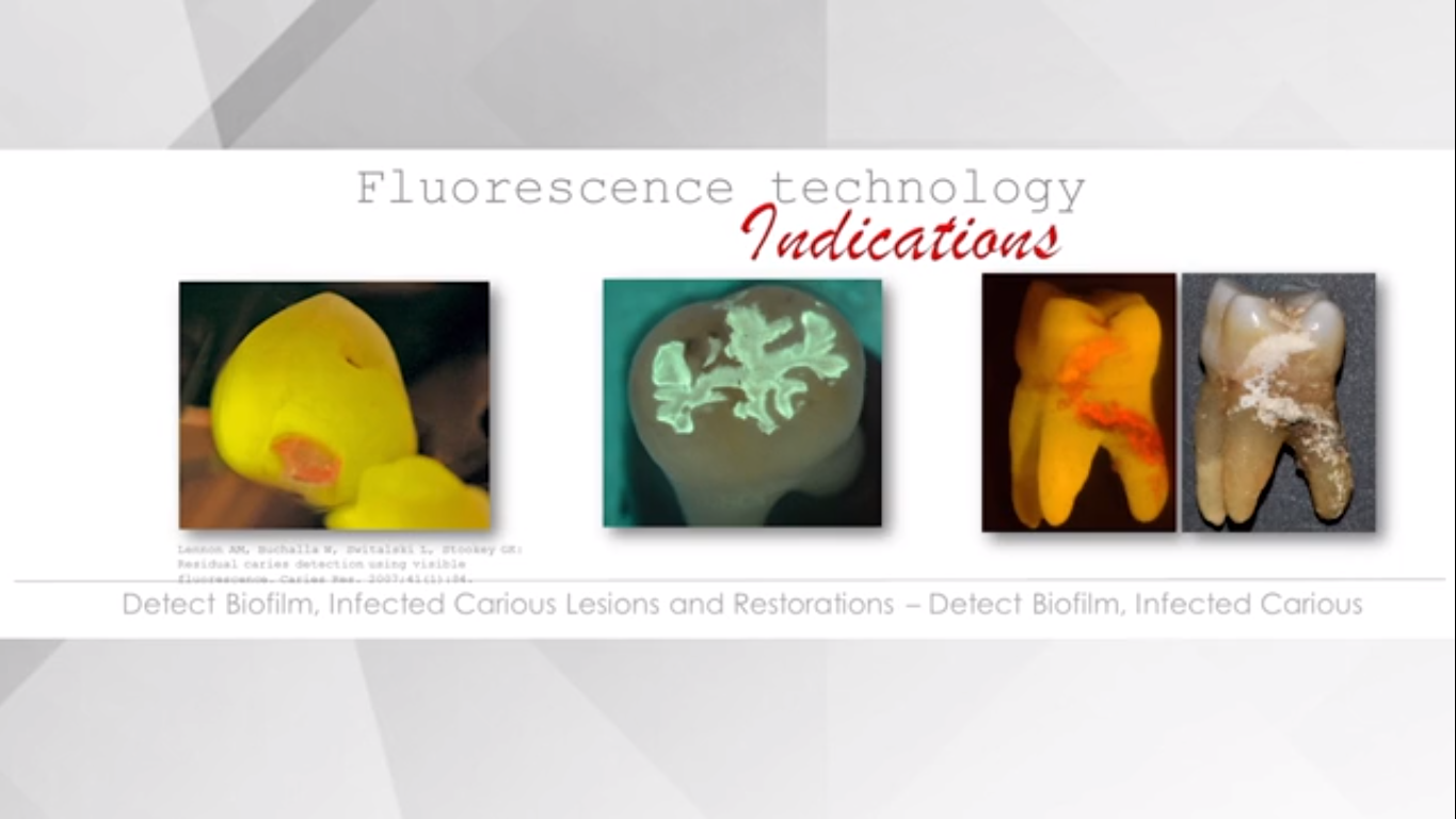
Fissures and cracks – Pain points and main challenges
Presbyopia starts to affect natural vision by the age of 401 and hence the naked eye, begins to lose considerable visual acuity. However, these visual deficiencies can be easily compensated thanks to optical devices2, though surprisingly few dentists are aware of their own visual limitations.3
One of the main steps in crack and fissure confirmation is visual examination, meaning each tooth should be thoroughly checked before placing a restoration to confirm or discard the presence of cracks.
Visual diagnosis has its limitations because of the resolution of the human eye. The resolving power of the unaided human eye is only 200 microns4 – which means it needs at least 200 microns between two points to recognize them as separate from each other. Everything with a diameter or width under 200 microns will be extremely difficult to see.
This invisible eye gap is a handicap when it comes to visual fissure and crack detection – and even more so, as dentists age. Therefore, the use of magnification devices is recommended to compensate visual deficiencies.5
“When I started using the microscope in 2003, magnification instantly grabbed my attention as clinical procedures improved dramatically. Put simply, thanks to detailed vision, optimal lighting conditions and comfort, I needed much less time to achieve much better results. Sincerely, I couldn’t imagine working without it.”
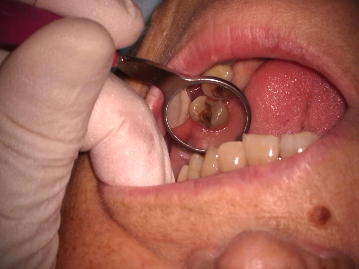
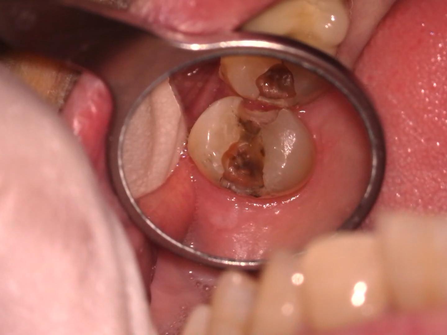
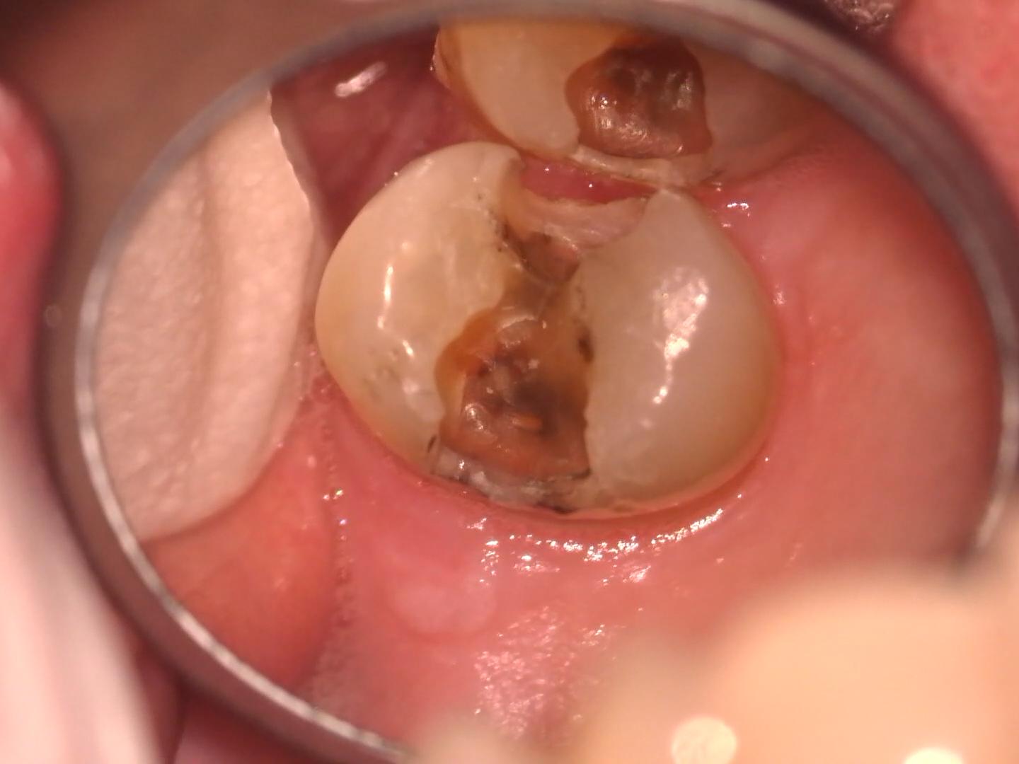
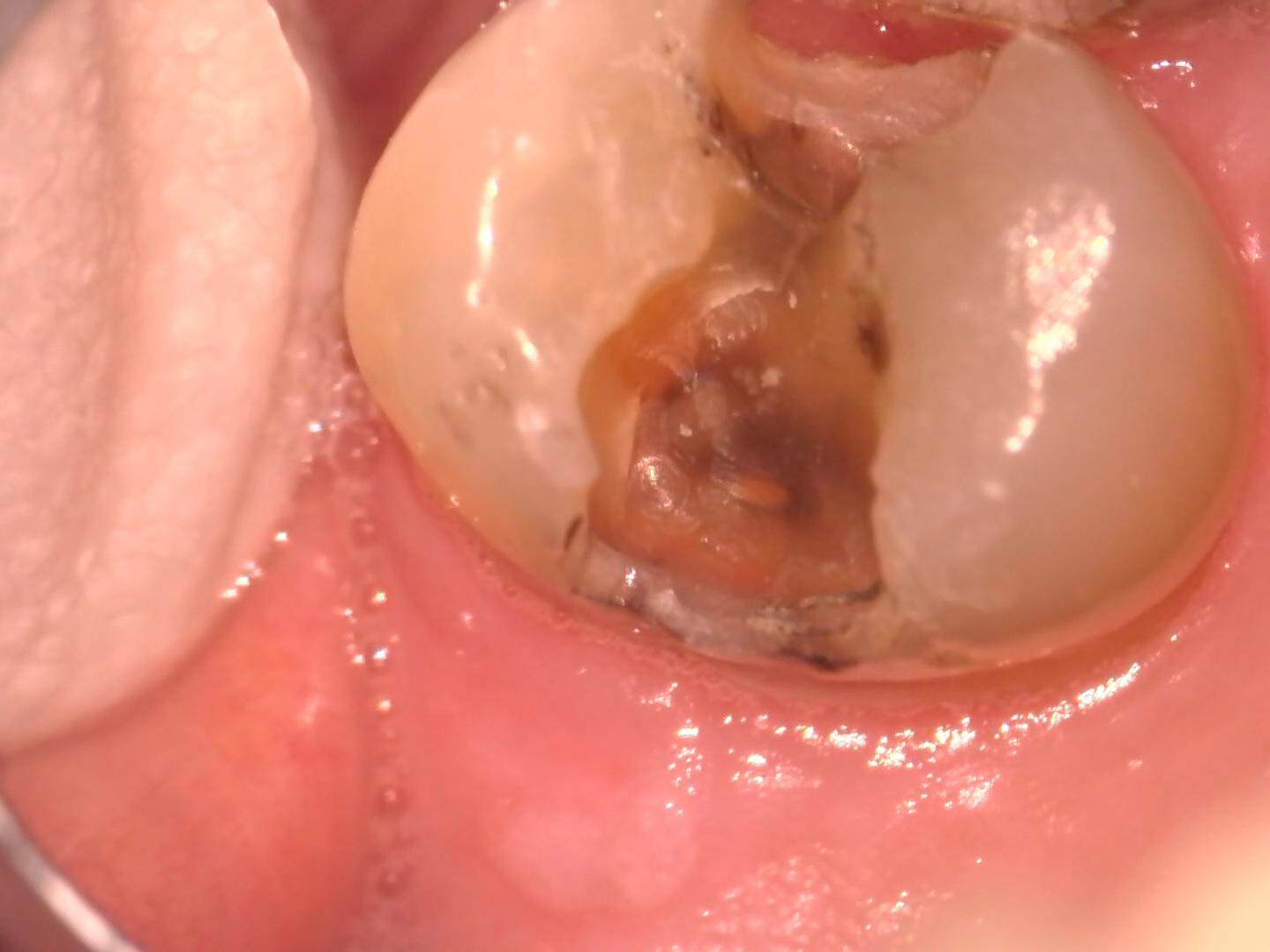
Crack diagnosis with naked eyes vs. magnification
Application pictures courtesy of Dr. Christian del Rey Schnitzler, Madrid, Spain.
Without magnification (naked eyes)
Application pictures courtesy of Dr. Christian del Rey Schnitzler, Madrid, Spain.
4x magnification
Application pictures courtesy of Dr. Christian del Rey Schnitzler, Madrid, Spain.
10x magnification
Application pictures courtesy of Dr. Christian del Rey Schnitzler, Madrid, Spain.
16x magnification
Application pictures courtesy of Dr. Christian del Rey Schnitzler, Madrid, Spain.
25x magnification
Magnification for better vision
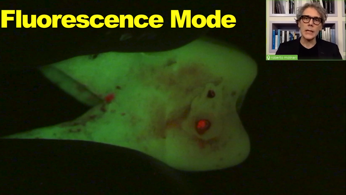
Magnification enhances the dentist’s ability to diagnose caries and cracks in teeth6, 7, 8, to distinguish better between colors, to differentiate between materials, tissues and substances and to detect interfaces.9
Dental microscopes – thanks to their magnification and co-axial illumination – are an especially great aid when it comes to crack and fracture detection, neither of which are visible to the naked eye nor palpable with an endodontic explorer.10 As a result, dentists can anticipate treatment long before coronal fractures and cracked teeth become symptomatic.9
For example, by using a microscope with 21x magnification, the resolution with which the eye sees increases up to 9.5 microns, allowing the eye to distinguish points separated by only 9.5 microns of distance. And therefore improving visual acuity to previous unreachable levels.
Burton JF, Bridgman GF. Presbyopia and the dentist: The effect of age on clinical vision. Int Dent J 1990;40:30312.
2
Eichenberger M, Perrin P, Neuhaus KW, Bringolf U, Lussi A. Visual acuity of dentists under simulated clinical conditions. Clin Oral Investig 2013;17:7259.
3
Eichenberger M, Perrin P, Ramseyer ST, Lussi A. Visual acuity and experience with magnification devices in Swiss dental practices. Oper Dent 2015;40:E1429.
4
Carr G, Murgel C. The use of operating microscopes in endodontics. Dent Clin N Am. 2004;54:191-214.
5
Perrin P, Ramseyer ST, Eichenberger M, Lussi A. Visual acuity of dentists in their respective clinical conditions. Clin Oral Investig 2014;18:20558.
6
ForgieAH, Pine CM, Pitts NB. The use of magnification in a preventive approach to caries detection. Quintessence Int 2002;33:136. 24.
7
Clark DJ, Sheets CG, Paquette JM. Definitive diagnosis of early enamel and dentin cracks based on microscopic evaluation. J Esthet Restor Dent 2003;15:391401.
8
Mamoun JS, Napoletano D. Cracked tooth diagnosis and treatment: An alternative paradigm. Eur J Dent 2015; 9:293303.
9
Mamoun JS. A rationale for the use of highpowered magnification or microscopes in general dentistry. General Dentistry 57 (2009): 18-26.
10
Slaton CC, Loushine RJ, Weller RN, Parker MH, Kimbrough WF, Pashley DH, et al. Identification of resected rootend dentinal cracks: A comparative study of visual magnification. J Endod 2003; 29:51922.
11
Carr G, Murgel C. The use of operating microscopes in endodontics. Dent Clin N Am. 2004; 54:191-214.


")
")