Suplemento
Mejora de la tecnología, consultas más rápidas y tratamientos personalizados
12 junio 2020
· 20 MIN LEER
![]({"xsmall":"https://www.zeiss.com/content/dam/med-hcp/reference-master/myzeiss/peer-insights/free/ophthalmic-diagnostics/272-improved-technologies/royce-chen.jpg/_jcr_content/renditions/original.image_file.100.100.file/royce-chen.jpg","small":"https://www.zeiss.com/content/dam/med-hcp/reference-master/myzeiss/peer-insights/free/ophthalmic-diagnostics/272-improved-technologies/royce-chen.jpg/_jcr_content/renditions/original.image_file.360.360.file/royce-chen.jpg","medium":"https://www.zeiss.com/content/dam/med-hcp/reference-master/myzeiss/peer-insights/free/ophthalmic-diagnostics/272-improved-technologies/royce-chen.jpg/_jcr_content/renditions/original./royce-chen.jpg","large":"https://www.zeiss.com/content/dam/med-hcp/reference-master/myzeiss/peer-insights/free/ophthalmic-diagnostics/272-improved-technologies/royce-chen.jpg/_jcr_content/renditions/original./royce-chen.jpg","xlarge":"https://www.zeiss.com/content/dam/med-hcp/reference-master/myzeiss/peer-insights/free/ophthalmic-diagnostics/272-improved-technologies/royce-chen.jpg/_jcr_content/renditions/original./royce-chen.jpg","xxlarge":"https://www.zeiss.com/content/dam/med-hcp/reference-master/myzeiss/peer-insights/free/ophthalmic-diagnostics/272-improved-technologies/royce-chen.jpg/_jcr_content/renditions/original./royce-chen.jpg","max":"https://www.zeiss.com/content/dam/med-hcp/reference-master/myzeiss/peer-insights/free/ophthalmic-diagnostics/272-improved-technologies/royce-chen.jpg/_jcr_content/renditions/original./royce-chen.jpg"})
Autor
Dr. Royce W. S. Chen
Centro Médico Irving, Universidad de Columbia (EE. UU.)
![]({"xsmall":"https://www.zeiss.com/content/dam/med-hcp/reference-master/myzeiss/peer-insights/free/ophthalmic-diagnostics/272-improved-technologies/jesse-jung.jpg/_jcr_content/renditions/original.image_file.100.100.file/jesse-jung.jpg","small":"https://www.zeiss.com/content/dam/med-hcp/reference-master/myzeiss/peer-insights/free/ophthalmic-diagnostics/272-improved-technologies/jesse-jung.jpg/_jcr_content/renditions/original.image_file.360.360.file/jesse-jung.jpg","medium":"https://www.zeiss.com/content/dam/med-hcp/reference-master/myzeiss/peer-insights/free/ophthalmic-diagnostics/272-improved-technologies/jesse-jung.jpg/_jcr_content/renditions/original.image_file.768.768.file/jesse-jung.jpg","large":"https://www.zeiss.com/content/dam/med-hcp/reference-master/myzeiss/peer-insights/free/ophthalmic-diagnostics/272-improved-technologies/jesse-jung.jpg/_jcr_content/renditions/original.image_file.1024.1024.file/jesse-jung.jpg","xlarge":"https://www.zeiss.com/content/dam/med-hcp/reference-master/myzeiss/peer-insights/free/ophthalmic-diagnostics/272-improved-technologies/jesse-jung.jpg/_jcr_content/renditions/original.image_file.1280.1280.file/jesse-jung.jpg","xxlarge":"https://www.zeiss.com/content/dam/med-hcp/reference-master/myzeiss/peer-insights/free/ophthalmic-diagnostics/272-improved-technologies/jesse-jung.jpg/_jcr_content/renditions/original./jesse-jung.jpg","max":"https://www.zeiss.com/content/dam/med-hcp/reference-master/myzeiss/peer-insights/free/ophthalmic-diagnostics/272-improved-technologies/jesse-jung.jpg/_jcr_content/renditions/original./jesse-jung.jpg"})
Autor
Dr. Jesse J. Jung
Especialista en vítreo y retina, East Bay Retina Consultants, Inc, EE. UU.
![]({"xsmall":"https://www.zeiss.com/content/dam/med-hcp/reference-master/myzeiss/peer-insights/free/ophthalmic-diagnostics/272-improved-technologies/sunil-shrivastava.jpg/_jcr_content/renditions/original.image_file.100.100.file/sunil-shrivastava.jpg","small":"https://www.zeiss.com/content/dam/med-hcp/reference-master/myzeiss/peer-insights/free/ophthalmic-diagnostics/272-improved-technologies/sunil-shrivastava.jpg/_jcr_content/renditions/original.image_file.360.360.file/sunil-shrivastava.jpg","medium":"https://www.zeiss.com/content/dam/med-hcp/reference-master/myzeiss/peer-insights/free/ophthalmic-diagnostics/272-improved-technologies/sunil-shrivastava.jpg/_jcr_content/renditions/original.image_file.768.768.file/sunil-shrivastava.jpg","large":"https://www.zeiss.com/content/dam/med-hcp/reference-master/myzeiss/peer-insights/free/ophthalmic-diagnostics/272-improved-technologies/sunil-shrivastava.jpg/_jcr_content/renditions/original.image_file.1024.1024.file/sunil-shrivastava.jpg","xlarge":"https://www.zeiss.com/content/dam/med-hcp/reference-master/myzeiss/peer-insights/free/ophthalmic-diagnostics/272-improved-technologies/sunil-shrivastava.jpg/_jcr_content/renditions/original./sunil-shrivastava.jpg","xxlarge":"https://www.zeiss.com/content/dam/med-hcp/reference-master/myzeiss/peer-insights/free/ophthalmic-diagnostics/272-improved-technologies/sunil-shrivastava.jpg/_jcr_content/renditions/original./sunil-shrivastava.jpg","max":"https://www.zeiss.com/content/dam/med-hcp/reference-master/myzeiss/peer-insights/free/ophthalmic-diagnostics/272-improved-technologies/sunil-shrivastava.jpg/_jcr_content/renditions/original./sunil-shrivastava.jpg"})
Autor
Dr. Sunil K. Srivastava
Especialista en retina, Cleveland Clinic, EE. UU.
RESUMEN
¿De qué manera la tecnología nueva y mejorada permite realizar consultas más rápidas y tratamientos personalizados?
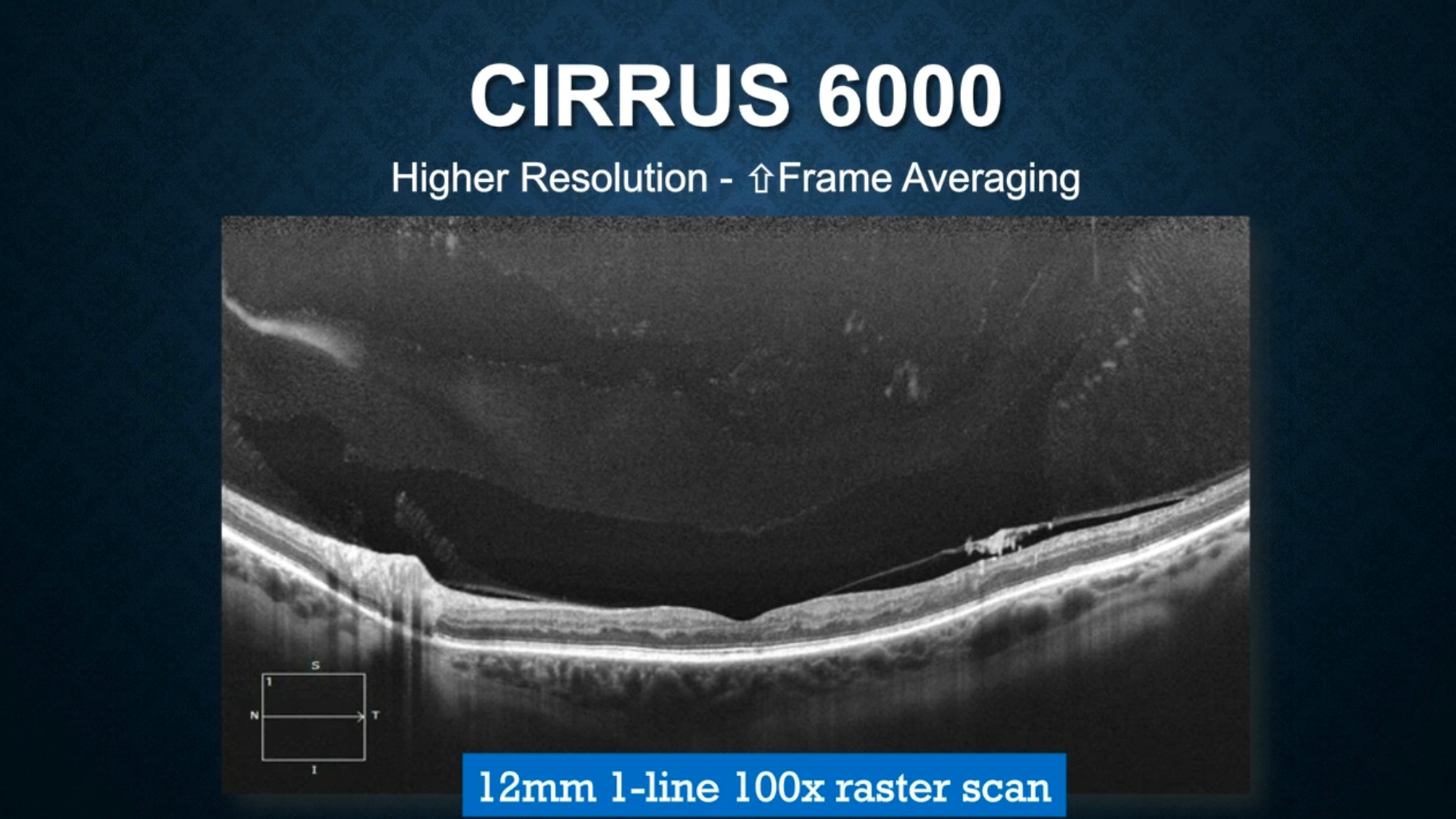
En este artículo, que se publicó originalmente en Retina Today, un grupo de expertos revisa dos herramientas de captura de imágenes diseñadas para mejorar el diagnóstico y el tratamiento de las enfermedades: la angiografía por OCT y la captura de imágenes de campo ultraamplio del fondo del ojo.