Supplément
Technologie améliorée, flux de tâches cliniques accélérés et traitements sur mesure
12 juin 2020
· 20 MIN LECTURE
![]({"xsmall":"https://www.zeiss.com/content/dam/med-hcp/reference-master/myzeiss/peer-insights/free/ophthalmic-diagnostics/272-improved-technologies/royce-chen.jpg/_jcr_content/renditions/original.image_file.100.100.file/royce-chen.jpg","small":"https://www.zeiss.com/content/dam/med-hcp/reference-master/myzeiss/peer-insights/free/ophthalmic-diagnostics/272-improved-technologies/royce-chen.jpg/_jcr_content/renditions/original.image_file.360.360.file/royce-chen.jpg","medium":"https://www.zeiss.com/content/dam/med-hcp/reference-master/myzeiss/peer-insights/free/ophthalmic-diagnostics/272-improved-technologies/royce-chen.jpg/_jcr_content/renditions/original./royce-chen.jpg","large":"https://www.zeiss.com/content/dam/med-hcp/reference-master/myzeiss/peer-insights/free/ophthalmic-diagnostics/272-improved-technologies/royce-chen.jpg/_jcr_content/renditions/original./royce-chen.jpg","xlarge":"https://www.zeiss.com/content/dam/med-hcp/reference-master/myzeiss/peer-insights/free/ophthalmic-diagnostics/272-improved-technologies/royce-chen.jpg/_jcr_content/renditions/original./royce-chen.jpg","xxlarge":"https://www.zeiss.com/content/dam/med-hcp/reference-master/myzeiss/peer-insights/free/ophthalmic-diagnostics/272-improved-technologies/royce-chen.jpg/_jcr_content/renditions/original./royce-chen.jpg","max":"https://www.zeiss.com/content/dam/med-hcp/reference-master/myzeiss/peer-insights/free/ophthalmic-diagnostics/272-improved-technologies/royce-chen.jpg/_jcr_content/renditions/original./royce-chen.jpg"})
Auteur
Dr Royce W.S. Chen
Irving Medical Center, Columbia University, États-Unis
![]({"xsmall":"https://www.zeiss.com/content/dam/med-hcp/reference-master/myzeiss/peer-insights/free/ophthalmic-diagnostics/272-improved-technologies/jesse-jung.jpg/_jcr_content/renditions/original.image_file.100.100.file/jesse-jung.jpg","small":"https://www.zeiss.com/content/dam/med-hcp/reference-master/myzeiss/peer-insights/free/ophthalmic-diagnostics/272-improved-technologies/jesse-jung.jpg/_jcr_content/renditions/original.image_file.360.360.file/jesse-jung.jpg","medium":"https://www.zeiss.com/content/dam/med-hcp/reference-master/myzeiss/peer-insights/free/ophthalmic-diagnostics/272-improved-technologies/jesse-jung.jpg/_jcr_content/renditions/original.image_file.768.768.file/jesse-jung.jpg","large":"https://www.zeiss.com/content/dam/med-hcp/reference-master/myzeiss/peer-insights/free/ophthalmic-diagnostics/272-improved-technologies/jesse-jung.jpg/_jcr_content/renditions/original.image_file.1024.1024.file/jesse-jung.jpg","xlarge":"https://www.zeiss.com/content/dam/med-hcp/reference-master/myzeiss/peer-insights/free/ophthalmic-diagnostics/272-improved-technologies/jesse-jung.jpg/_jcr_content/renditions/original.image_file.1280.1280.file/jesse-jung.jpg","xxlarge":"https://www.zeiss.com/content/dam/med-hcp/reference-master/myzeiss/peer-insights/free/ophthalmic-diagnostics/272-improved-technologies/jesse-jung.jpg/_jcr_content/renditions/original./jesse-jung.jpg","max":"https://www.zeiss.com/content/dam/med-hcp/reference-master/myzeiss/peer-insights/free/ophthalmic-diagnostics/272-improved-technologies/jesse-jung.jpg/_jcr_content/renditions/original./jesse-jung.jpg"})
Auteur
Dr Jesse J. Jung
Spécialiste vitréo-rétinien, East Bay Retina Consultants, Inc., États-Unis
![]({"xsmall":"https://www.zeiss.com/content/dam/med-hcp/reference-master/myzeiss/peer-insights/free/ophthalmic-diagnostics/272-improved-technologies/sunil-shrivastava.jpg/_jcr_content/renditions/original.image_file.100.100.file/sunil-shrivastava.jpg","small":"https://www.zeiss.com/content/dam/med-hcp/reference-master/myzeiss/peer-insights/free/ophthalmic-diagnostics/272-improved-technologies/sunil-shrivastava.jpg/_jcr_content/renditions/original.image_file.360.360.file/sunil-shrivastava.jpg","medium":"https://www.zeiss.com/content/dam/med-hcp/reference-master/myzeiss/peer-insights/free/ophthalmic-diagnostics/272-improved-technologies/sunil-shrivastava.jpg/_jcr_content/renditions/original.image_file.768.768.file/sunil-shrivastava.jpg","large":"https://www.zeiss.com/content/dam/med-hcp/reference-master/myzeiss/peer-insights/free/ophthalmic-diagnostics/272-improved-technologies/sunil-shrivastava.jpg/_jcr_content/renditions/original.image_file.1024.1024.file/sunil-shrivastava.jpg","xlarge":"https://www.zeiss.com/content/dam/med-hcp/reference-master/myzeiss/peer-insights/free/ophthalmic-diagnostics/272-improved-technologies/sunil-shrivastava.jpg/_jcr_content/renditions/original./sunil-shrivastava.jpg","xxlarge":"https://www.zeiss.com/content/dam/med-hcp/reference-master/myzeiss/peer-insights/free/ophthalmic-diagnostics/272-improved-technologies/sunil-shrivastava.jpg/_jcr_content/renditions/original./sunil-shrivastava.jpg","max":"https://www.zeiss.com/content/dam/med-hcp/reference-master/myzeiss/peer-insights/free/ophthalmic-diagnostics/272-improved-technologies/sunil-shrivastava.jpg/_jcr_content/renditions/original./sunil-shrivastava.jpg"})
Auteur
Dr Sunil K. Srivastava
Spécialiste de la rétine, clinique de Cleveland, États-Unis
RÉSUMÉ
La capacité des technologies nouvelles et améliorées à accélérer les flux de tâches cliniques et à personnaliser les traitements
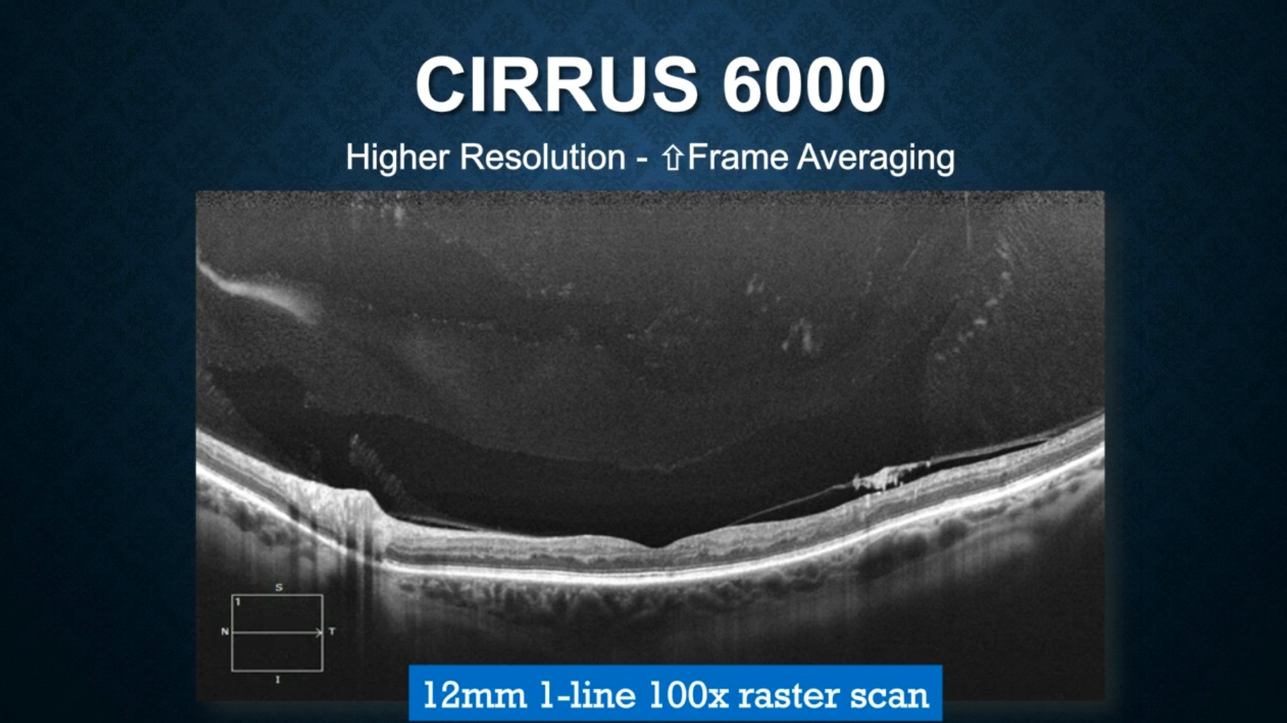
Dans ce supplément, publié à l'origine dans Retina Today, un panel d'experts passe en revue deux outils d'imagerie – l'OCT-angiographie et l'imagerie de fond d'œil ultra grand champ – destinés à améliorer le diagnostic et la prise en charge des pathologies.