

Microscopy Solutions for Sample Preparation
For Further Examinations such as Genetic StudiesMany disciplines – such as developmental biology, molecular biology, embryology, plant sciences and neurobiology – work with model organisms that often need special handling or preparation before further examinations such as genetic studies.
C. elegans (a nematode), zebrafish Danio rerio and the fruit fly Drosophila melanogaster are widely used, respectively, in worm labs, zebrafish labs and fly facilities. These model organisms need to be routinely sorted, picked, counted, manipulated, dissected, imaged and monitored during their development stages (e.g. egg, embryo, larva …). Stereo or dissecting microscopes are indispensable tools for these tasks.
Model organisms can be bacteria, fungi, plants or animals. Ideally, model organisms should exhibit certain characteristics:
- Share many genes with humans
- Small size and easy to grow in the lab
- Visible congenital traits
- Sequenced genome available
- Easy genetic manipulation
- Genetically tractable
- Short generation times and life cycle
Examples of common model organisms
-
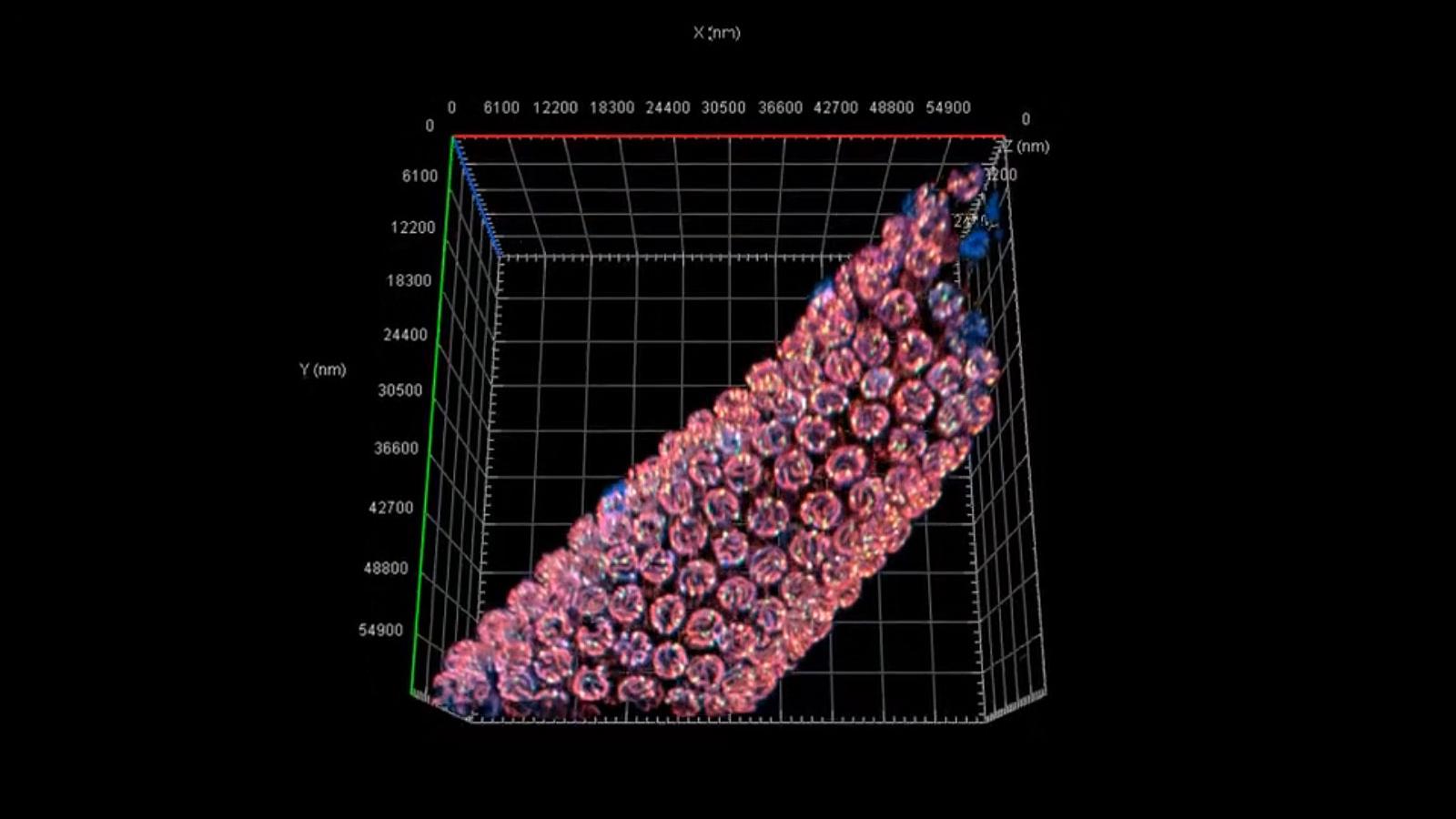
Caenorhabditis elegans germline.
 Drosophila melanogaster
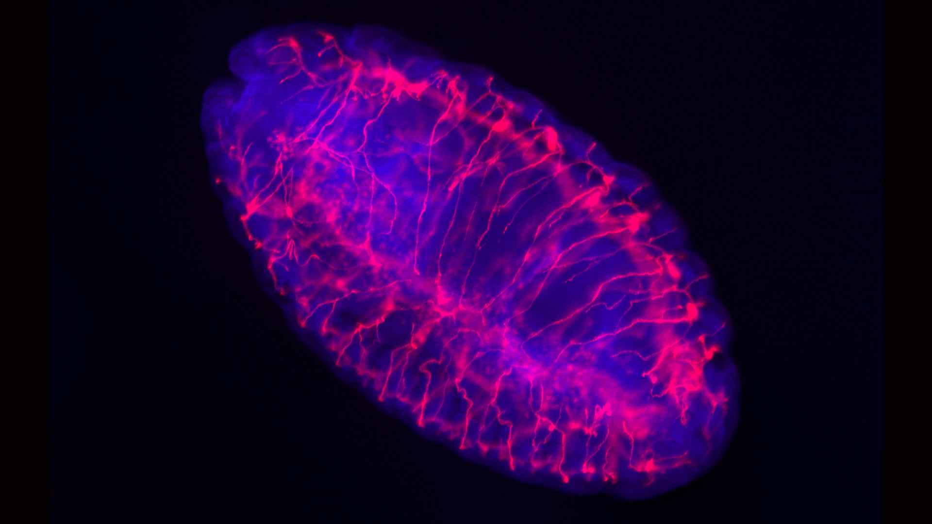 Zebrafish
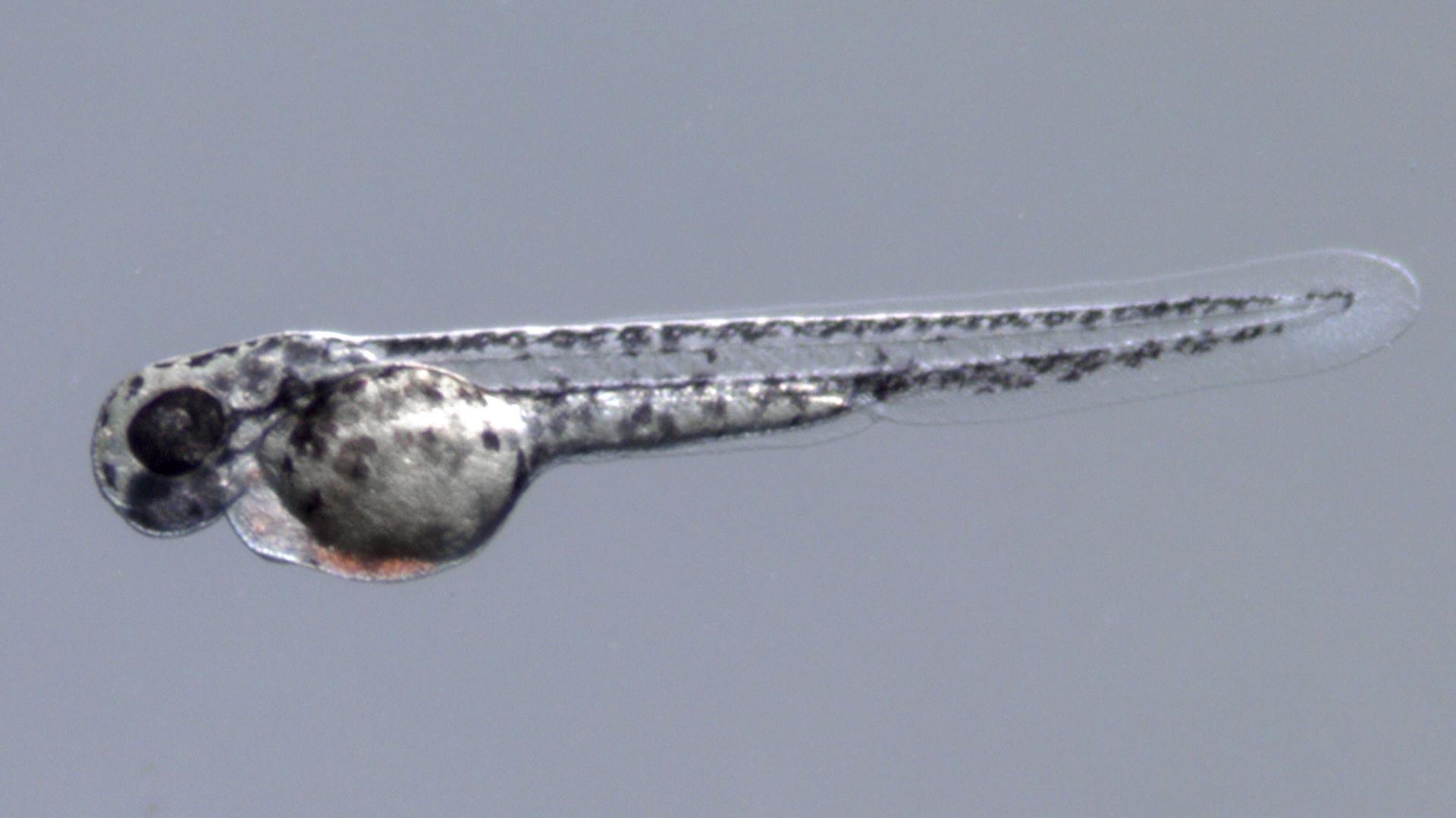 Xenopus laevis, Xenopus tropicalis
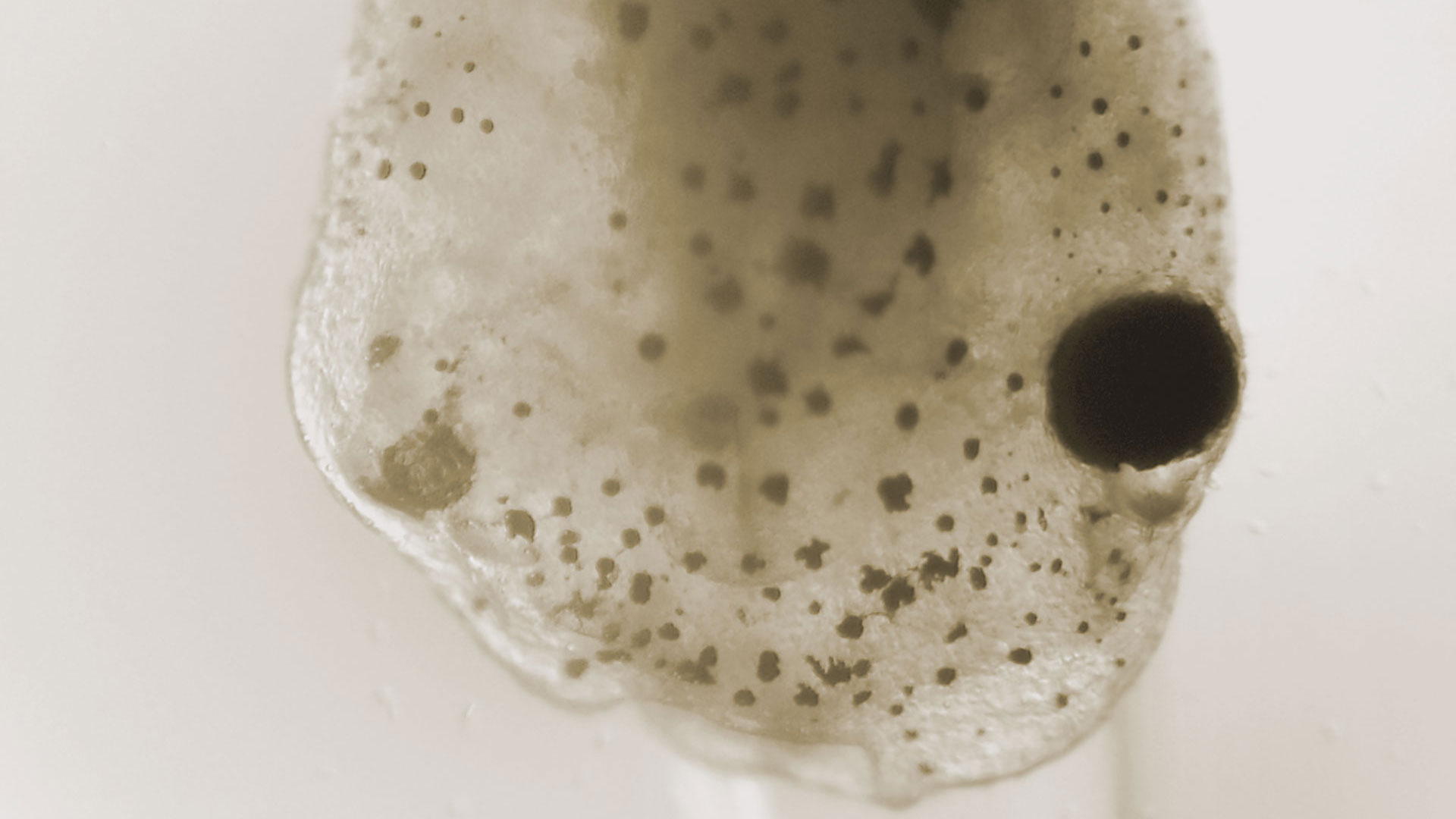 Field of Study
• Developmental biology
• Molecular biology
• Physiology
• Pharmacology
• Toxicology• Genetics
• Developmental biology
• Neuroscience• Embryology
• Developmental biology
• Molecular biology
• Toxicology
• Neurobiology• Embryology
• Developmental Biology
• Cell Biology
• NeurobiologyCharacteristics• Transparent throughout lifetime
• Rapid generation time (3 days)
• Easy to manipulate• Easy to grow and maintain
• Short generation time (8-14 days)
• 4 chromosome pairs
• Easy to manipulate• Share of 70% of genes with humans
• Easy to breed
• Transparant
• Easy to manipulate• Embryos easily accessible
• 10000 oocytes
• Large oocytes/embryos
• Eeasy to manipulate -
Mus musculus
 Rattus norvegicus
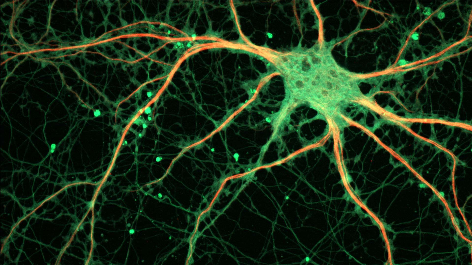 Axolotl
 Arabidopsis thaliana
 Field of Study
• Neurobiology
• Genetics
• Genomics
• Pharma
• Clinical Research• Neuroscience
• Toxicology
• Genetics
• Gnomics
• Physiology• Regenerative medicine
• Developmental biology• Developmental Biology
• Cell Biology
• Molecular Biology
• Genetics
• Plant PhysiologyCharacteristics• Relatively short generation time (~10 weeks)
• 99% homolog to humans
• Disease model
• Genetically tractable
• Easy to manipulate• Disease model
• Source for primary neurons
• Larger organsCan regenerate its
• Tail
• Limbs
• Parts of its brain…• Small genome
• 5 chromosome pairs
• Many mutants
• Easy to manipulate


Microscope Requirements
There are two types of stereo microscopes – Greenough and Common Main Objective (CMO) – each with its own special characteristics. The CMO-type provides certain advantages over the Greenough-type, particularly when it comes to illumination, fluorescence capabilities, digital image documentation and ergonomics. Greenough stereo microscopes, however, are more compact, highly integrated and economical in price.
Scientists and technicians can be supported in their daily routines by a 3D stereoscopic view combined with a large free-working distance and ergonomic features. These microscopes are perfect for mechanical manipulation, sorting, and general sample preparation. Stereo or zoom microscopes are also used preferentially for fluorescence screening of modified genotypes (for example, through CRISPR/Cas9 mediated mutations) in transgenic studies using markers such as the green (GFP) or red (RFP) fluorescent protein. Combining a large field of view with high resolution will speed up this process significantly.
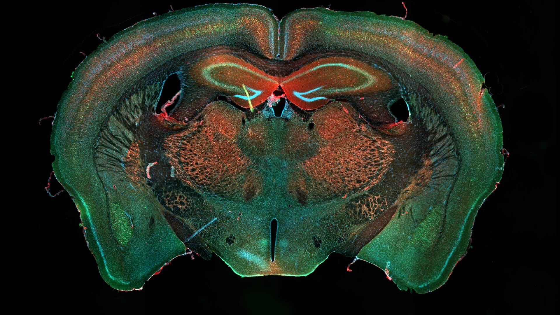
Dissecting microscopes are also widespread in neuroscience – say, when dealing with larger vertebrate animals such as rats, which often serve as a source of primary cell cultures.
Application Examples


