Laser preparation of two H-Bar lamellas in a 3 mm wide copper semicircular grid standing upright.
Technology Solutions for Metals

Nanoscale 3D Imaging of Buried Features

Rapidly Extract and Analyze Features of Interest

The properties of metals and alloys are controlled by their microstructure at all points along the production process - from casting, forging. and extrusion through to machining. welding, fabrication, and service. The microstructure is three-dimensional and may not be uniform in all directions. Features of interest have length, breadth, and height and may be subsurface, necessitating the ability to access buried features and analyze in 3D at high resolution. Modifying and milling your samples may also be required.

Use of Crossbeam FIB-SEM with femtosecond laser for a range of new analytical possibilities to fully understand metals samples in three dimensions.

  • Structure
  • Composition
  • Orientation
Nanoscale 3D Imaging of Buried Features

Rapidly Extract and Analyze Features of Interest

Using a ZEISS Crossbeam focused ion beam scanning electron microscope (FIB-SEM) with femtosecond laser opens a range of new analytical possibilities to fully understand your samples in three dimensions.

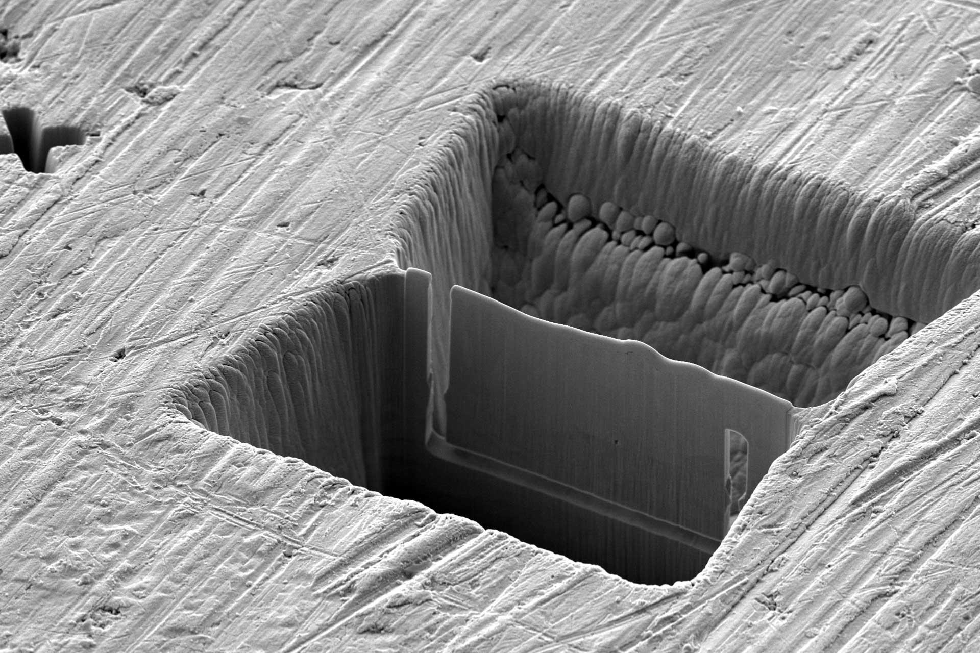
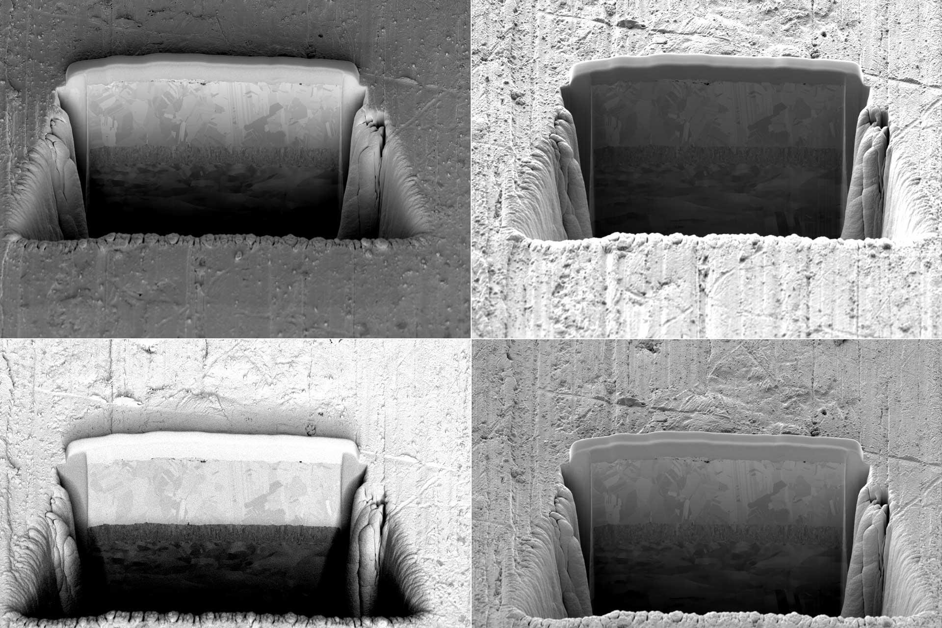
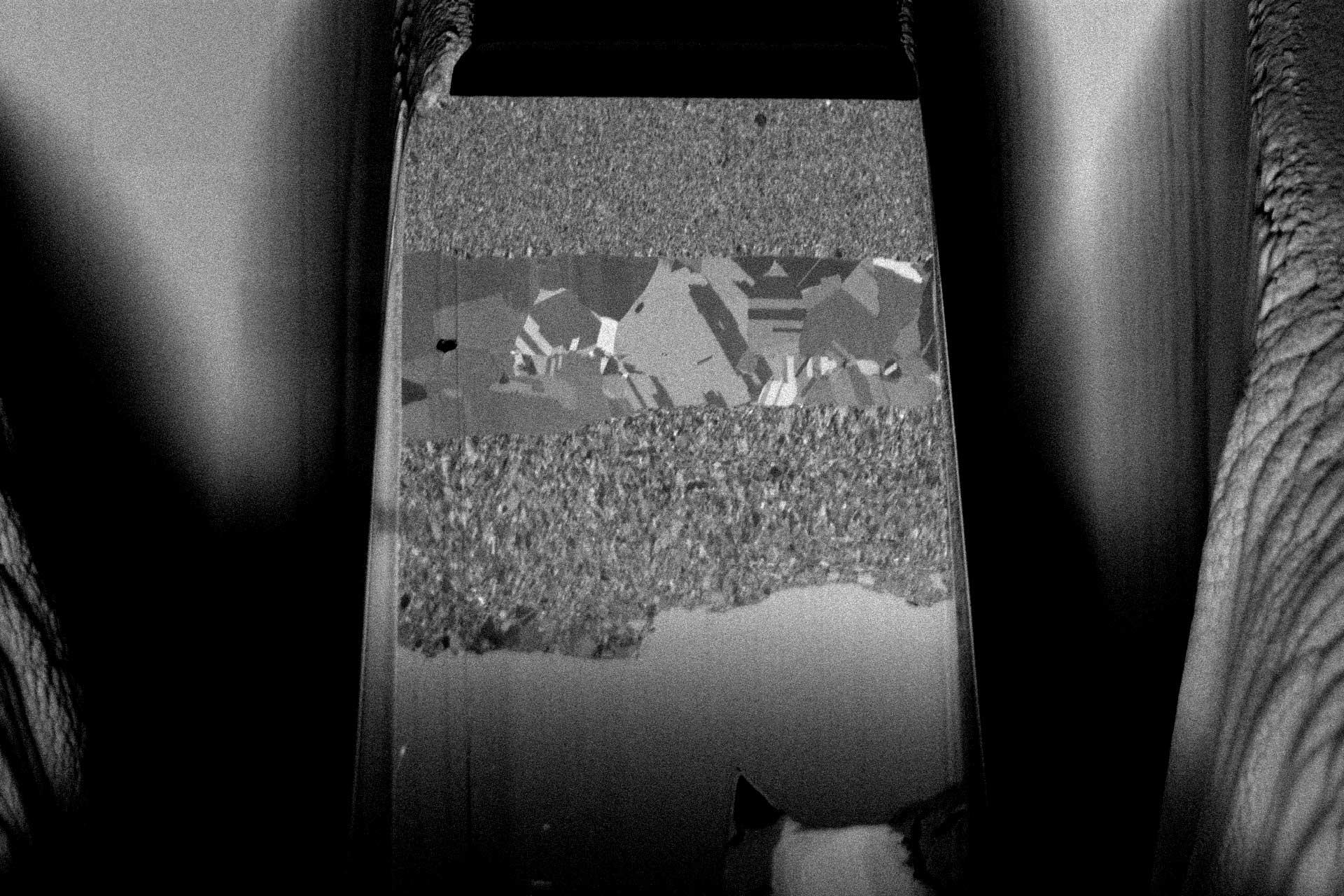
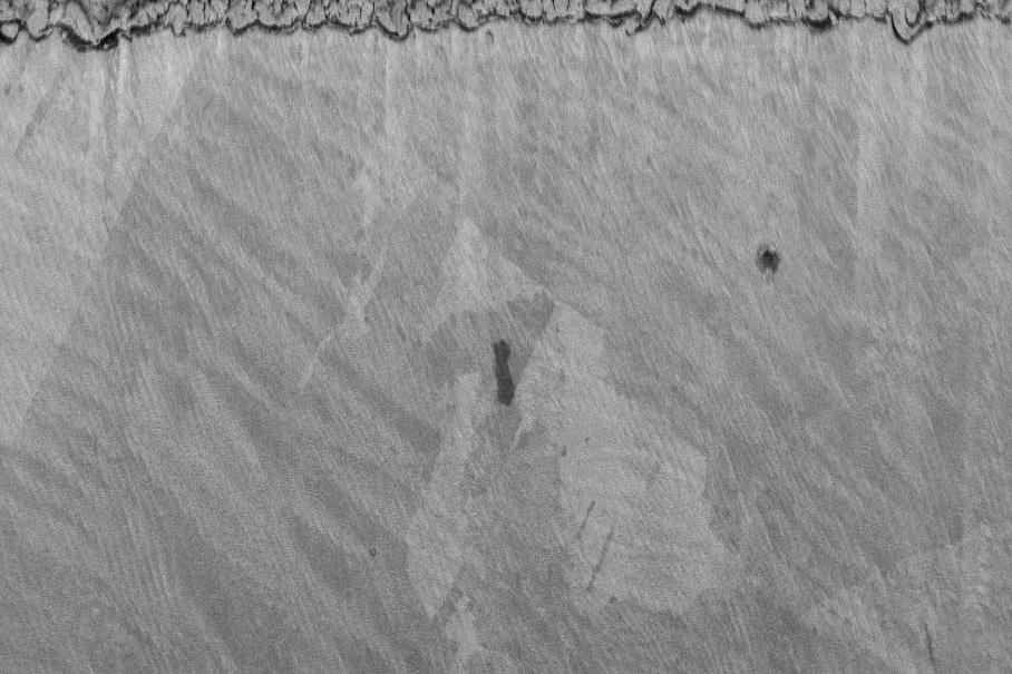
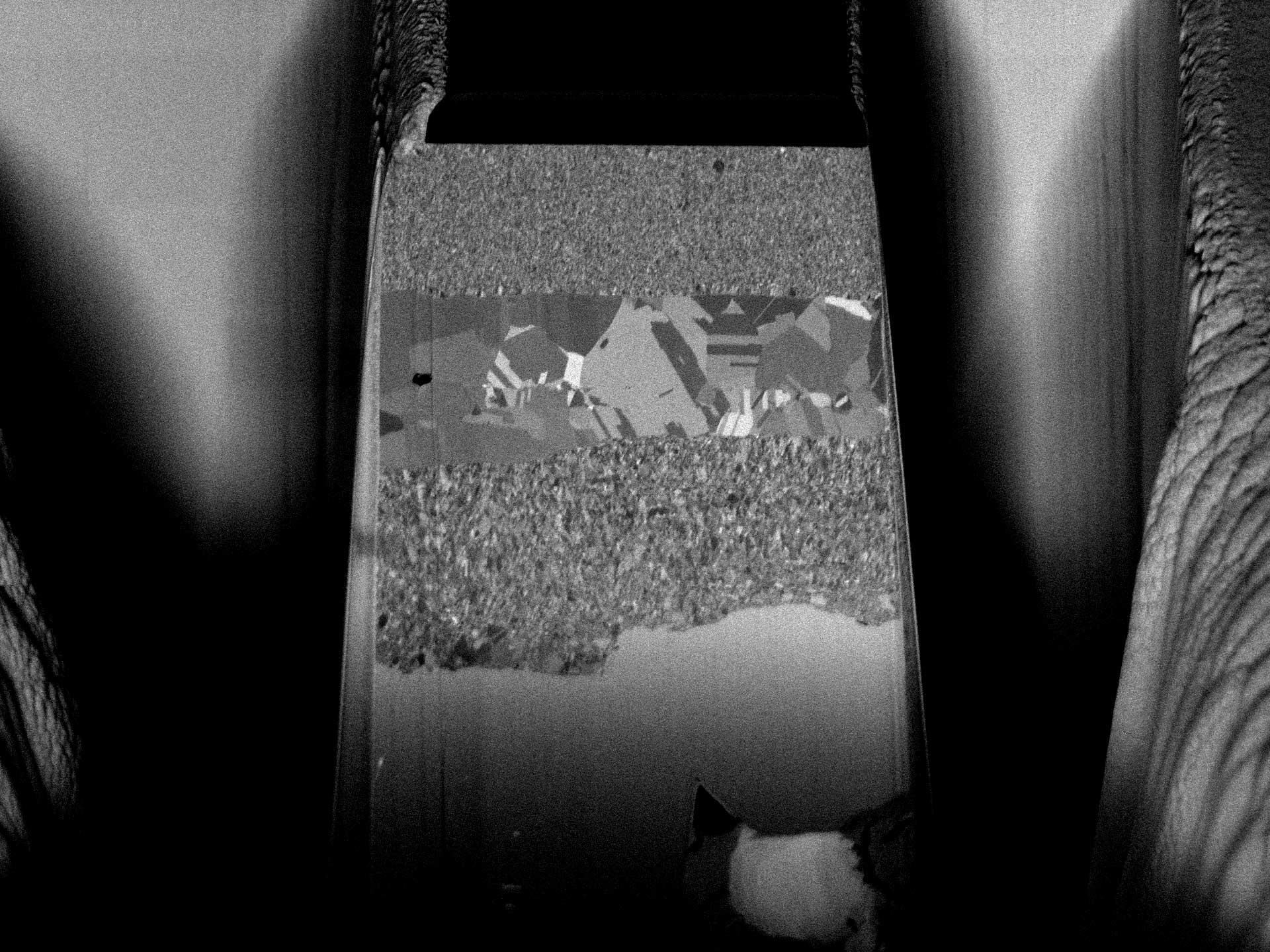
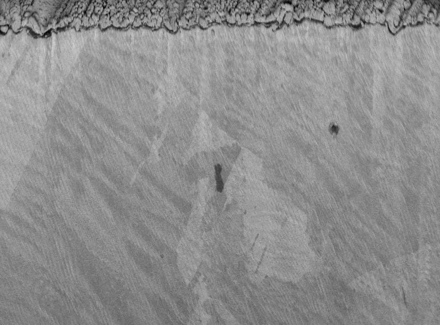
  • Prepare highly polished cross-sections through regions of interest
  • Deposit layers to protect your samples during subsequent analysis
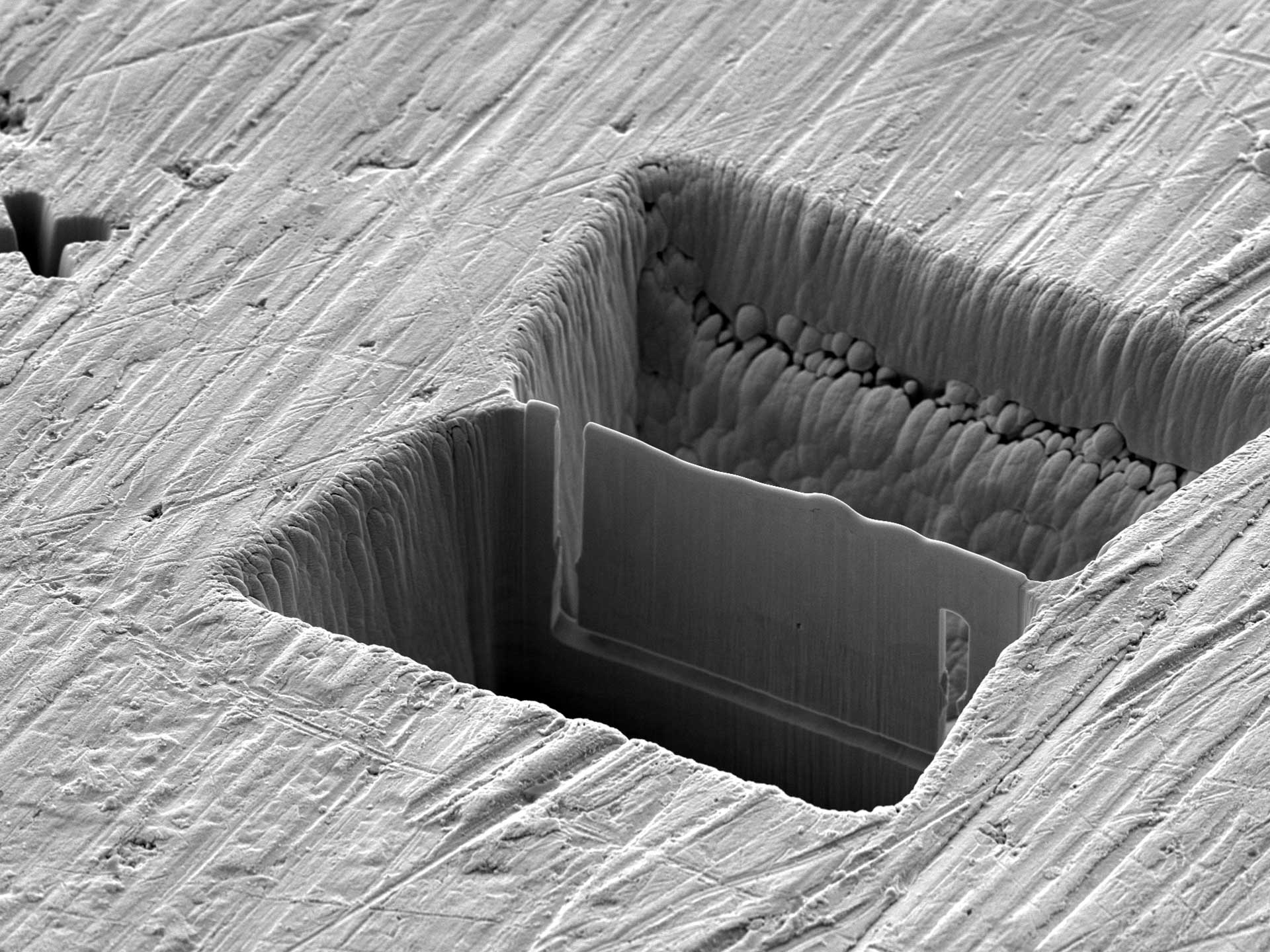
  • Extremely rapidly mill and remove regions of your sample to access deeply buried features
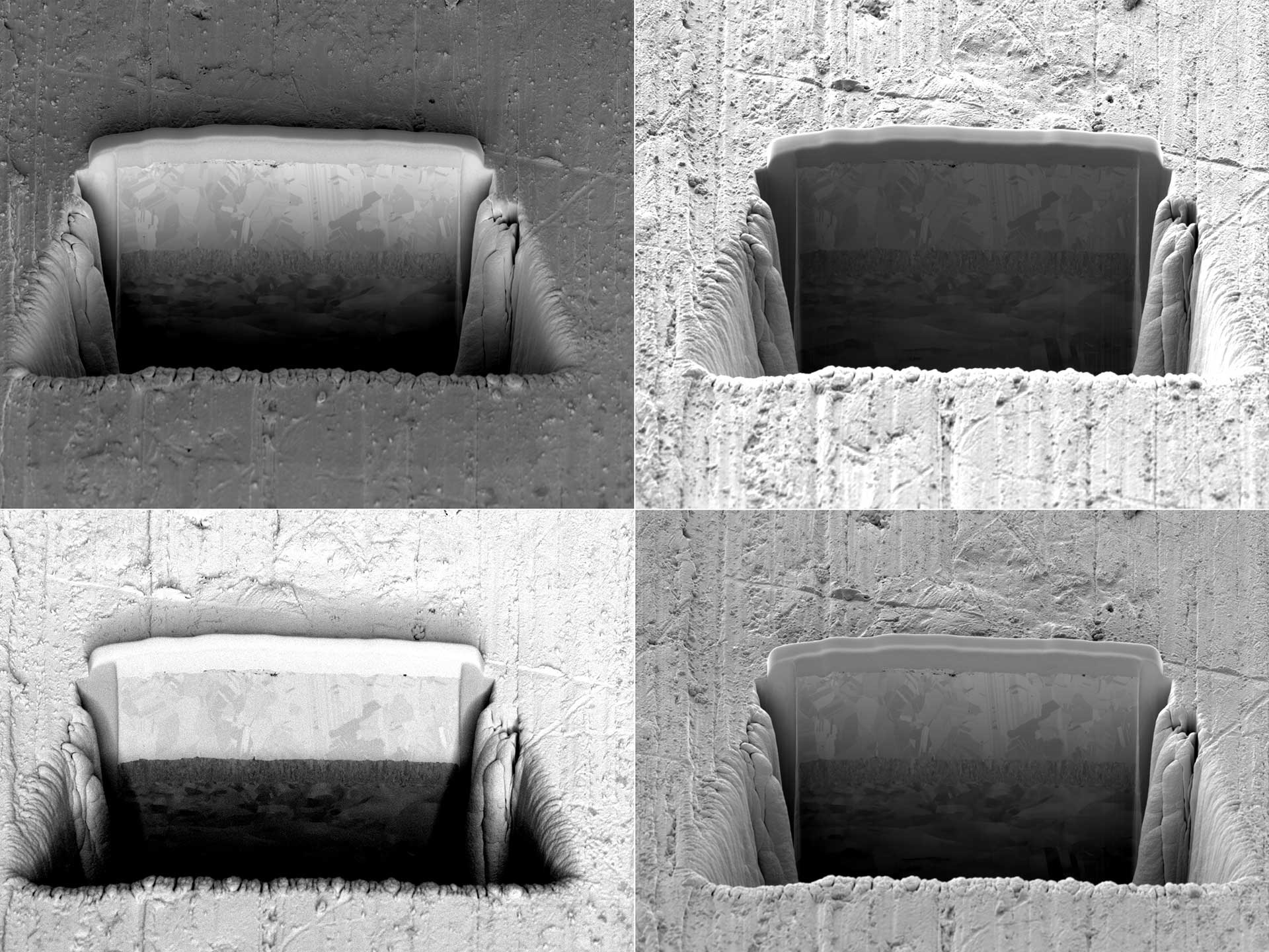
  • Quickly and reliably prepare lamellae for transmission electron microscopy
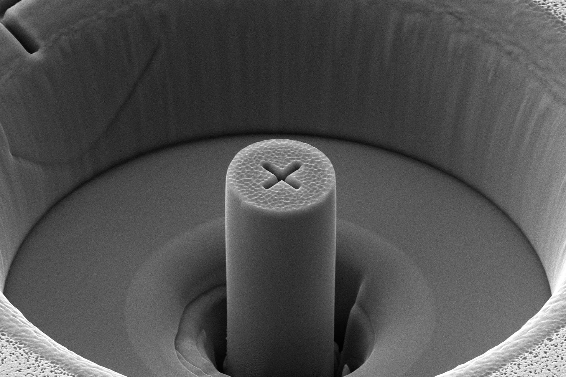
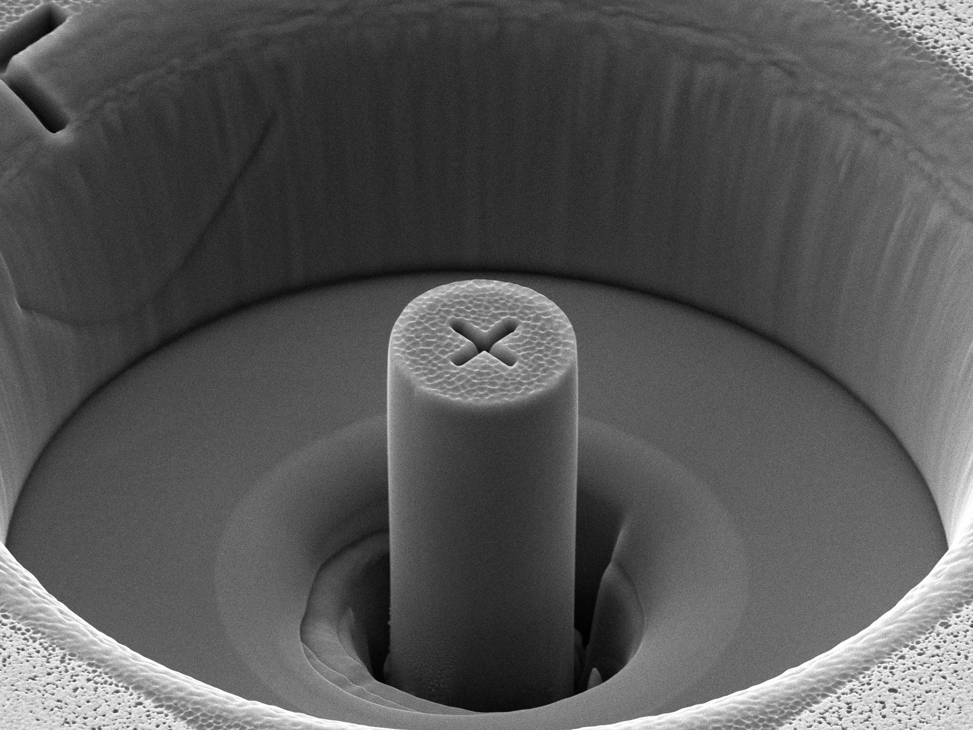
  • Micromachine and prepare pillars, textured surfaces, or any other features on the micro or nanoscale
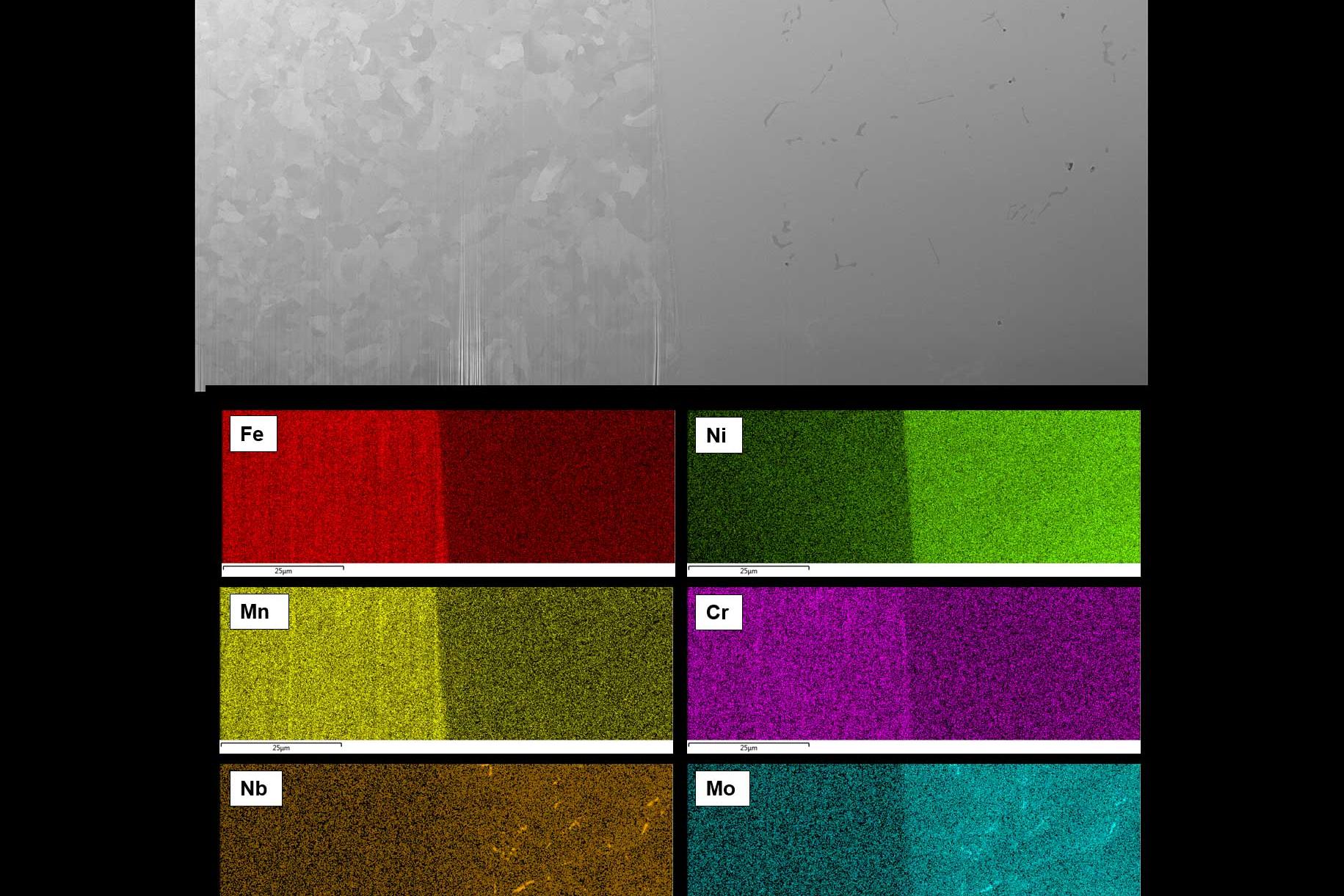
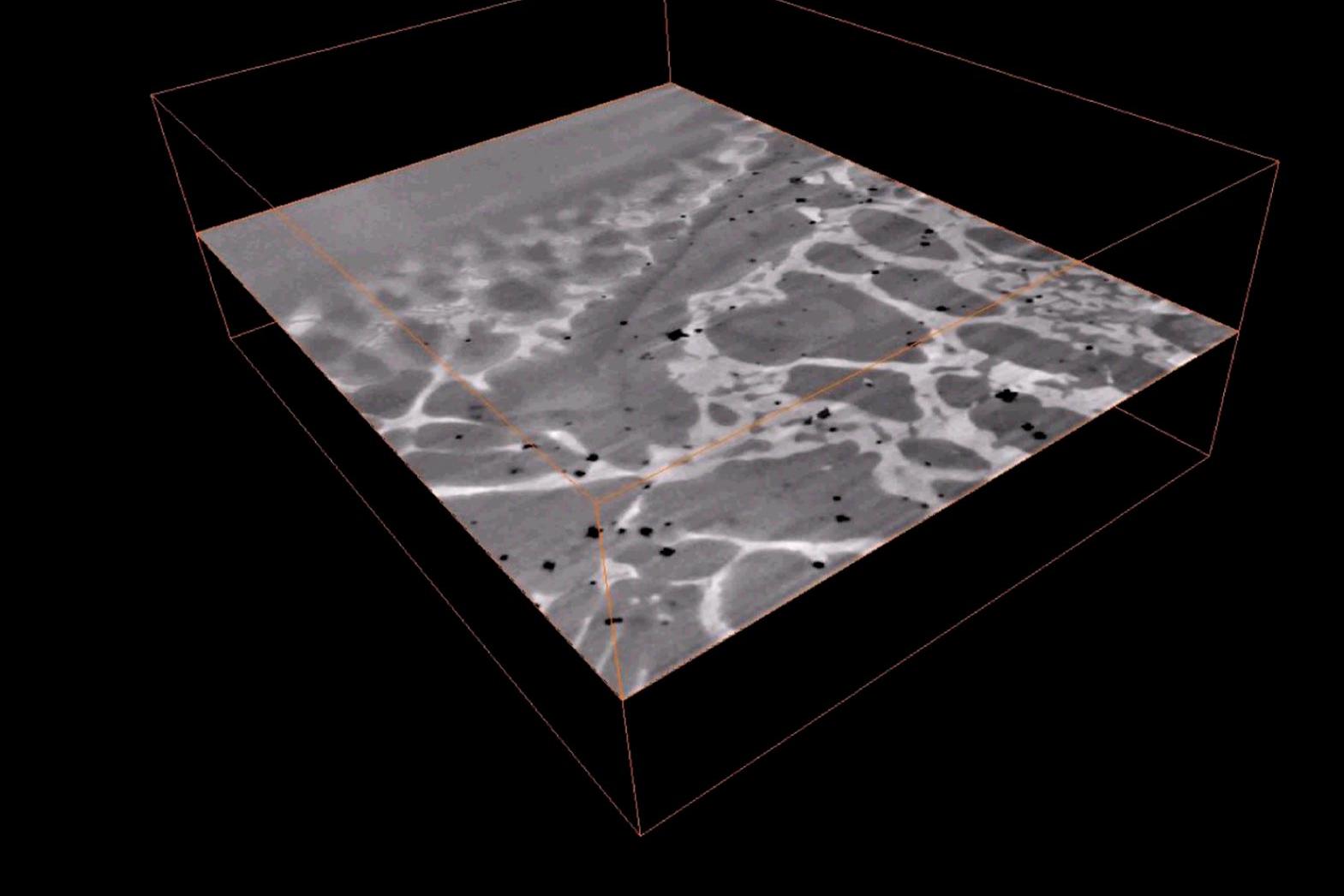
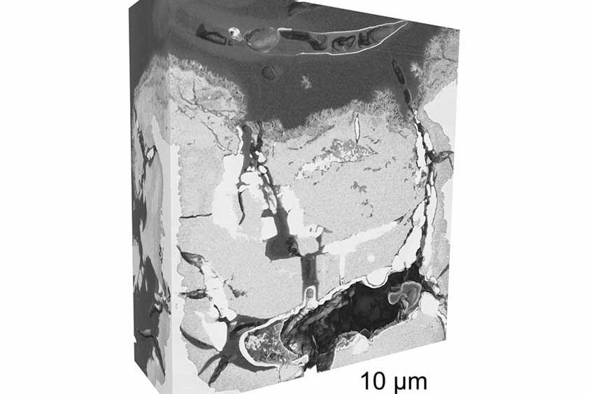
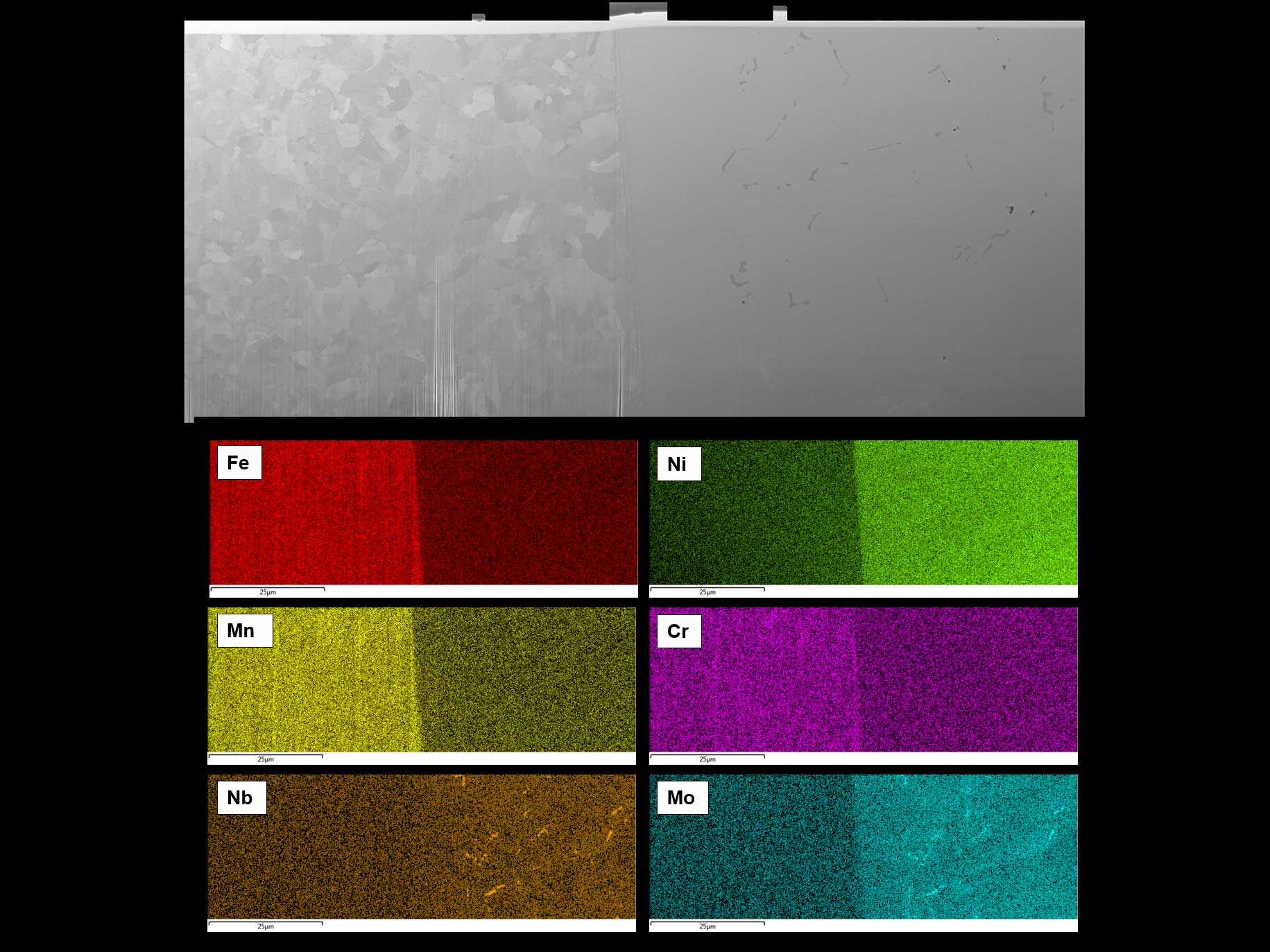
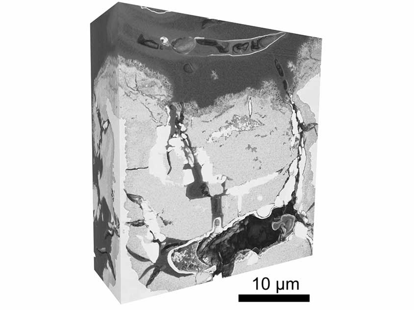
  • Serial slice and image through features of interest: build up a complete picture in 3D by taking images, EDS maps, or EBSD maps of every slice
  • Correlate with data from ZEISS X-ray microscopes to precisely locate and analyze buried features
  • Assess advanced steels, dissimilar joints, inclusions, precipitates, dual-phase alloys, and other complex microstructures

Applications for Nanoscale 3D Imaging of Buried Features

  • Compositional mapping using EDS.

    Serial Slicing through Dissimilar Weld – 8630 Steel and Alloy 625

    Compositional mapping using EDS.

    FIB-SEM data courtesy of ZEISS labs@location at Plymouth Electron Microscopy Centre (University of Plymouth), supported by the European Regional Development Fund.
    Sample courtesy of TWI Ltd.


    Serial Slicing through Dissimilar Weld

    8630 Steel and Alloy 625

    Compositional mapping using EDS.
    FIB-SEM data courtesy of ZEISS labs@location at Plymouth Electron Microscopy Centre (University of Plymouth), supported by the European Regional Development Fund.
    Sample courtesy of TWI Ltd.

  • Lamella of a Silver/Nickel/Copper-Layered System

    Lamella of a Silver/Nickel/Copper-Layered System – Ready for Lift-Out

    Fabricated with automatic sample preparation on ZEISS Crossbeam.

    Lamella of a Silver/Nickel/Copper-Layered System

    Ready for Lift-Out

    Fabricated with automatic sample preparation on ZEISS Crossbeam.

  • Interface of Laser Weld

    NiTi and Stainless Steel Wires

    Reconstruction of different phases at transition.
    ZEISS FIB-SEM imaging, courtesy of P. Burdet, EPFL, Switzerland

  • Cross-Section of a Steel Sample Imaged in Quad Mode

    Prepared with a ZEISS FIB-SEM simultaneously acquiring 4 different signals.
    Top left: Inlens SE; top right: SE2; bottom left: Inlens EsB; bottom right: Inlens SE mixed with SE2.
    Sample courtesy D. Willer, MPA Stuttgart, DE


    Cross-Section of a Steel Sample

    Imaged in Quad Mode

    Prepared with a ZEISS FIB-SEM simultaneously acquiring 4 different signals.
    Top left: Inlens SE; top right: SE2; bottom left: Inlens EsB; bottom right: Inlens SE mixed with SE2.
    Sample courtesy D. Willer, MPA Stuttgart, DE


  • Detailed Morphology of Intergranular Corrosion

    Detailed Morphology of Intergranular Corrosion – Magnesium Alloy

    Resolution of 8 nm/voxel. The region of interest was located using X-ray microscopy in the ZEISS Xradia Versa and Ultra series, and then moved to ZEISS Atlas software to perform high resolution FIB-SEM tomography in this region to enhance multiscale analysis.
    Sample courtesy of the University of Manchester UK; image courtesy of Philip Withers (University of Manchester UK)
     

    Detailed Morphology of Intergranular Corrosion

    Magnesium Alloy

    Resolution of 8 nm/voxel. The region of interest was located using X-ray microscopy in the ZEISS Xradia Versa and Ultra series, and then moved to ZEISS Atlas software to perform high resolution FIB-SEM tomography in this region to enhance multiscale analysis.
    Sample courtesy of the University of Manchester UK; image courtesy of Philip Withers (University of Manchester UK)
     

  • Sectioning through Non-metallic Protective Layers

    Sectioning through Non-metallic Protective Layers - NdFeB Magnet

    ZEISS Crossbeam FIB ona polymer protective layer with two copper layers with different grain sizes.

    Sectioning through Non-metallic Protective Layers

    NdFeB Magnet

    ZEISS Crossbeam FIB ona polymer protective layer with two copper layers with different grain sizes.

  • Parallel-Sided Cylinder with a Central Alignment Mark

    Parallel-Sided Cylinder with a Central Alignment Mark – Fabricated “Top-Down”

    Using ZEISS Crossbeam 540 with Atlas 5 NPVE Advanced. Field of view 40 μm.
    Image courtesy David Unrau, Fibics Incorporated, Ottawa, Canada

    Parallel-Sided Cylinder with a Central Alignment Mark

    Fabricated “Top-Down”

    Using ZEISS Crossbeam 540 with Atlas 5 NPVE Advanced. Field of view 40 μm.
    Image courtesy David Unrau, Fibics Incorporated, Ottawa, Canada

  •  200 µm-Wide Laser Polished Cross-Section

    200 µm-Wide Laser Polished Cross-Section – Alloy 600 (Nickel)

    Without FIB polishing. Microstructural details like grains, inclusions and pores  are directly visible, showing the quality of the laser cut; processed in under 250s.
    Image courtesy Tim Schubert, Aalen University, DE

    200 µm-Wide Laser Polished Cross-Section

    Alloy 600 (Nickel)

    Without FIB polishing. Microstructural details like grains, inclusions and pores are directly visible, showing the quality of the laser cut; processed in under 250s. Image courtesy Tim Schubert, Aalen University, DE

Downloads

Visit the ZEISS Download Center for available translations and further manuals.

Contact ZEISS Microscopy

Contact

Form is loading...

/ 4
Next Step:
  • Step 1
  • Step 2
  • Step 3
Contact us
Required Information
Optional Information

If you want to have more information on data processing at ZEISS please refer to our data privacy notice.