

ZEISS Lattice SIM 5
Live Imaging with Uniform Super-Resolution in All Spatial DimensionsZEISS Lattice SIM 5 has been optimized for capturing subcellular structures and their dynamics. Powered by the Lattice SIM technology and the SIM² image reconstruction algorithm, ZEISS Lattice SIM 5 provides you with outstanding super-resolution capabilities down to 60 nm in both living and fixed cells. Additionally, you can choose SIM Apotome imaging mode and a low-magnification objective to achieve fast overview images of your sample before zooming into super-resolution details.
Capture Dynamic Processes and Finest Subcellular Structures
Equipped with the ZEISS Lattice SIM illumination pattern and the SIM² image reconstruction algorithm, ZEISS Lattice SIM 5 raises structured illumination microscopy (SIM) to a new level. You will always achieve the best possible results, even when using lower light exposures to protect living specimens. Double the conventional SIM resolution and discriminate the finest subcellular structures that are no more than 60 nm apart. The light-efficient Lattice SIM technology provides the gentlest imaging of living and fixed specimens, giving you not only double spatial resolution compared to classic SIM, but also high temporal resolution with up to 255 fps.
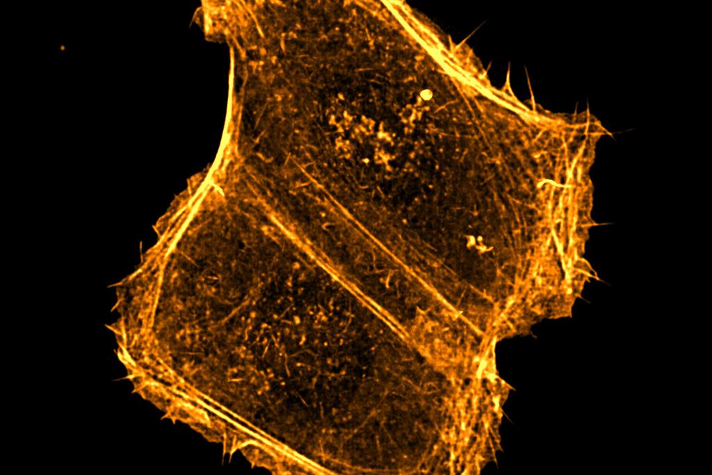
Caption: Actin dynamics in a U2OS cell expressing LifeAct-GFP imaged with the Lattice SIM 3D Leap mode and reduced phases. Objective: Plan-Apochromat 63× / 1.4 Oil
Optimize to the Needs of Living Samples
The flexibility of ZEISS Lattice SIM 5 allows you to balance the needs of your experiment by prioritizing resolution, speed, or by finding the right balance in between. Use the photon budget to enhance lateral resolution well below 100 nm or reduce the number of required raw images to boost acquisition speed and gentleness. ZEISS Lattice SIM 5 has a number of options for reducing raw images which allows you to select for the best acquisition settings that target your desired spatial and temporal resolution.
Caption: Time lapse imaging of the endoplasmic reticulum in a Cos-7 cell reveals highly dynamic structural changes. Sample courtesy of Miyawaki Lab, RIKEN Institute, Japan.
 and actin (Phalloidin Alexa Fluor 561, orange)")
 and actin (Phalloidin Alexa Fluor 561, orange)")
Get More Reliable Experiment Results
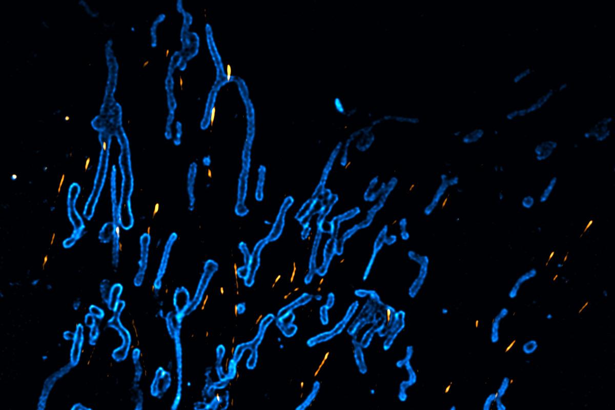
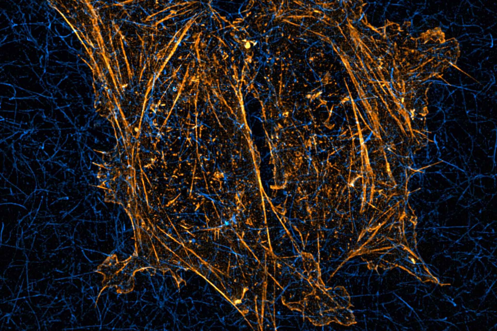
Cos-7 cells stained for microtubules (anti-tubulin Alexa Fluor 488, cyan) and actin (Phalloidin Alexa Fluor 561, orange)
Get More Reliable Experiment Results
ZEISS Lattice SIM 5 comes with outstanding out-of-focus light suppression, giving you the sharpest sectioning in widefield microscopy, even for highly scattering samples. The SIM² image reconstruction uses a special SIM point spread function to robustly reconstruct all structured-illumination-based acquisition data of your ZEISS Lattice SIM 5 with minimal image artifacts – for both living and fixed samples. Rest assured knowing that you are basing your experimental conclusions on reproducible data generated from a powerful and proven algorithm.
Caption: Cos-7 cells stained for microtubules (anti-tubulin Alexa Fluor 488, cyan) and actin (Phalloidin Alexa Fluor 561, orange)
The Technology behind ZEISS Lattice SIM 5


Lattice SIM
Your 3D Super-Resolution Technique for Live ImagingIn Lattice SIM, the sample is illuminated with a lattice spot pattern instead of grid lines as in conventional SIM. Due to its intrinsic two-dimensionality, the lattice pattern requires only translational repositioning but no rotation. This leads to a dramatic increase in imaging speed. In addition, the lattice pattern provides higher contrast to allow a more robust image reconstruction. Since the sampling efficiency is doubled compared to classic SIM, half as much light exposure is needed making Lattice SIM a preferred live cell imaging technique.


Widefield imaging
The image resolution is physically limited due to the diffraction limit. Additionally, the image quality suffers from out-of-focus blur and background signal.


Classic SIM imaging
To generate higher frequencies, the sample is illuminated with a grid pattern and imaged at different rotational and translational positions of this pattern. The processed image has twice the resolution in all three dimensions.


Lattice SIM imaging
The sample is illuminated with a lattice spot pattern instead of grid lines. Compared to classic SIM, sampling efficiency is two times higher. The lattice pattern gives higher contrast and is more robust for processing.


Reconstructed image
After acquisition, the resulting super-resolution image is calculated. With Lattice SIM, you can image longer with less bleaching and maintain image quality at higher frame rates.


SIM² Image Reconstruction
Double Your SIM ResolutionSIM² is a groundbreaking image reconstruction algorithm that increases the resolution and sectioning quality of structured illumination microscopy data. SIM² is compatible with all SIM imaging modes and fully integrated in ZEISS ZEN software.
Unlike conventional reconstruction algorithms, SIM² is a two-step image reconstruction algorithm. First, order combination, denoising, and frequency suppression filtering are performed. All the effects resulting from these digital image manipulations are translated into a digital SIM point spread function (PSF). The subsequent iterative deconvolution uses this PSF. Similar to the advantages of using experimental PSF for deconvolution of hardware-based microscopy data, the SIM² algorithm is superior to conventional one-step image reconstruction methods in terms of resolution, sectioning, and robustness.


SIM Apotome
Flexible Optical SectioningChoose the SIM Apotome acquisition mode to achieve fast overview images before zooming into super-resolution details. SIM Apotome uses structured illumination to give you fast optical sectioning of larger volumes with crisp contrast and high resolution in all dimensions.
SIM Apotome in combination with the SIM² reconstruction algorithm allows you to further adjust the gentleness of fast live-cell imaging with high contrast and resolution. Or use your new optical sectioning speed to increase productivity when acquiring large sample areas or large volumes at different magnifications.
Boost the Speed of SIM Imaging Even Further
Increase the temporal resolution and productivity for 2D and 3D imaging by using the speed enhancement modes.2D Burst mode
Get full temporal informationBurst mode processing uses the rolling window approach to let you observe processes in your living samples at up to 255 fps. Since Burst mode is a post-acquisition step, you have the flexibility to use it with previously acquired data sets. You decide how much temporal resolution is required for your data analysis.
3D Leap mode
Digital sectioning at a new levelFor demanding fast imaging in 3D, the Leap mode acquisition enables you to reduce your imaging time and lower the light exposure on your sample. This works by imaging only every third plane, for three-times higher volume imaging speed and three-times fewer light exposures.
Simultaneous Two-Color Imaging
Investigation of living samples very often focuses on interactions of different proteins or organelles. Simultaneous imaging of the involved structures is key to proper understanding of these highly dynamic processes. ZEISS Lattice SIM 5 can be equipped with two cameras in parallel and perform true simultaneous two-color imaging within your entire field of view.


Resolve the Details Hiding in the Depth
High-Quality Sectioning and Super-Resolution in Thick SamplesThe Lattice SIM illumination pattern exhibits both higher contrast and deeper sample penetration as compared to classical SIM. Achieve super-resolution images along with high-quality sectioning even in thick or scattering samples.
A novel clearing and embedding technology developed by Prof. Tang and his team (Hsiao et al., Nature Communications 2023) combined with the robust Lattice SIM illumination pattern and excellent image reconstruction technology enabled imaging throughout an entire mouse intestine section of ~200 µm thickness. Networks of blood vessels and nerves can be visualized with finest details even at this depth.
Observe Life’s Finest Details
Live imaging at high spatiotemporal resolutionZEISS Lattice SIM 5 combines high speed imaging with incredible light efficiency, low photon dosage and sensitivity. You can observe cellular, subcellular, and even sub-organelle structures in living specimens in 2D and 3D over time.
Mitochondria are highly dynamic organelles that constantly undergo fusion and fission events to ensure proper distribution of ATP across the cell. In order to do their job, they are known to interact with many other subcellular compartments including microtubules, which they ride on to get to their destinations, or the ER, which wraps around mitochondria to initially constrict their diameter before fission events.


