

ZEISS Lattice SIM 3
Fast Optical Sectioning of Developing Organisms and Tissue MicrostructuresZEISS Lattice SIM 3 is specifically designed to meet the imaging requirements of multicellular organisms and tissue sections. This system exploits the full potential of the SIM Apotome technology: fast optical sectioning at superior quality, large fields of view with access to smaller regions of interest, near-isotropic resolution, and the gentlest super-resolution imaging possible.
 and nuclei (NucRed Live 647).")
 and nuclei (NucRed Live 647).")
Capture Entire Model Organisms and Tissue Sections
ZEISS Lattice SIM 3 fully leverages SIM Apotome technology, to provide the most outstanding optical sectioning at large fields of view with near-isotropic resolution. ZEISS Lattice SIM 3 is your system of choice for fast imaging of larger volumes, such as 3D model organisms, embryos, organoids, or tissue sections. Whether you work with living or fixed samples, ZEISS Lattice SIM 3 provides access to structured illumination microscopy of multicellular organisms with superior penetration depth.
Caption: Spheroid stained for mitochondria (MitoTracker Green) and nuclei (NucRed Live 647).


Acquire Super-Resolution Images as Fast and Gentle as Widefield Images
Choose between the standard SIM Apotome imaging mode for the highest available resolution or the imaging mode with reduced phases for slightly lower resolution but significantly increased speed and gentleness. Combine SIM Apotome with the Leap mode to significantly speed up super-resolution acquisition. SIM Apotome makes even lossless acquisition possible, meaning for every reconstructed image just one raw image is needed.
Caption: Spheroid invading collagen matrix; cells are expressing Lifeact-tdTomato; color-coded depth projection.
Go from a Large-Field Overview to the Super-Resolution Details
For large sample experiments, ZEISS Lattice SIM 3 offers the most advantageous combination of a large field of view and super-resolution imaging. SIM Apotome mode in combination with SIM² image reconstruction enables lateral super-resolution down to 140 nm with superior optical sectioning and sensitivity. Additionally, imaging in Lattice SIM mode with a ZEISS 25× multi-immersion objective and subsequent SIM² processing provides similar lateral resolutions with larger fields of view and more flexible adaptation to the refractive index of your sample.
Caption: Murine brain imaged in SIM Apotome and Lattice SIM modes over a Z stack range of 170 µm. Overview image: Plan Neofluar 10×. Volume rendering: LD LCI Plan-Apochromat 25× / 0.8 Imm Corr. Sample courtesy of Herms Lab (MCN, University of Munich, Germany).
The Technology behind ZEISS Lattice SIM 3


SIM Apotome
Optical Sectioning with Exceptional QualityLive cell imaging with a widefield system often suffers from out-of-focus blur or background signal. These effects can decrease contrast and resolution. ZEISS Lattice SIM 3 fully leverages the benefits of the SIM Apotome technology, enabling structured illumination microscopy for low-magnification objectives to give you fast and gentle optical sectioning for your multicellular samples.
The SIM Apotome acquisition mode in combination with the SIM² reconstruction algorithm allows you to further adjust the gentleness of fast live-cell imaging with high contrast and resolution. Or use your new optical sectioning speed to increase productivity when acquiring large sample areas or large volumes at different magnifications.


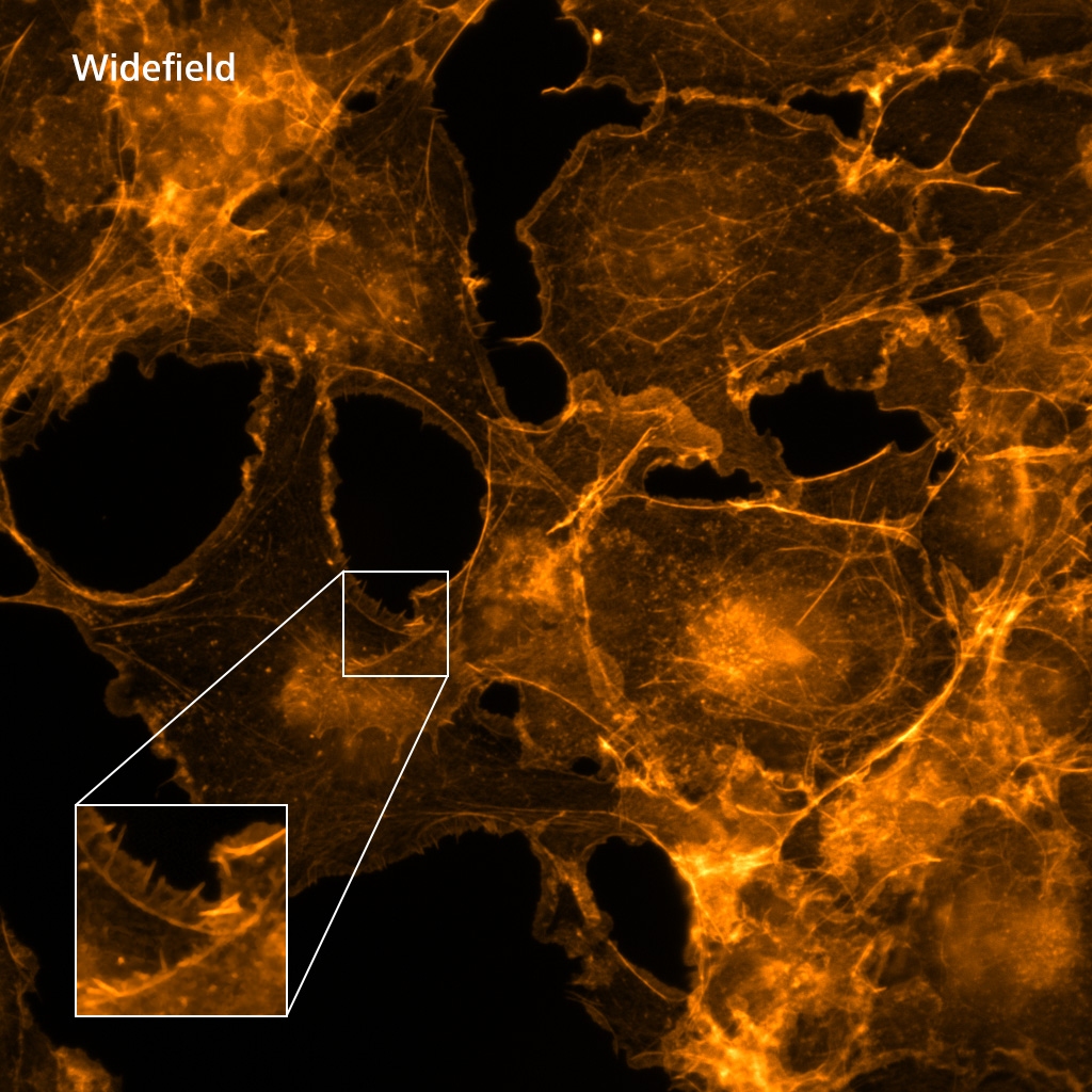
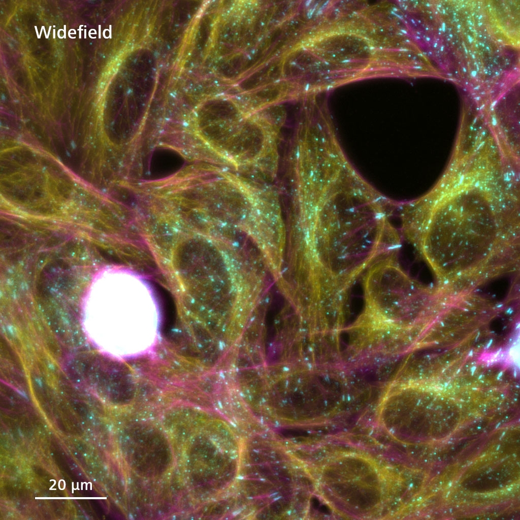
Widefield image
The image quality suffers from out-of-focus blur and background signal. (The signal from the focal plane is encircled by a white dashed line.)


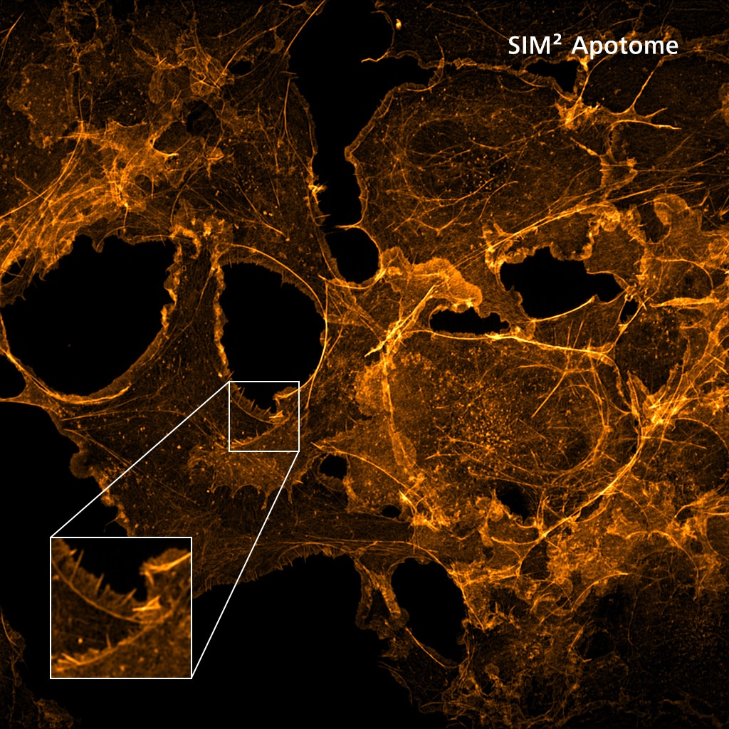
SIM Apotome acquisition
A grid pattern is used to illuminate and rapidly modulate the fluorescence signals in the focal plane at 3 or 5 different grid positions.


Reconstructed optical section
After acquiring images with different grid positions, these frames are combined into a resulting image which contains only information from the focal plane.
Balance Your Need for Speed and Resolution
Higher imaging speeds and decreased light exposures are a constant demand in imaging experiments. The robustness and flexibility of ZEISS Lattice SIM 3 structured illumination patterns plus the image reconstruction software allow a significant reduction to the number of phase images required for SIM Apotome acquisition mode, and, importantly, this only causes a slight decrease in the resolution of the final images. SIM Apotome acquisition can be operated at 3 phase images per frame, increasing the imaging speed by 66 %. The increased speed is also advantageous for fast screening of large sample areas, such as tissue sections.
In combination with the Leap mode, you can further decrease the number of phase images per final frame to enable the gentlest super-resolution imaging possible.

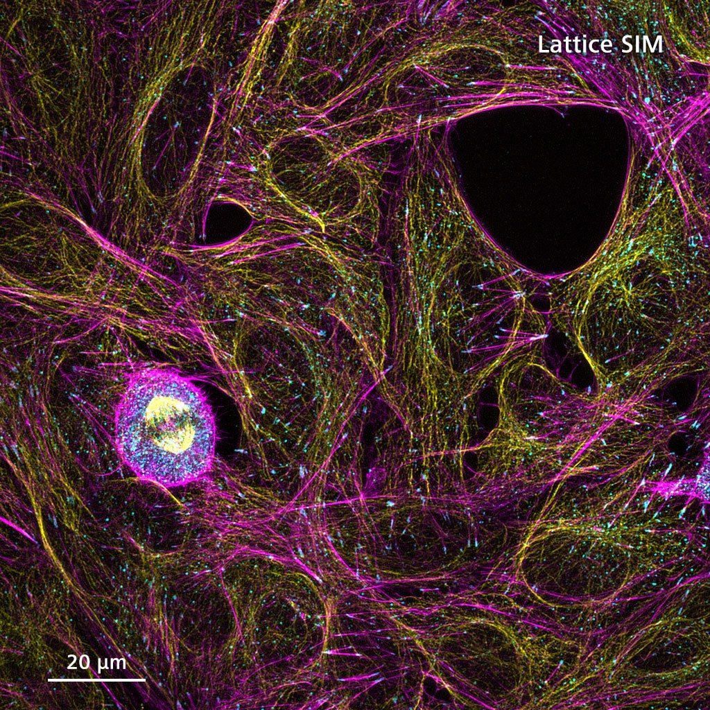

Lattice SIM
Your 3D Super-Resolution TechniqueZEISS Lattice SIM 3 also includes the Lattice SIM imaging mode optimized for use with a special 25x multi-immersion objective. The sample area is illuminated with a lattice spot pattern instead of grid lines. The lattice pattern provides higher contrast to allow deeper sample penetration and, in combination with SIM², robust image reconstruction with super-resolution down to 140 nm.
Boost the Speed of SIM Imaging Even Further
Increase the temporal resolution and productivity for 2D and 3D imaging by using the speed enhancement modes.2D Burst mode: Get full temporal information
Burst mode processing uses the rolling window approach to let you observe processes in your living samples at up to 255 fps. Since Burst mode is a post-acquisition step, you have the flexibility to use it with previously acquired data sets. You decide how much temporal resolution is required for your data analysis.
3D Leap mode: Digital sectioning at a new level
For demanding fast imaging in 3D, the Leap mode acquisition enables you to reduce your imaging time and lower the light exposure on your sample. This works by imaging only every third plane, for three-times higher volume imaging speed and three-times fewer light exposures.
, CD8 cells (yellow) and Leishmania parasites (magenta)")
, CD8 cells (yellow) and Leishmania parasites (magenta)")
Region of interests of a skin tissue section stained for cell nuclei (cyan), CD8 cells (yellow) and Leishmania parasites (magenta). Objective: 25× / 0.8 multi-immersion. Image courtesy: Helen Ashwin, Department of Biology, University of York, UK.
Super-Resolution Imaging in Immunology
Zoom into the DetailsImmunofluorescence of tissue sections is commonly used in immunological research to investigate distribution of and interactions between pathogens and immune cells, all with the aim to develop novel therapies for pathogenic diseases. For compelling results, it is not only crucial to image complete sections as to not miss relevant areas but also to image with enough resolution to identify and quantify individual events.
In the application example shown here, skin tissue sections were imaged to investigate distribution of CD8 cells relative to Leishmania parasite infection sites. The enlarged area is a digital zoom-in only, meaning that it is possible to zoom into any region of the overview image and quantify cell nuclei, CD8 cells and Leishmania parasites.


Digital zoom into the image above. Parasites can be visualized and quantified in each cell of the section. Image courtesy: Helen Ashwin, Department of Biology, University of York, UK.


Top: Lower half of Drosophila slice stained for nervous system and synapses (Anti-HRP, orange). Objective: Plan-Neofluar 10× / 0.8 Air.
Bottom: Also stained for synaptotagmins (Anti-synaptotagmin, cyan). Objective: LD LCI Plan-Apochromat 25× Imm Corr, imaged with SIM Apotome and Lattice SIM for comparison. Image courtesy of Prof. Sean Sweeney, University of York, UK.
Super-Resolution Imaging in Neuroscience
Understand How Neurons Respond to Damage, Disease and Metabolic ChangeThe synaptic structure and in particular the actives zones where synaptic vesicles are released are key players in signal transmission and proper function of neurons. The imaging of active zones require resolution beyond what can be achieved by standard confocal microscopy.
The lab of Prof. Sean Sweeney investigates a novel mutant that is a regulator of neuronal survival and metabolic responses. Nervous system and synapses are co-labelled with synaptotagmins to observe the general structure of the synapse and distribution of the presynaptic vesicles. Super-resolution microscopy helps identify and quantify the differences in synapse structure and composition of the active zones.
Super-Resolution Imaging of Living Yeast
Gentle and Fast with Near-Isotropic ResolutionLive yeast cells are among the most challenging samples in fluorescence microscopy. They are extremely light sensitive and smaller than most used cell lines. Furthermore, yeast cells grow in suspension; they can move freely in the culture dish and are of spherical shape, without clearly defined orientation. Tackling all these challenges requires extremely gentle and fast imaging combined with high resolution in all spatial dimensions.
SIM² Apotome is the perfect tool to image live yeast cells with super-resolution, yet fast and gentle enough to observe the cells over extended periods of time. The example clearly demonstrates this unique capabilities. Various subcellular compartments (surface marker, endosomes, vacuole, endoplasmic reticulum) were tagged with superfolder GFP and imaged for 12 hours.
Image Large Volumes
Get Finest Detail Even at DepthSIM Apotome combined with reduced phases and Leap mode allows you to image large volumes extremely fast and efficiently. Crunch through large volumes fast by recording only one raw image per final reconstructed image. Select regions of interest, switch objectives and use Lattice SIM to gain super-resolution images with a lateral resolution down to 140 nm within the context of the whole sample.
A novel clearing and embedding technology developed by Prof. Tang and his team (Hsiao et al., Nature Communications 2023) combined with the advantages of SIM Apotome acquisition and excellent image reconstruction technology enabled us to image an entire mouse intestine section of 3 mm × 4 mm and ~200 µm thickness within a couple of minutes. Networks of blood vessels and nerves can be visualized with finest details even at depth.


