AI Microscopy Solutions
Enhancing Image Acquisition, Analysis and Service
Discover the seamless integration of AI into microscopy, and unleash the potential of your research like never before.


The Future of Microscopy with AI-Powered Solutions
Artificial Intelligence (AI) has been a game-changer in many sectors, and microscopy is no exception. ZEISS AI Microscopy Solutions leverage the power of artificial intelligence to revolutionize the way you acquire images, analyze and interpret microscopic data and manage the performance of your microscope.


Integrating AI into Microscopy
Our AI microscopy solutions are designed to enhance precision, speed, and scalability in your research, offering unprecedented insights into the microscopic world. Whether you are in the field of life sciences, materials science, geosciences, electronics or education and routine, our AI microscopy solutions are here to transform your microscopic exploration into an experience of the future.


FAQs
-
AI-based image analysis in microscopy utilizes artificial intelligence, including machine learning and deep learning, to enhance the analysis of microscopic images. It involves training algorithms to recognize patterns and structures, improving the accuracy and efficiency of tasks such as sample detection, image segmentation, denoising, object classification, and 3D reconstruction. This technology accelerates and automates image analysis in microscopy for more effective scientific research and diagnostics.
-
In microscopy, deep learning involves the training of artificial neural networks to independently analyze microscopy images by learning patterns and structures from datasets annotated by experts. This training enables the networks to perform tasks such as image segmentation, object detection, and classification with increased precision and efficiency. For a deeper understanding of the distinctions between machine learning and deep learning, you can refer to our AI eBook.
-
The integration of AI in microscopy guarantees the acquisition and analysis of multidimensional image data at high throughputs and in a reproducible manner—fundamental for scientific discovery. These technologies automate routine manual tasks, liberating valuable time for researchers to design more experiments and innovate at an accelerated pace.
-
a. Whether seeking new cancer therapies through subcellular observation in a fluorescence microscope, designing battery materials via microstructure analysis with SEM, or optimizing hydrocarbon yield through mineralogy study with an X-ray microscope, AI provides insights from both small and large datasets at higher throughputs. This versatility underscores the transformative impact of AI in accelerating scientific discoveries across numerous domains in microscopy.
-
Traditional image analysis relies on predefined rules, such as simple thresholding, to extract information from images. For instance, using a fixed threshold to segment images based on pixel intensity. However, these rule-based approaches can be unreliable when images deviate from ideal conditions, leading to irreproducible results. In contrast, AI-based analysis goes beyond specific rules, as AI systems learn hidden patterns from diverse datasets. This enables them to generalize and maintain robustness, even when imaging conditions change, ensuring more reliable and adaptable results in microscopy.
-
AI is progressively addressing everyday challenges, and this trend extends to microscopy, where every aspect of the workflow stands to benefit. As generative AI continues to evolve, serving as a valuable assistant, we anticipate more natural language-driven interactions with microscopy systems. Imagine a user loading a multi-well plate with numerous experiments and simply asking, "Please image these samples at 10X and summarize the insights from all the wells." This vision foresees a seamless transition from sample to insights, eliminating the need for manual setup, data transfer, preprocessing, segmentation, and classification. The future of microscopy with AI promises heightened productivity, even in handling massive experiments involving tens of thousands of iterations.
-
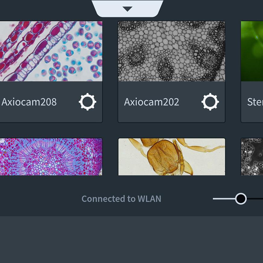
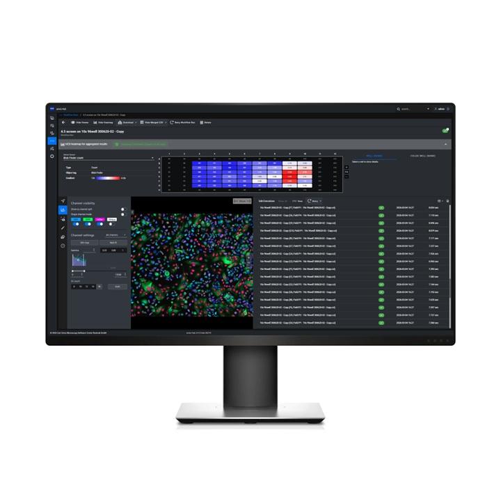
For those proficient in Python coding, implementing AI-based solutions in microscopy involves utilizing open-source libraries. However, this requires expertise in handling large datasets and a deep understanding of managing image patches and reconstructing them to their original size. Fortunately, ZEISS offers a range of AI tools through a user-friendly, no-code interface accessible to everyone. In both ZEN and arivis software ecosystems, users can leverage tools for denoising, segmentation, classification, and more using AI. For example, the arivis Cloud platform provides infrastructure for training custom AI models in image segmentation, usable either on the cloud or locally in arivis Pro or ZEN software packages, ensuring consistent results across platforms.
-
Whether it is ZEN, ZEN Blue, arivis Pro, arivis Hub, or arivis Cloud, every ZEISS software incorporates AI tools for image analysis. This ensures that users benefit from advanced capabilities, making each software package a powerful tool for enhancing productivity.
-
Deep learning (DL) is a subset of machine learning (ML), but when people mention machine learning, they typically refer to conventional ML, where predefined digital filters are applied to training data. This conventional approach involves feature engineering, where someone assembles these filters, often working with a limited number of features, typically up to a few tens. This limitation can make it challenging for these algorithms to capture the complexity present in some images. In contrast, deep learning is a feature learning technique that optimizes millions of parameters by learning from vast training data, making it more robust against changing image conditions. In essence, the key distinction lies in the few engineered features of ML versus the multitude of learned features in DL.