ZEISS Celldiscoverer 7
Adaptable Automation for Advanced WorkflowsIf your research requires explorative high-content imaging, you are often faced with a trade-off between the desired image quality and the need to capture large amounts of data efficiently. ZEISS Celldiscoverer 7 is your research companion for collecting statistically relevant data, giving you easy access to high-quality imaging, adaptability to demanding experiments, and stable long-term operation.


Easy Access to High-Quality Imaging
You don't have to become a microscopy expert to acquire high-quality data. Simply insert your sample carrier and let ZEISS Celldiscoverer 7 make all the necessary settings. Automatic calibration routines ensure optimal conditions and reproducible results. The system finds and keeps the focus after detecting the sample carrier and its optical properties. Spherical aberrations are corrected automatically to always deliver best contrast and resolution. For demanding long-term imaging, Celldiscoverer 7 gives you automated water immersion and integrated incubation to keep your cells happy with just the right environmental conditions. Without manual adjustments, even if you operate the microscope remotely, you get unbiased data that meet your highest expectations.


Adaptable to Demanding Experiments
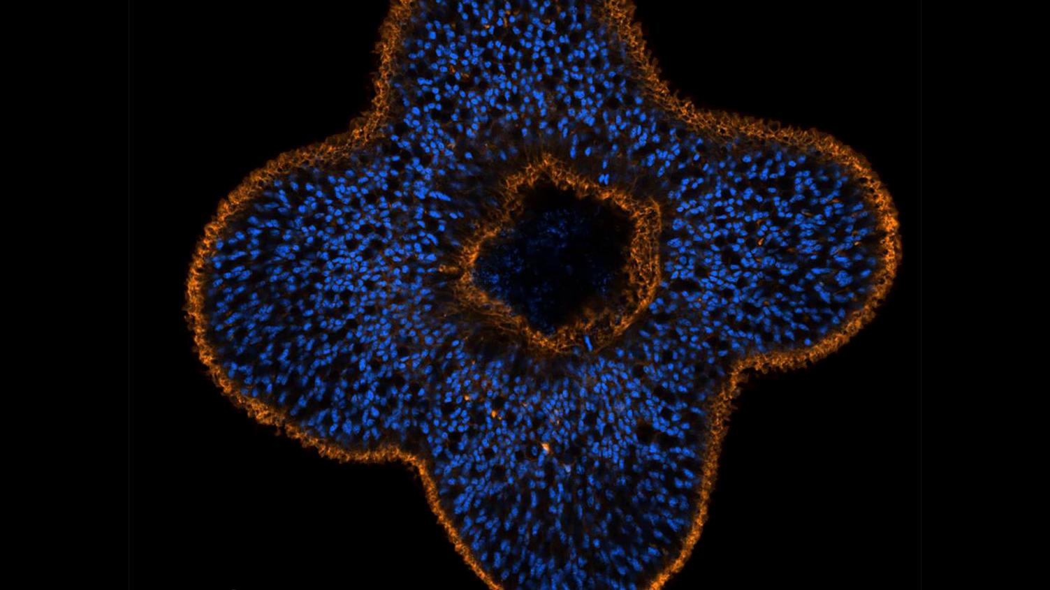
Optimized for explorative high-content applications, Celldiscoverer 7 gives you experimental freedom with a range of imaging modes that can be freely combined to tailor data acquisition to individual requirements. Go for fast widefield imaging to perform live cell experiments and rapid time-lapse recordings. Enable label-free imaging in multi-well plates without artifacts. Enjoy a wide spectral range for multi-fluorescence experiments and add unique transmitted light contrast methods. For stunning 3D data, add confocal imaging with LSM 910 and Airyscan. Combine all these possibilities in customized workflows and let the built-in intelligence of Celldiscoverer 7 work for the benefit of your research.
Image caption: Rat cortical primary culture. Sample courtesy of H. Braun, LSM Bioanalytik GmbH, Magdeburg, Germany


Reliable in Multi-User Environments
In facilities with multiple users, the robustness of an imaging system is key for its acceptance and efficient use. Once a workflow has been set up with Celldiscoverer 7, it can be executed reproducibly again and again, even if the system has been used for other experiments in the meantime – no manual recalibration is needed. The robust design of the system enables stable long-term operation and prevents unwanted user intervention – damaged objective lenses are a thing of the past. Predictive service offers lasting and optimal instrument performance for increased system uptime and reliable results from automated imaging. Predictive analytics allow to determine the optimal time point for the next service visit, e.g., when a component is about to reach the wear limit.
ZEISS Celldiscoverer 7 Product Family
")
")
|
")
")
|
|
|---|---|---|
|
Imaging Modes |
|
|
|
Widefield imaging |
✓ |
✓ |
|
Confocal imaging with LSM 910 |
– |
✓ |
|
LSM Plus |
– |
optional |
|
Airyscan 2 |
– |
optional |
|
Airyscan 2 Multiplex |
– |
optional |
|
Airyscan jDCV |
– |
optional |
|
Automated Workflows and Advanced Methods |
|
|
|
Guided Acquisition |
✓ |
✓ |
|
Bio Applications |
✓ |
✓ |
|
ZEN Intellesis – AI based image segmentation |
✓ |
✓ |
|
ZEN Connect – connect all multi-modal data |
✓ |
✓ |
|
(Automated) Photomanipulation |
– |
✓ |
|
Optical sectioning |
– |
✓ |
|
Simultaneous widefield and confocal imaging |
– |
✓ |
|
External Cameras |
|
|
|
ZEISS Axiocam 807 mono |
optional |
– |
|
ZEISS Axiocam 820 mono |
optional |
– |
|
ZEISS Axiocam 712 mono R2 |
optional |
– |
|
Photometrics Prime 95B |
optional |
– |
|
Hamamatsu Orca Flash 4.0 V3 |
optional |
– |
High-Quality Data Acquisition Made Easy
Explore ZEISS Celldiscoverer 7


Automatic Adaptation to Your Samples and Sample Carriers
Live cell imaging requires objectives with high numerical apertures. Those objectives will only deliver high contrast and sensitivity if their optics can adapt to variations in bottom thickness or to the material of different sample carriers. You are free to use Petri dishes, chamber slides, multi-well plates, plastic or glass, thin or thick vessel bottoms, low-skirt or high-skirt plates. Automatic sample recognition detects all relevant vessel features while loading your sample. Autocorr adjusts the correction ring of the objective to compensate for spherical aberrations.


ZEISS Celldiscoverer 7 water immersion
Autoimmersion: There Is No Life Without Water
In cell biology or screening applications, your samples mostly consist of water and / or will be mounted in aqueous solutions. Celldiscoverer 7 combines an outstanding 50x water immersion objective with rapid automated immersion supply and removal. An elastic silicon membrane simultaneously seals the sample chamber to avoid unnecessary airflow while protecting the system from potential liquid spillage. You can perform unbiased live cell experiments at 37 °C over several days or carry out extensive scanning processes on multi-well plates.


Find Focus – Keep Focus
Use the hardware-based Find Focus function to automatically focus your sample and find your region of interest quickly with just a single click. Select Definite Focus to maintain the focal position throughout your live cell experiment, whether it takes a few seconds or several days. Or combine both methods: Celldiscoverer 7 can automatically create focus maps for multiple positions in long-term time-lapse experiments.
The Adaptive Lens Guard protects the objective from collisions with your sample vessel or hardware components, automatically maximizing the available scanning area. Sample carrier holders are designed so that the entire sample can be imaged even at maximum magnification, e.g., in chamber slides.


LED module with up to 7 individually controlled excitation lines for maximum flexibility in the choice of dyes
LED module with up to 7 individually controlled excitation lines for maximum flexibility in the choice of dyes
LED Technology for Live Cell Imaging
Celldiscoverer 7 brings you all the advantages of LED technology for efficient widefield illumination with low phototoxicity, fast switching times and homogeneous illumination. Real-time and long-term stabilization with performance optimization ensure comparability of images. The LED module includes up to 7 individually controlled excitation lines for maximum flexibility in the choice of dyes, from deep blue to far red. You always get enough excitation power to shorten exposure times and to speed up your image acquisition.
LSM 910 with Airyscan 2
Automated Confocal 3D ImagingLife happens in 3D. Add LSM 910 with Airyscan 2 and get the best of both worlds: ease of use and automation from a fully integrated microscope platform and the superb confocal image quality and flexibility of LSM 9 family. Super-resolution 3D imaging with up to 1.9x resolution improvement becomes possible. Easily separate multiple labels with spectral imaging. Analyze dynamic processes in living samples with photomanipulation for FRAP, FRET or related techniques. Precisely connect widefield and confocal images. Fast mixed-mode acquisition simplifies and speeds up your workflow and gives you unique insights into your sample.


Enhanced Throughput with Robotic Loader Integration
Maximize the efficiency of your imaging workflow with Celldiscoverer 7's optional robotic sample loader, provided by our trusted partner, Peak Analysis & Automation (PAA). This advanced automation solution seamlessly integrates with the system to significantly increase throughput, enabling high-volume sample processing without compromising on precision or quality. The robotic loader is designed to handle all SBS multi-well plate formats. By automating the loading and unloading processes, researchers can focus on data analysis and interpretation, while Celldiscoverer 7 takes care of the repetitive imaging tasks, streamlining your research operations and accelerating discovery. Additional modules, such as liquid handling can be integrated into the whole workflow.


Comprehensive Image Analysis with arivis Software
Unlock the full potential of your imaging data with the powerful arivis Pro image analysis software, fully capable to complement Celldiscoverer 7. This versatile software suite offers robust tools for analyzing a wide range of imaging data, from 2D snapshots to complex 3D and 4D datasets. With its intuitive interface and advanced algorithms, arivis enables precise quantification and visualization of cellular structures and dynamics, providing deeper insights into your research. Whether you are conducting high-content screening or detailed morphological studies, arivis ensures that you can efficiently process and interpret your Celldiscoverer 7 data, transforming raw images into meaningful scientific information.
In addition to arivis Pro, arivis Cloud is an online tool designed to train neural networks for challenging image segmentation tasks. These neural networks can be seamlessly integrated into arivis Pro, enhancing its capabilities for precise image analysis. Furthermore, arivis Hub combines online storage with powerful online processing functionalities, allowing arivis Pro pipelines to run in parallel across multiple datasets. This integration ensures that you can manage and analyze large volumes of imaging data efficiently, leveraging the full suite of arivis software solutions to maximize the potential of your Celldiscoverer 7.


")
")


")
")
 cells")
 cells")