

Imaging Large Model Organisms
Fixed or LivingWhen imaging large model organisms, it can be difficult to achieve high resolution imaging of minute details deep inside your samples. The specimen may be too thick or dense for your current imaging system.Or you may be challenged with the unsurmountable length of time required to image the entire organism at your desired resolution.
If you are creating time lapse videos of living, large organisms, you will need to minimize phototoxicity, photodamage or illumination-induced biological effects and artefacts.


Extended Time Lapse Imaging of Developing Organisms
Traditionally, intense illumination light was required to excite fluorophores deep within living model organisms. This light can be toxic to the sample, bleach the fluorescent labels and possibly cause illumination-induced behavior – making extended time lapse imaging impossible to document how organisms naturally develop. ZEISS systems – whether confocal, light sheet or the automated system for live sample – ZEISS Celldiscoverer 7 – require minimal excitation light, ensuring extremely gentle imaging that enables you to image your developing organism from hours to days.
 forearm")
 forearm")
 forearm")
 forearm")
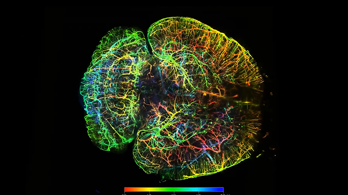
Salamander (axolotl) forearm, optically cleared, imaged with light sheet microscopy. Image courtesy of Wouter Masselink & Elly Margaret Tanaka, IMP – Research Institute of Molecular Pathology, Austria
Salamander (axolotl) forearm, optically cleared, imaged with light sheet microscopy. Image courtesy of Wouter Masselink & Elly Margaret Tanaka, IMP – Research Institute of Molecular Pathology, Austria
Achieve New Depth with Cleared Samples
Optical tissue clearing has enabled imaging much deeper into large specimens. Light sheet microscopy, such as with Lightsheet 7, or confocal microscopy, such as with the LSM 9 family, enable you to achieve crisp, high resolution 3D imaging of your cleared frogs, mice or other large model organisms in a surprisingly short amount of time.


Visualization of various organs in a resin-embedded mouse embryo imaged with X-ray microscopy. Sample courtesy of Massachusetts General Hospital, USA
Visualization of various organs in a resin-embedded mouse embryo imaged with X-ray microscopy. Sample courtesy of Massachusetts General Hospital, USA
3D Non-destructive Histological Imaging
X-ray microscopy offers non-destructive, 3D histological imaging of entire model organisms. Tissues and individual organs such as eye, brain or kidney can be studied histologically, without dissection, inside the organism on a cellular level. Depending on your application, ZEISS Xradia Context microCT enables fast scanning of large samples, ZEISS Xradia Versa is adaptable for versatile applications and ZEISS Xradia Ultra provides the highest 3D resolution.


Related Products


-
1
* The images shown on this page represent research content. ZEISS explicitly excludes the possibility of making a diagnosis or recommending treatment for possibly affected patients on the basis of the information generated with an Axioscan 7 slide scanner.