Discover a New Light Sheet Microscope
For Multiview Imaging of Living and Cleared Specimens

ZEISS Microscopy
Abstract
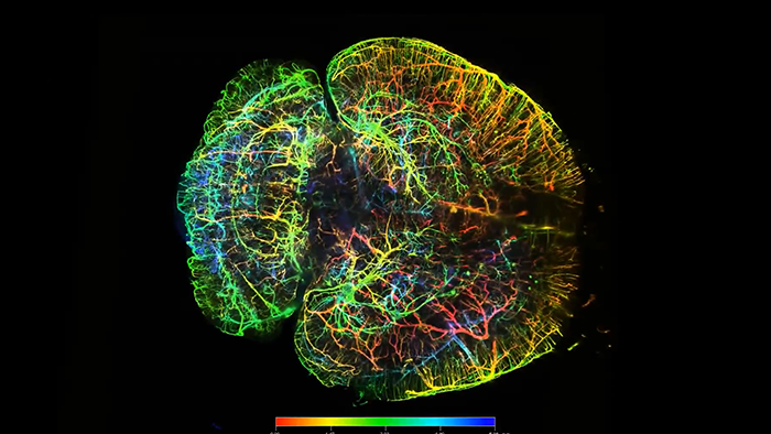
Light Sheet Fluorescence Microscopy (LSFM) is a powerful alternative to traditional epi-illumination techniques, especially for 3D imaging within whole live organisms and large tissue explants. By selectively illuminating the observed optical section with a thin sheet of light, photo-damage and bleaching are reduced to a minimum, making light sheet microscopy ideal for nondestructive imaging of fragile samples. Due to the speed and efficiency of image acquisition, LSFM is particularly well suited for volume imaging of samples that have been rendered transparent through various tissue-clearing methods.
This webinar will include a brief introduction to light sheet microscopy and a short overview of optical clearing techniques. We will demonstrate how common challenges of cleared tissue imaging can be solved with features such as flexible sample mounting and the ability to accommodate different optical clearing agents. We will also explore several tools for micro- to meso-scale imaging, including a variety of long working distance objectives, that make light sheet microscopy an extremely versatile technique for imaging cleared tissues.