Why full cell volume imaging in thick tissues matters for decisions in research and drug development
Understanding cancer requires more than thin tissue sections. At Harvard Medical School’s Laboratory of Systems Pharmacology, researchers combine imaging innovation, computational analysis, and clinical collaboration to uncover how diseases emerge and evolve in human tissue. Their mission is clear:
The Laboratory of Systems Pharmacology (LSP)'s mission is to drive innovation in healthcare through the understanding of diseases.
Thin 5 micron sections often fragment cells and obscure how immune, stromal and tumor cells truly interact. By extending cyclic immunofluorescence (CyCIF) to thicker tissues from FFPE specimens and incorporating high resolution 3D imaging, Clarence and his colleagues aim to provide more reliable biological insight for both research and drug development.
2026,
LSP
The Laboratory of Systems Pharmacology (LSP) combines imaging, computation, and clinical collaboration to study human disease.
Scientific mission and cross-sector relevance
At the LSP, cyclic immunofluorescence evolved from monolayer plate-based experiments into tissue-scale analysis of biopsies across cancers and infectious disease.
Academia – whole cell, membrane level insight refines hypotheses on cell states and spatial context.
Industry – more trustworthy phenotyping and contact mapping strengthens interpretation and reduces risk.
Why whole cell imaging changes what we can know
A key realization in Clarence’s work was understanding to what extent biological interpretation of cells is distorted when using thin sections. Traditional 2D slices capture only fragments of cells, which affects marker interpretation and cell–cell interaction mapping.
He describes a pivotal moment when thick section 3D CyCIF revealed different staining patterns across the 50 plex reconstruction of a set of human clinical samples. Immune cells, tumor cells and their neighbors appeared with intact shapes and membrane contacts – features previously hidden in 2D.
We routinely receive overwhelmingly positive feedback from our clinical collaborators who went on this journey with us to make 50-plex 3D CyCIF possible […] A real breakthrough was seeing how visually different these proteins looked in thick patient samples compared to thin sections.
3D high-resolution imaging reveals:
polarized and punctate protein distribution patterns within cells
continuous membrane–membrane contacts between neighboring cells
cell shape (microvilli, filopodia, dendritic processes, etc) and texture
complete cell volumes important for cell type classification
rare populations such as T PEX cells with higher accuracy
Moving to 30–50 µm tissue with confocal sectioning preserves full cell volumes and supports:
precise membrane contacts
intracellular protein distribution and texture
lineage and state interpretation
greater confidence for both academic and industry applications
2026,
LSP
High‑plex imaging data helps researchers identify biologically relevant patterns and regions for further analysis.
How clinicians and researchers working together in 3D
3D imaging starts long before a scan is taken. Clinical collaborators annotate regions of interest such as early cancer or vertical growth phase on H&E sections based on their expert medical knowledge. This targeted approach ensures that only the most biologically meaningful tissue regions receive high‑resolution 3D coverage with minimal risk of false positives.
Academia – exploration of diverse cell states in a resource-efficient manner.
Industry – reproducible, focused acquisition aligned with quality controlled workflows.
Why studying disease in 3D matters
Researchers at Harvard Medical School explain how high‑plex imaging helps reveal biology in its native tissue context.
Third-party Content Blocked
The video player is blocked due to your cookie preferences. To change the settings and play the video, please click the button below and consent to use of "Functional" tracking technologies.
2026,
LSP
Iterative imaging workflows combine multiplex staining, optical sectioning and computational analysis to generate high‑dimensional 3D datasets.
Building workflows that balance discovery and reliability
The thick section CyCIF process uses iterative cycles of staining, imaging, and bleaching using all open-source reagents and protocols. Each round includes a nuclear marker for reliable 3D registration. Tile based segmentation and reconstruction handle datasets of 200–900 terabytes (depending on size of region to be imaged) across dozens of channels.
Academia – flexibility to develop antibody panels of novel markers and incorporate different image analytical methods.
Industry – standardized protocols for reproducible signal quantification to reduce interpretive risk.
Clarence emphasizes that achieving sufficient contrast in 35–50 micron FFPE tissue required optical sectioning, high-NA oil immersion objectives, sensitive detectors, and flexible software to define regions of interest and align cycles.
We maintain that we had to prioritize contrast over ease of use and speed […]. We needed a microscope system that could perform with low fluorescence signals through sensitive optics as well as sophisticated denoising algorithms.
Quality practices that strengthen confidence
The team’s approach reflects a quality mindset shaped by collaboration:
validated and custom-designed antibody panels for tumor, immune, and stromal markers
nuclear and cytoplasmic dyes for cross cycle alignment and segmentation respectively
collaboration with clinical teams for specimen relevance
open protocols and publicly available registered data
For the LSP, open data is a scientific responsibility. Because the method is resource intensive, the team sees public availability of complex data to help the broader community explore questions that exceed the capacity of any single lab.
2026,
LSP
Whole‑cell segmentation enables researchers to quantify cellular structure, morphology and spatial relationships in three dimensions.
2026,
LSP
Whole‑cell segmentation enables researchers to quantify cellular structure, morphology and spatial relationships in three dimensions.
Sharing knowledge to accelerate spatial biology
Throughout development, Clarence works with microscopy specialists and computational biologists to refine contrast, reduce crosstalk, integrate second harmonic generation or adopt new segmentation tools like uSegment3D and u-Unwrap3D.
Academia –teaching and methodological transparency.
Industry – validated, repeatable workflows for scale.
It also reflects the LSP’s belief that thick section imaging must become accessible beyond a small number of expert labs.
2026,
LSP
Confocal optical sectioning enables researchers to visualize cellular structures deep within thick tissue while preserving spatial context.
2026,
LSP
Confocal optical sectioning enables researchers to visualize cellular structures deep within thick tissue while preserving spatial context.
Imaging capabilities that enable thick section 3D CyCIF
For reliable 3D in 30–50 µm FFPE tissue, the team highlights needs such as:
contrast optimized optical sectioning
sensitive under low signal conditions
high NA objectives optimized for sample and coverslip
clinician-selected samples and ROIs
software that supports flexible cycle alignment
stability with handling large files
These capabilities help labs adopt or replicate thick section multiplexing even with most challenging specimens – relevant to both grant funded groups and ROI driven environments.
2026,
LSP
High‑plex 3D imaging preserves spatial context, revealing how different cell populations are organized within intact human tissue volumes.
2026,
LSP
High‑plex 3D imaging preserves spatial context, revealing how different cell populations are organized within intact human tissue volumes.
Applications that reveal cellular truth in human tissue
Examples that Clarence and his colleagues highlight include:
intact immune and tumor cells with extended processes in melanoma tissue
PD1–PDL1 punctate distribution at actual membrane contacts
vascular segments spanning depth ranges hard to interpret in 2D
rare cell populations such as T PEX cells in preserved spatial niches
multicellular communities where several immune cells interact to engage a single tumor cell effectively
Spatial biology in action
Explore how high‑plex imaging reveals cellular organization, vascular structures and tissue microenvironments in human samples.
Third-party Content Blocked
The video player is blocked due to your cookie preferences. To change the settings and play the video, please click the button below and consent to use of "Functional" tracking technologies.
High‑plex 3D imaging preserves spatial context, revealing how different cell populations are organized within intact human tissue volumes.
Cellular organization in melanoma tissue
Third-party Content Blocked
The video player is blocked due to your cookie preferences. To change the settings and play the video, please click the button below and consent to use of "Functional" tracking technologies.
Confocal optical sectioning enables researchers to visualize vascular features across depth while preserving their spatial relationship to surrounding tissue.
Visualizing vascular structures in 3D
Third-party Content Blocked
The video player is blocked due to your cookie preferences. To change the settings and play the video, please click the button below and consent to use of "Functional" tracking technologies.
Three‑dimensional imaging helps reveal how immune and tumor cells are arranged within complex tissue microenvironments.
Mapping cellular communities in melanoma
Third-party Content Blocked
The video player is blocked due to your cookie preferences. To change the settings and play the video, please click the button below and consent to use of "Functional" tracking technologies.
Confocal optical sectioning enables researchers to visualize vascular features across depth while preserving their spatial relationship to surrounding tissue.
Visualizing vascular structures in 3D
Third-party Content Blocked
The video player is blocked due to your cookie preferences. To change the settings and play the video, please click the button below and consent to use of "Functional" tracking technologies.
Three‑dimensional imaging helps reveal how immune and tumor cells are arranged within complex tissue microenvironments.
Mapping cellular communities in melanoma
Advances in whole‑cell imaging are opening new opportunities to study disease in its native biological context.
A future shaped by high-plex whole cell volume insight
The LSP’s work reflects a shift in spatial biology: from incomplete fragments toward whole cell volumes, context rich in increasing our understanding of disease. This enables both new scientific questions and more informed decisions in translational environments.
Now that we've demonstrated that we can measure full cell volumes and their surrounding neighbors in 3D, we can begin to apply these measurements to look at underreported cell types and immune structures in a broader set of disease settings.
In Brief
3D CyCIF captures intact whole-cell volumes instead of fragmented thin sections, supporting more reliable phenotyping and immune–tumor interaction mapping.
Standard 4–5 µm tissue sections often contain incomplete cells, which can distort marker interpretation and obscure cell–cell contacts. By imaging thicker 30–50 µm FFPE tissue in 3D, researchers can analyze polarized and punctate protein patterns, cell shape, membrane contacts and spatial neighborhoods in their preserved tissue context. This supports stronger biological interpretation for academic research and translational drug discovery.
Clinical collaborators help identify biologically relevant tissue regions, while researchers apply 3D CyCIF imaging and computational analysis to study those regions in depth.
In the Harvard LSP workflow, clinical collaborators refer patient samples and annotate regions of interest, such as early cancer or vertical growth phase, based on H&E sections and medical expertise. This allows the imaging team to focus high-resolution 3D acquisition on the most informative tissue areas instead of scanning entire specimens. The result is a more targeted, efficient and biologically meaningful workflow for studying human disease.
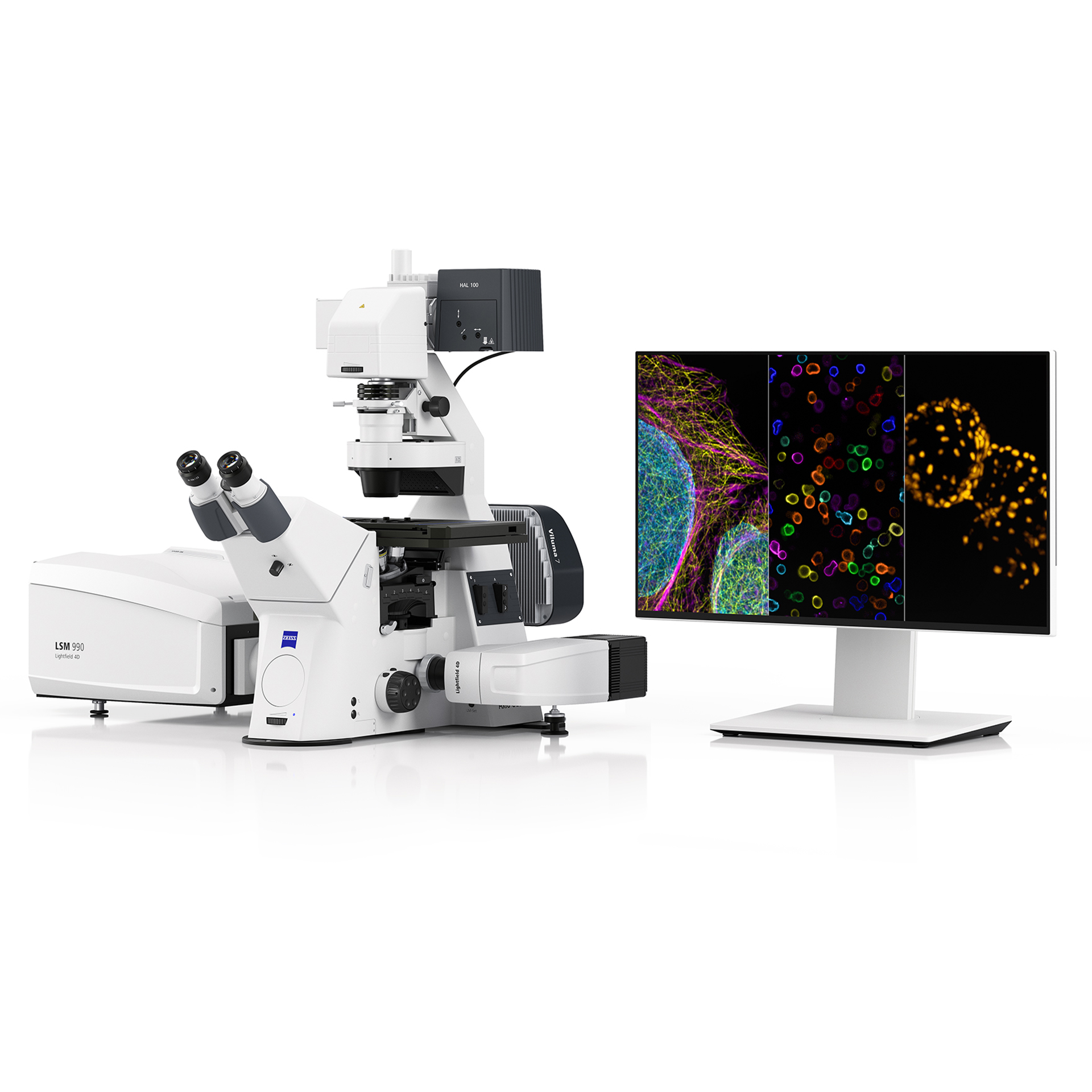
Thick-section 3D CyCIF requires confocal optical sectioning, high-NA oil immersion objectives, sensitive detection and software that supports ROI selection and cycle alignment.
Researchers at Harvard Medical School's Laboratory of Systems Pharmacology reported that successful imaging of 35–50 µm FFPE tissue depended on optical sectioning, high‑NA oil immersion objectives, sensitive optics and flexible software for ROI selection and cycle alignment. In this project, high‑plex 3D CyCIF imaging was performed using a ZEISS confocal imaging platform, supporting visualization of whole‑cell volumes, protein distribution and membrane‑level contacts in human tissue.