Lightfield technology reveals how airway epithelial cells repair alveoli
When a severe viral lung infection occurs, it can damage the tiny air sacs in the lungs (alveoli), leading to acute respiratory distress syndrome (ARDS), a condition that severely impairs breathing. Normally, the lungs repair themselves using stem cells in the airways and alveoli, such as airway epithelial cells, bronchioalveolar stem cells, and alveolar type 2 epithelial cells, which restore normal lung function.
In cases of severe injury, the body produces KRT5+ basal-like cells to rapidly rebuild damaged areas. However, these cells are inefficient at gas exchange, reducing lung function despite structural repair. This hinders full recovery and normal breathing. The exact role of KRT5+ basal-like cells in lung repair remains unclear – whether they merely occupy space or actively disrupt recovery.
Tiantian Lu (left) and Professor Ying Xi (right) in their lab at ShanghaiTech University.
Uncovering airway epithelial differentiation into alveolar epithelial cells
A collaborative team led by Professor Xi Ying (ShanghaiTech University), Professor Ren Tao (Shanghai Sixth People's Hospital), and Professor Zhao Jincun (Guangzhou Medical University/Guangzhou Lab) are investigating how severe respiratory viral infections cause diffuse alveolar damage and progress to ARDS.
Their recent study reveals that dysplastic KRT5+ basal-like cells in lung tissue promote T cell retention, which inhibits alveolar repair mediated by airway epithelial cells after influenza virus clearance. These findings suggest potential therapeutic strategies to enhance alveolar regeneration following viral pneumonia.
After severe alveolar damage, airway epithelial cells activate and migrate to injury sites, attempting to differentiate into alveolar epithelial cells for repair.
Inhibition of repair
Abnormally proliferating KRT5+ basal-like cells facilitate long-term T cell retention in lung tissue. These resident T cells secrete IFNγ, which significantly impairs epithelial cells' ability to regenerate alveoli.
Therapeutic potential
Neutralizing CD4+ or CD8+ T cells with antibodies markedly improved epithelial cell differentiation into functional type II alveolar epithelial cells, accelerating lung tissue regeneration.
Our research provides a crucial theoretical foundation for developing novel therapies to promote post-injury lung regeneration and repair after viral pneumonia.
ZEISS Lightfield 4D in action
CD4+ T cell knockout promotes migration of pulmonary epithelial cells from airways following influenza virus infection
Third-party Content Blocked
The video player is blocked due to your cookie preferences. To change the settings and play the video, please click the button below and consent to use of "Functional" tracking technologies.
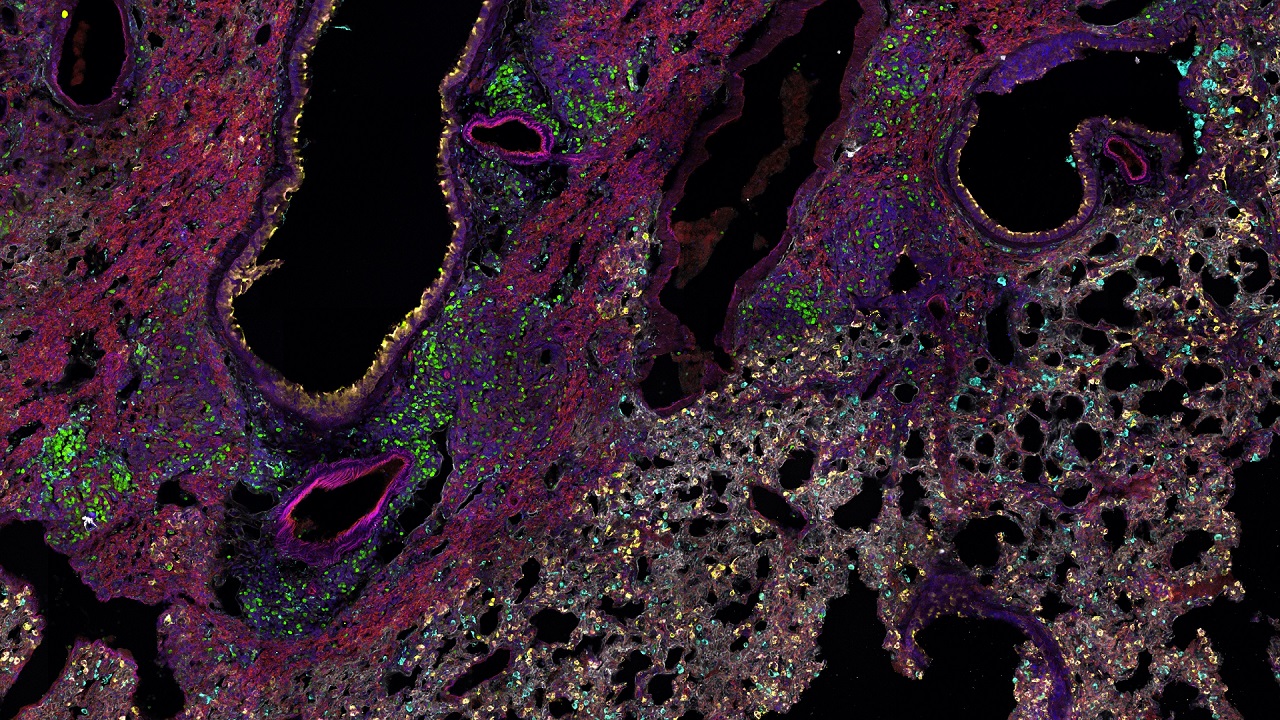
CD4+ T cell knockout promotes migration of pulmonary epithelial cells from airways following influenza virus infection. Green fluorescence indicates Scgb1a1-creER; R26-mTmG mouse epithelial cell lineage markers, with arrows pointing to epithelial cells migrating away from the airway. Acquired with ZEISS Lightfield 4D.
Cells were imaged every 30min over a total duration of 48h across 360 positions. To avoid excessively large file sizes, the acquisition was paused once after 24h and then resumed for the remaining imaging period.
Real-time insights into a key lung repair process
Traditional imaging methods struggle to capture rapid physiological changes in three dimensions, limiting their ability to fully represent dynamic biological processes. Leveraging ZEISS Lightfield 4D technology, researchers successfully observed a critical lung repair process in real time within mouse models. This advanced imaging approach enabled precise visualization of the multidimensional dynamics of airway epithelial cell differentiation into alveolar epithelial cells. By eliminating the reliance on static tissue sections taken at single time points, light-field technology reconstructs the entire dynamic repair process, providing visually tangible and comprehensive insights into cellular differentiation.
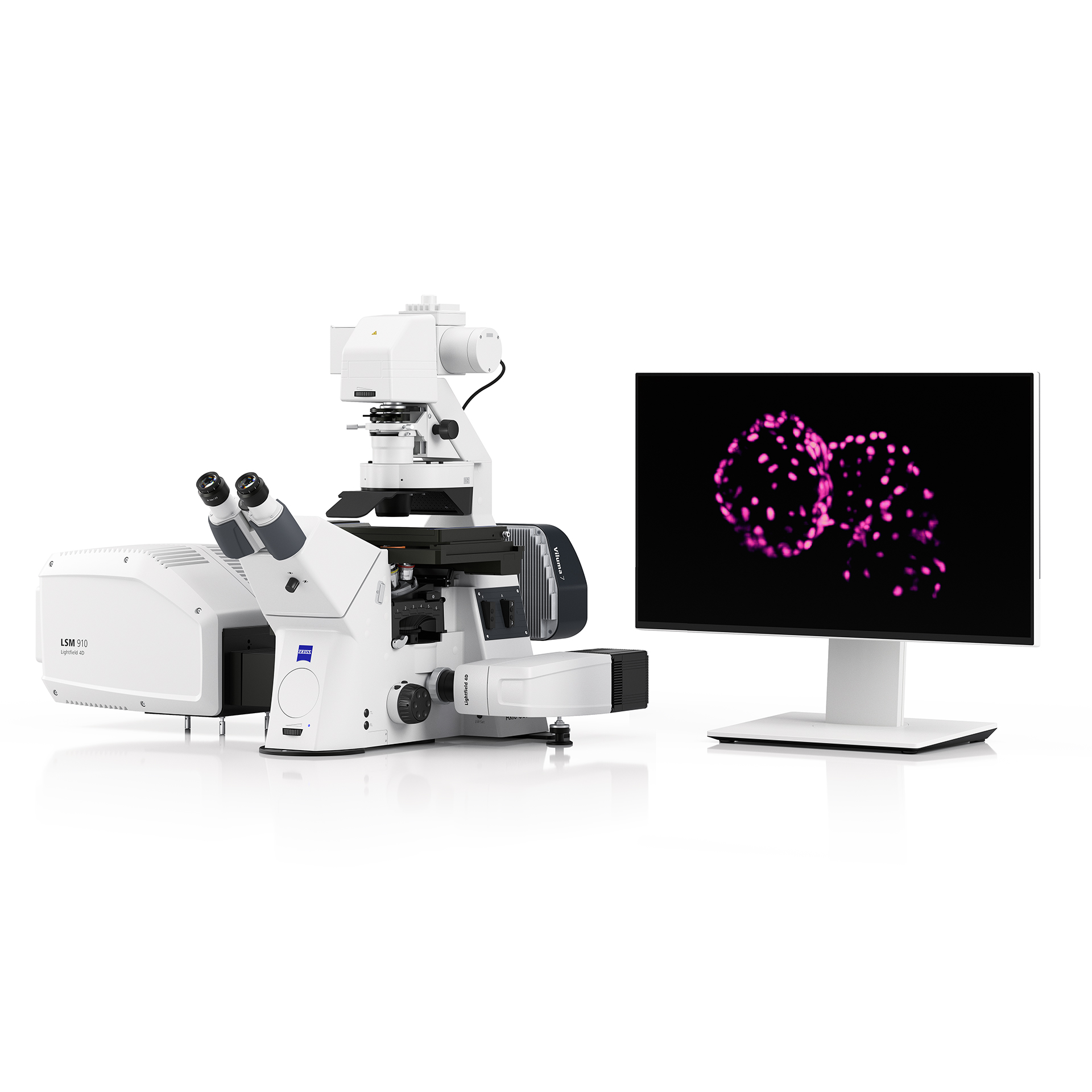
ZEISS LSM 910 with Lightfield 4D enabled high temporal resolution, live-cell, volume imaging of thick sections of mouse lung tissue.
The research team had previously explored other fluorescence imaging methods. However, imaging live cells in 300-μm-thick samples remained challenging and required careful consideration of factors such as speed (temporal resolution), phototoxicity, and focal plane stability.
The need for three-dimensional data acquisition at multiple points simultaneously further emphasized the importance of speed. Ultimately, ZEISS LSM 910 with Lightfield 4D technology enabled the understanding of this critical process.
New hope for post-viral lung repair
This research reveals a critical mechanism underlying impaired lung regeneration: the "basal-like cells-T cells-IFNγ" axis. By identifying IFNγ and T cell residency pathways, such as CXCR3 and integrin α4β7, as key contributors to post-viral lung dysfunction, the study opens new avenues for targeted therapies.
Modulating these pathways could enhance lung repair, improve gas exchange efficiency, and accelerate recovery in patients with severe viral infections. Future investigations may focus on developing drugs or interventions that inhibit IFNγ signaling or regulate T cell activity, offering hope for more effective treatments for conditions like ARDS and long-term post-viral lung damage.
In brief
Airway epithelial cells are activated and migrate to injury sites in the lungs after severe alveolar damage. They attempt to differentiate into alveolar epithelial cells, specifically type II alveolar epithelial cells, to restore normal lung function. However, their repair process can be inhibited by factors such as the retention of T cells in lung tissue and the secretion of IFNγ, which impairs their ability to regenerate alveoli.
ZEISS Lightfield 4D technology enables real-time imaging of dynamic biological processes, such as airway epithelial cell differentiation into alveolar epithelial cells. Traditional volumetric imaging methods rely on comparatively slow sequential z-stack acquisition, which can make imaging of fast dynamic processes in 3D inaccessible, forcing researchers to resort to analysis of fixed tissue sections. However, Lightfield 4D captures entire volumes instantaneously and with high time resolution, now enabling 3D capture of the fast dynamic processes of lung repair, now providing comprehensive insights. It overcomes challenges like phototoxicity, focal plane stability, and limits in temporal resolution, making it ideal for observing rapid physiological changes in 3D.
Therapies can enhance lung repair by targeting mechanisms that inhibit regeneration. Neutralizing CD4+ or CD8+ T cells with antibodies reduces their retention in lung tissue, which in turn decreases the secretion of IFNγ, a cytokine that impairs epithelial epithelial cells' ability to regenerate alveoli. Blocking IFNγ signaling directly improves alveolar regeneration and tissue repair. Additionally, therapies that promote the differentiation of airway epithelial cells into type II alveolar epithelial cells accelerate lung tissue regeneration and restore gas exchange efficiency. Future research may focus on developing drugs or biologics that target these pathways, offering new therapeutic options for conditions like ARDS and long-term post-viral lung damage.