Digitization
Use Case

Digitization

Unlock the full potential of your collections with Zeiss.
Our cutting-edge solutions enable detailed digital reproductions, ensuring your artifacts and documents are preserved and accessible worldwide.


Empowering Technology Through Preservation Digitization represents a revolutionary step in preserving and sharing historical artifacts and documents. Utilizing advanced microscopy, institutions can create high-resolution reproductions that are both detailed and accessible globally. This process not only safeguards the physical items from deterioration but also democratizes access to treasures that might otherwise be confined to the physical constraints of museum walls. ZEISS microscopes are instrumental in this process, offering unparalleled precision and versatility. They allow for detailed examination and capture of the minutest features, essential for accurate digital representations

With the X-ray microscope, we can look at the entire structure at multiple scales and still with a very high resolution.

Keith Duncan Director, X-ray Imaging Facility, The Donald Danforth Plant Science Center

Complete your lab

with our new Solution Bundles

Unveiling the Potential of Digitization with our new Solution Bundles.

Microscopy & Digitization

ZEISS MultiSEM 505/506

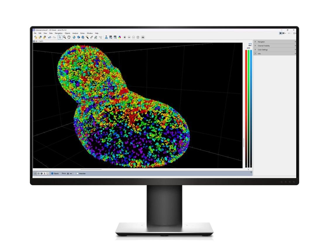
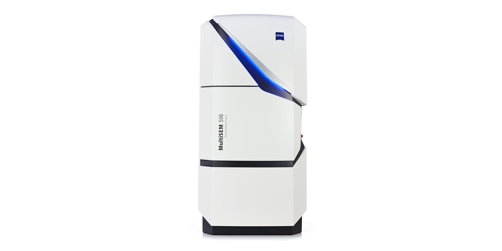
The World’s Fastest Scanning

Electron Microscopes

Unleash the acquisition speed of up to 91 parallel electron beams – to image samples in the centimeter scale at nanometer resolution. This unique scanning electron microscope is designed for continuous, reliable 24/7 operation. Simply set up your high-throughput data acquisition workflow and MultiSEM will acquire high-contrast images automatically.

Digitization FAQs

  • ZEISS provides a comprehensive range of microscopes for plant biology, including:

    • Stereo and widefield microscopes for whole plant and tissue imaging
    • Laser scanning confocal and multiphoton microscopes for high-resolution, 3D imaging of plant cells and structure
    • Light-Sheet Microscopy for fast, gentle imaging of large specimens with minimal photobleaching, ideal for live plant imaging
    • Super-Resolution Microscopy for visualizing structures at a resolution beyond the diffraction limit of light, revealing fine details in plant cells
    • X-ray Tomography for non-destructive 3D imaging of internal plant structures, preserving the specimen's integrity
    • Volume Electron Microscopy (vEM) techniques for ultrastructural insights into plant tissues in 3D
  • ZEISS offers solutions, such as Light-Sheet Microscopy and Lattice Light-Sheet Microscopy, that allow researchers to observe dynamic processes in real-time. This capability is crucial for studying how plants respond to environmental stressors, such as drought or salinity, by visualizing cellular changes and adaptations as they occur.

  • Yes, ZEISS microscopes, particularly the LSM 980, utilize spectral imaging capabilities that enable the simultaneous imaging of multiple fluorescent markers while effectively separating autofluorescence. This is essential for studying complex cellular interactions in plant tissues without interference from naturally fluorescing compounds.

  • How does ZEISS support long-term time-lapse imaging of plant specimens?

    ZEISS microscopy solutions, like Light-Sheet Microscopy, are designed for long-term time-lapse imaging by providing a stable environment with controlled conditions. This allows researchers to monitor dynamic processes, such as cell division and organ development, over extended periods while maintaining sample viability.
  • How can ZEISS software help with plant image analysis?

    Data analysis is crucial for extracting meaningful insights from complex plant imaging datasets. ZEISS offers advanced software solutions, such as arivis Cloud and arivis Pro, which utilize machine learning for efficient cell segmentation and counting, phenotypic trait quantification and 3D reconstruction of plant structures. These automated, AI-driven tools streamline image analysis, enabling researchers to focus on their findings and discoveries.

    ZEISS arivis Software Ecosystem:


  • ZEISS offers a variety of microscopy solutions, such as super-resolution microscopy and fluorescence microscopy, to enable detailed visualization of chloroplast structure and function, pigment composition and stress responses and photosynthetic efficiency and energy transfer. By capturing high-resolution images and dynamic processes, researchers can uncover the mechanisms of photosynthesis and how plants optimize energy production under varying environmental conditions.

Comprehensive Solutions and Capabilities

Discover Our Application Hub

Explore applications to discover tailored solutions for your unique laboratory needs and elevate your research capabilities.