
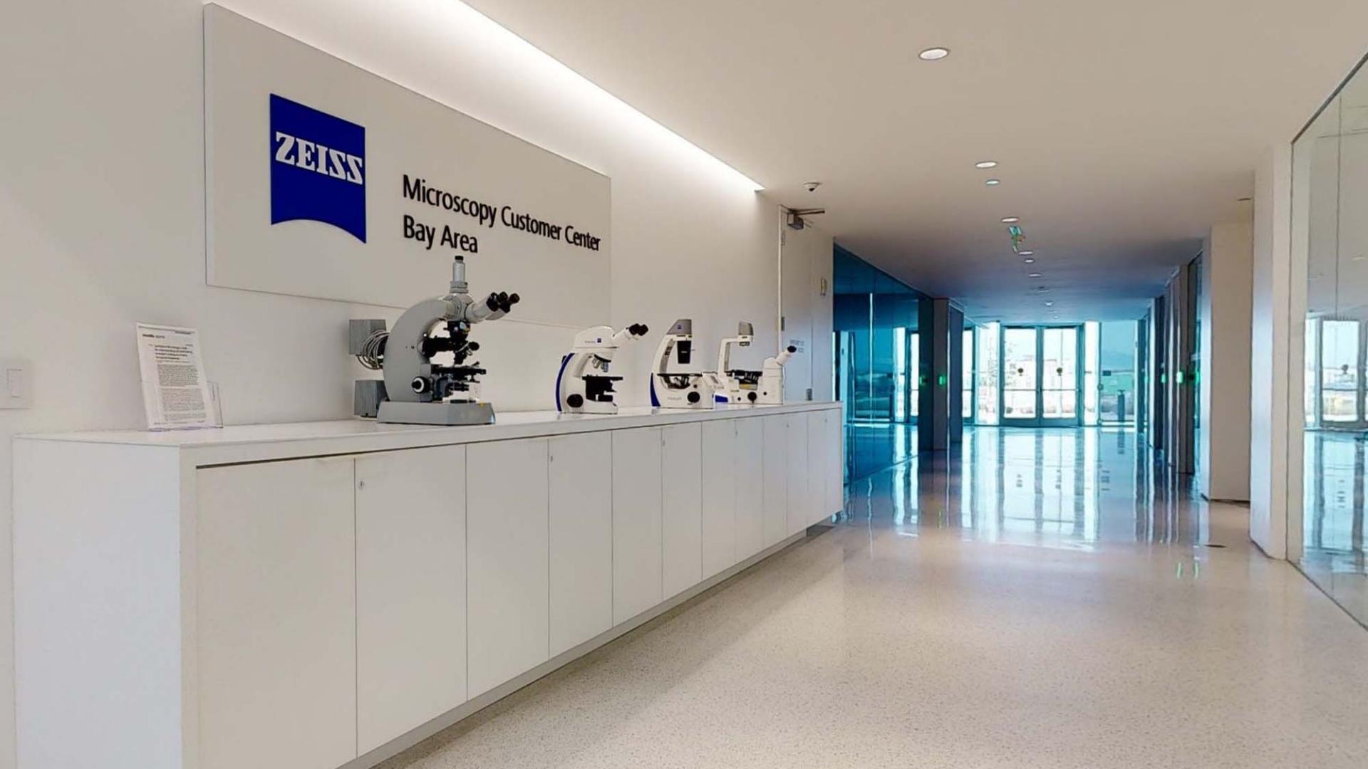
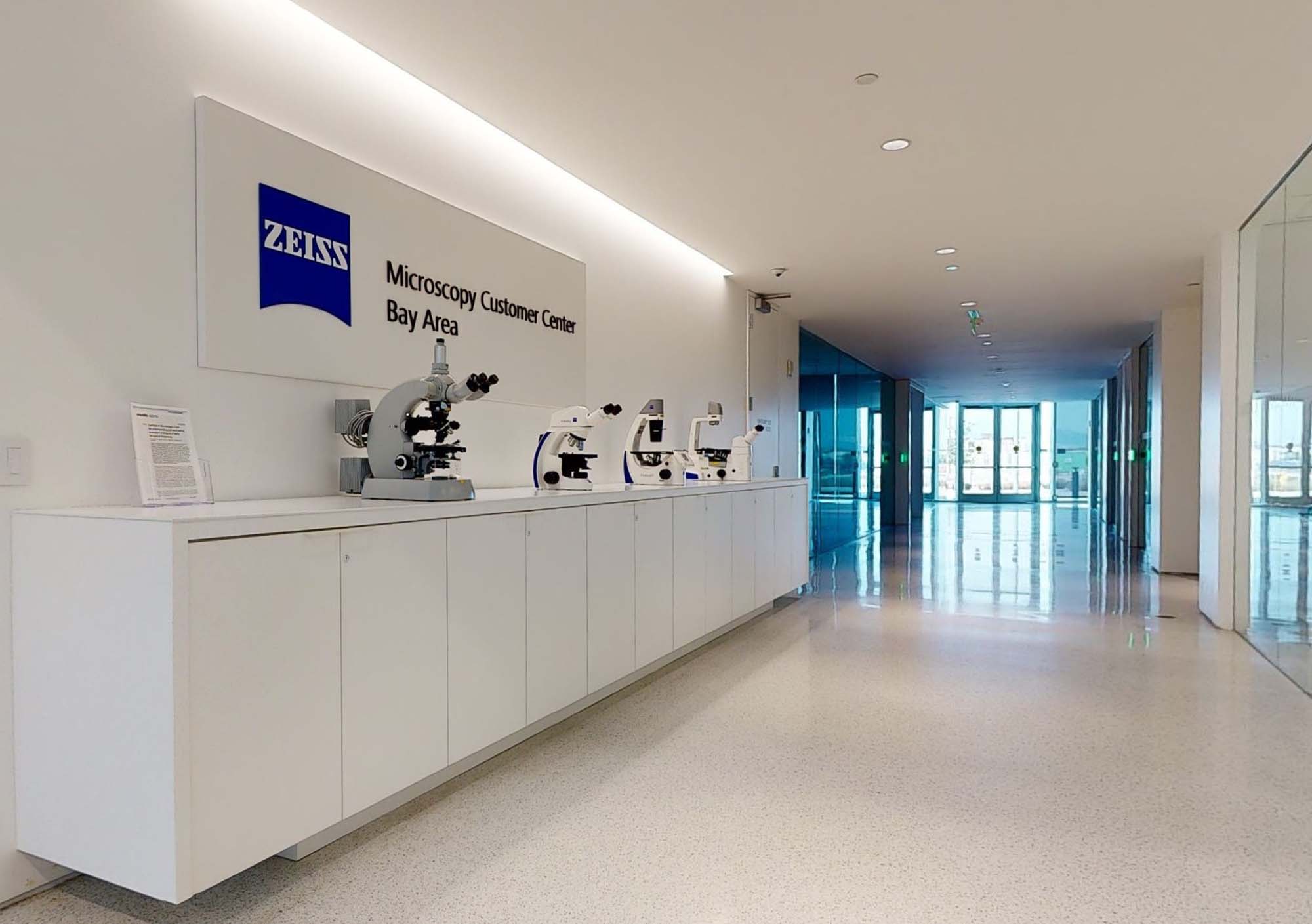
ZEISS Microscopy Customer Center
Bay Area

ZEISS Microscopy Customer Center
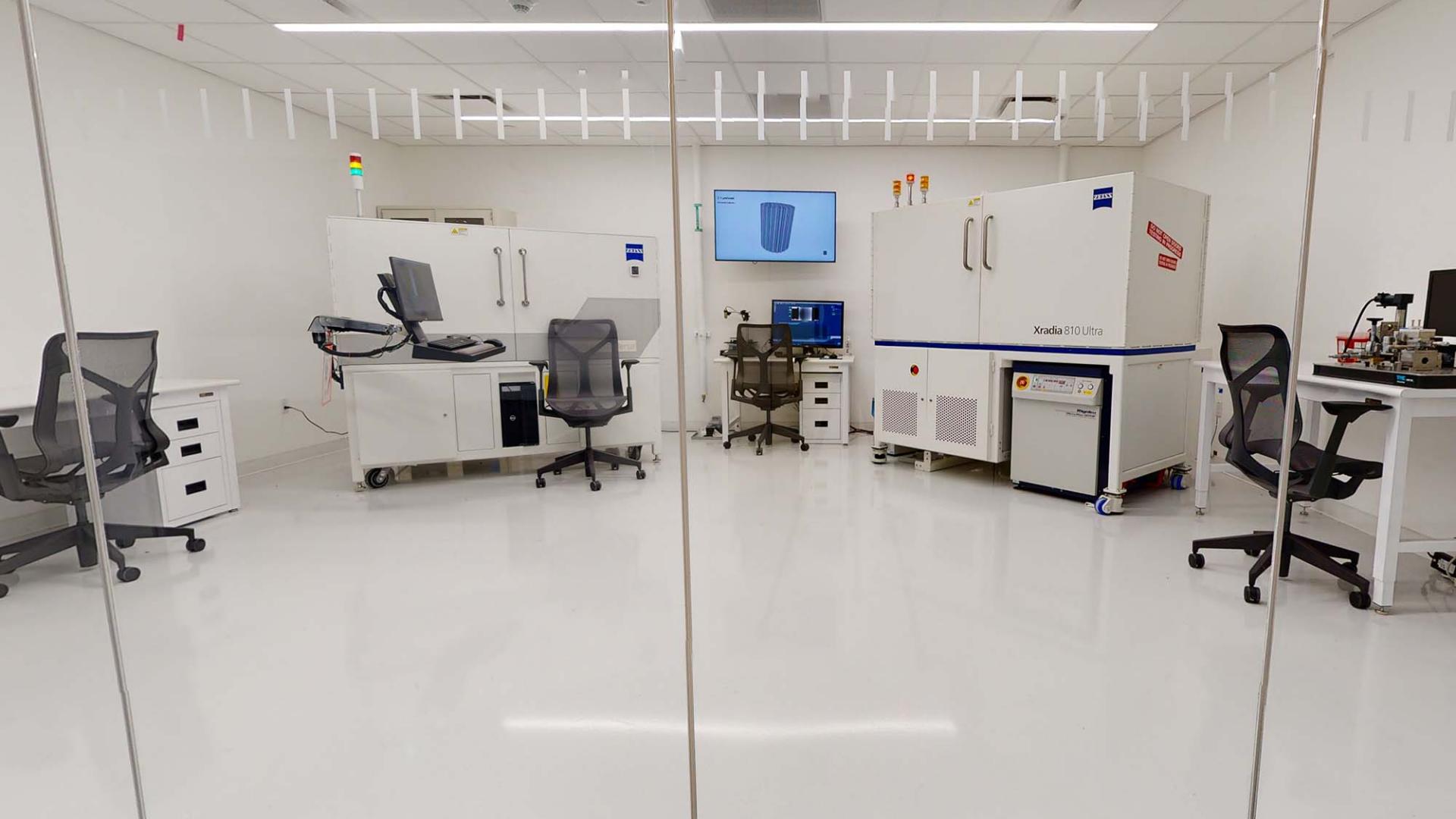
The ZEISS Microscopy Customer Center gives academic and industry customers across North America both in-person and virtual opportunities to interact with the latest ZEISS electron, light, and X-ray microscopes. Part of the ZEISS Research Microscopy Solutions business, the Customer Center is supported by resident application experts in life science, materials research, and electronics who review customers' requirements and applications to provide them with the best possible solutions. Customers also have access to a sophisticated sample preparation laboratory that supports the materials research, electronics and life science markets.
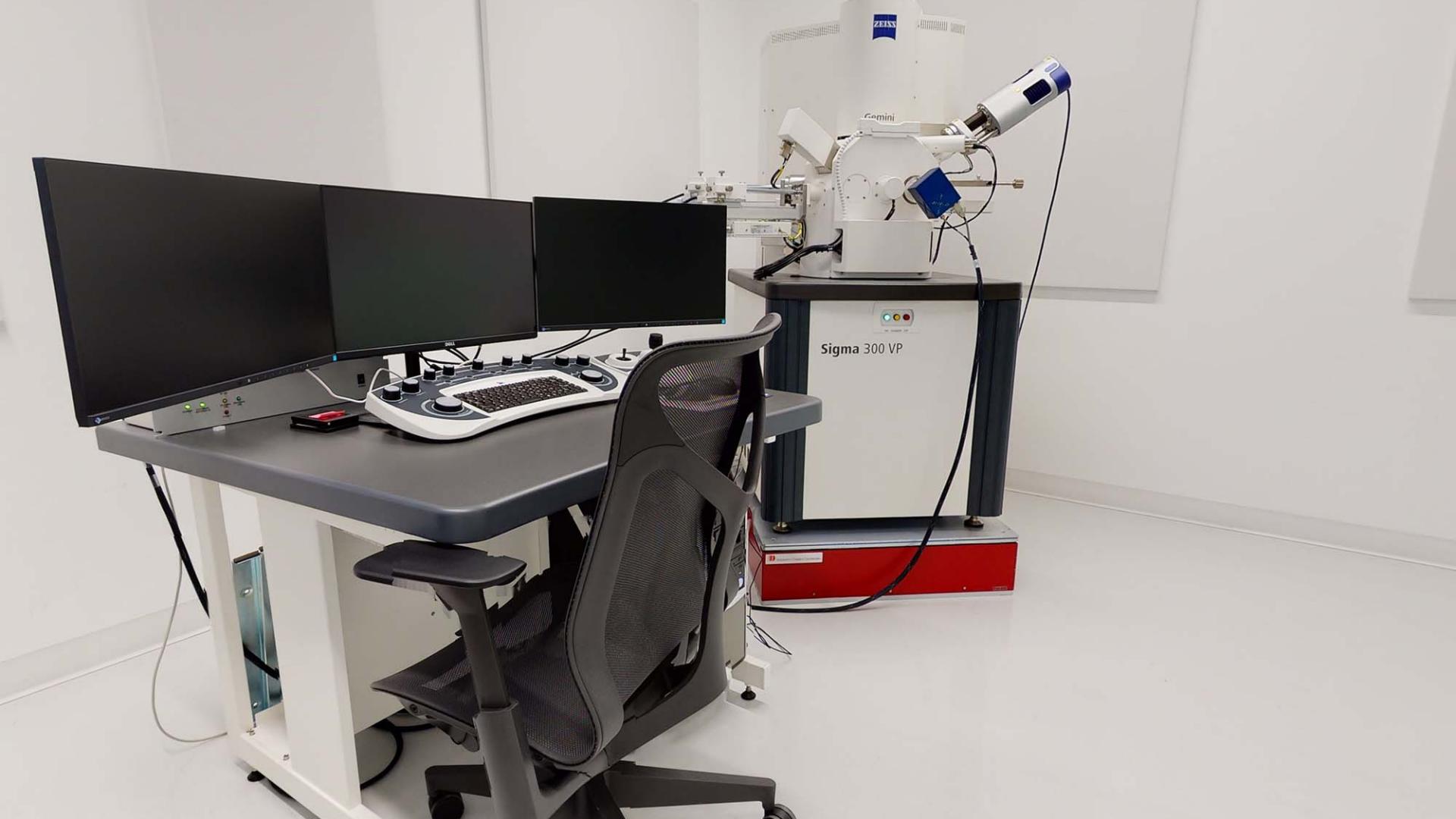
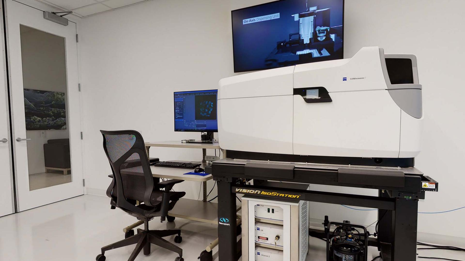
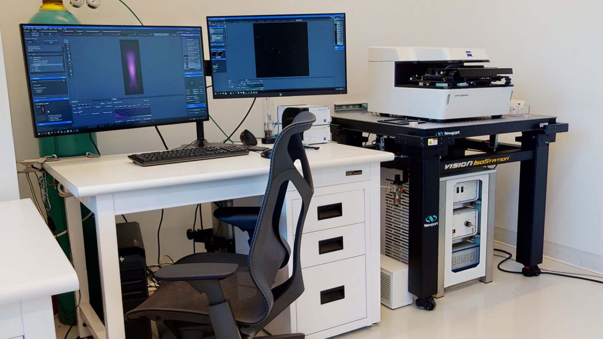
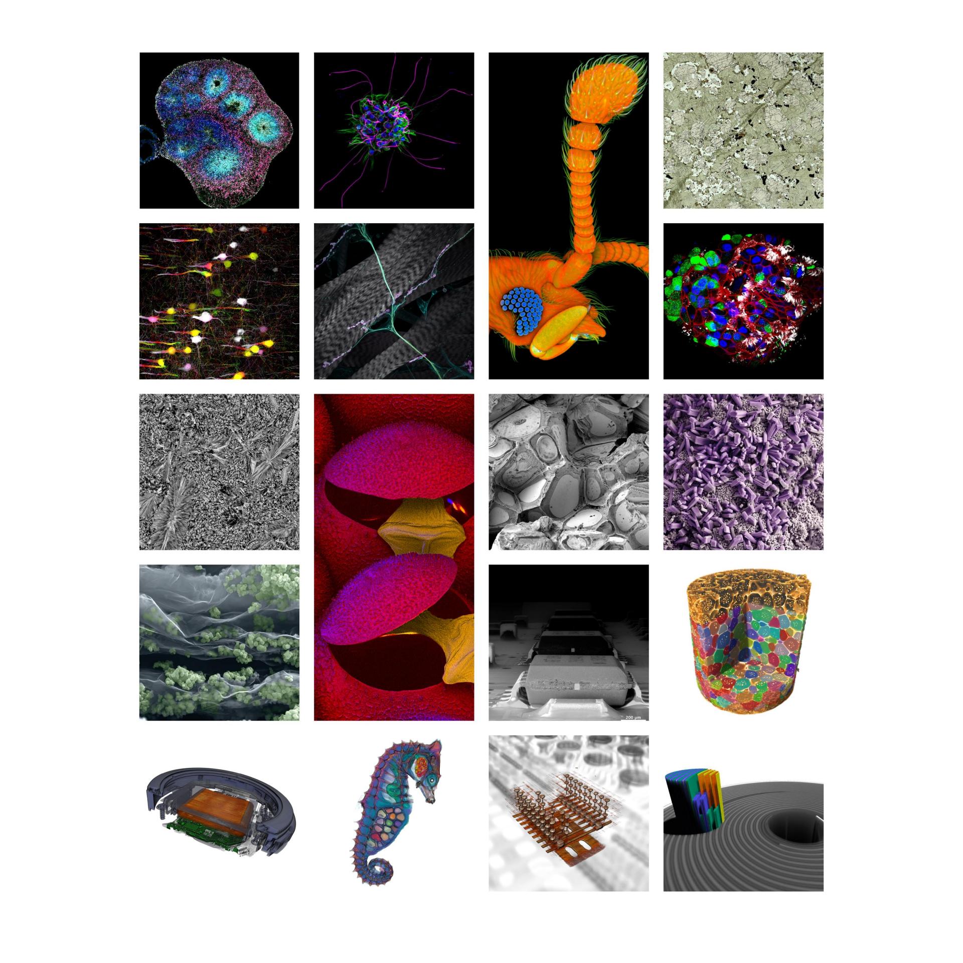
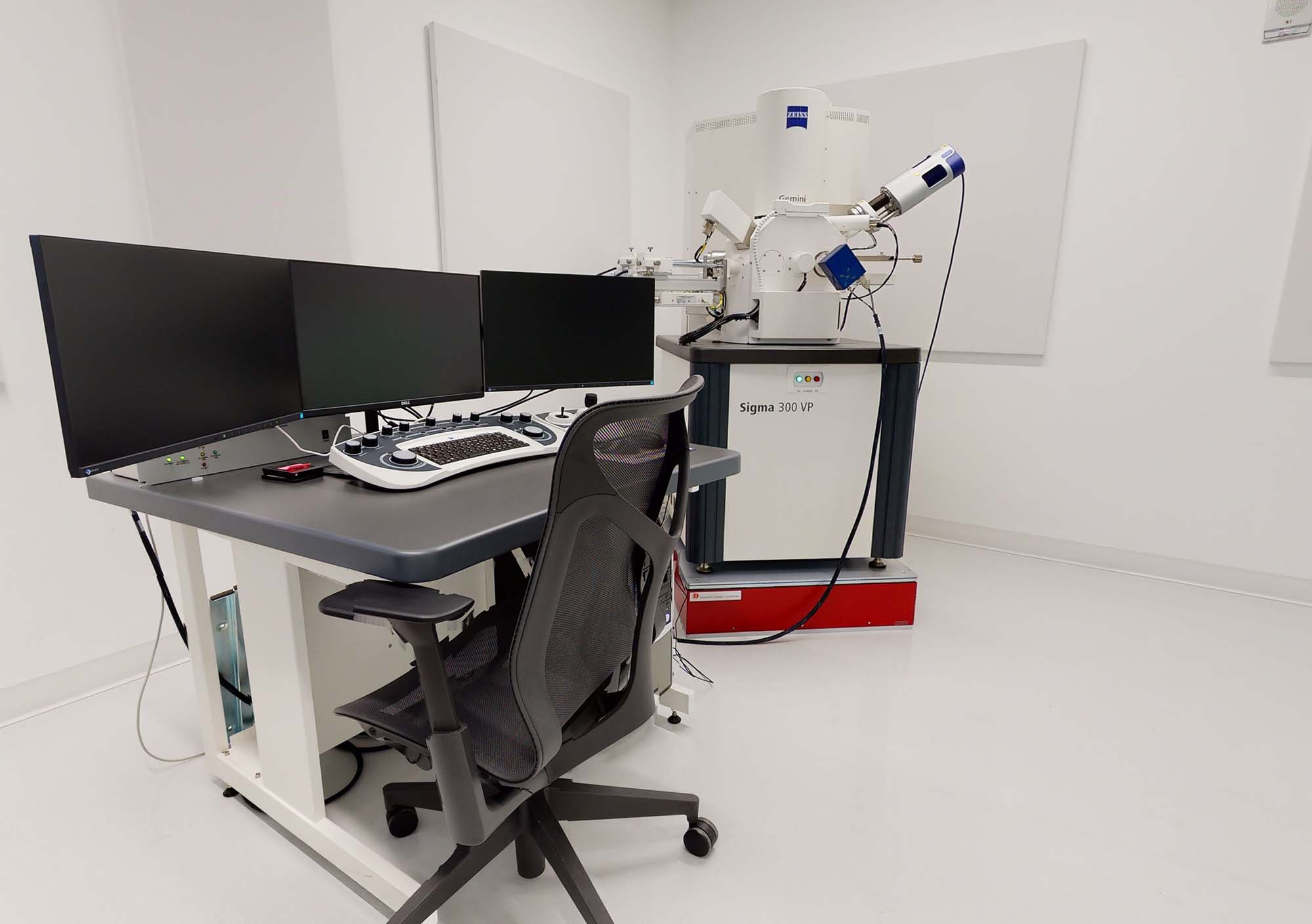
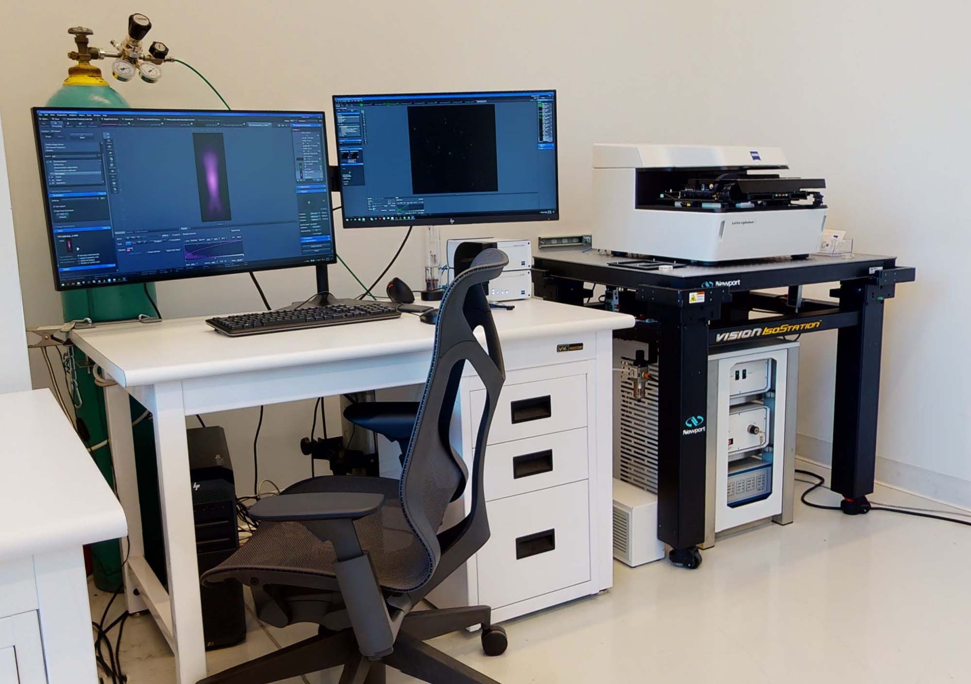
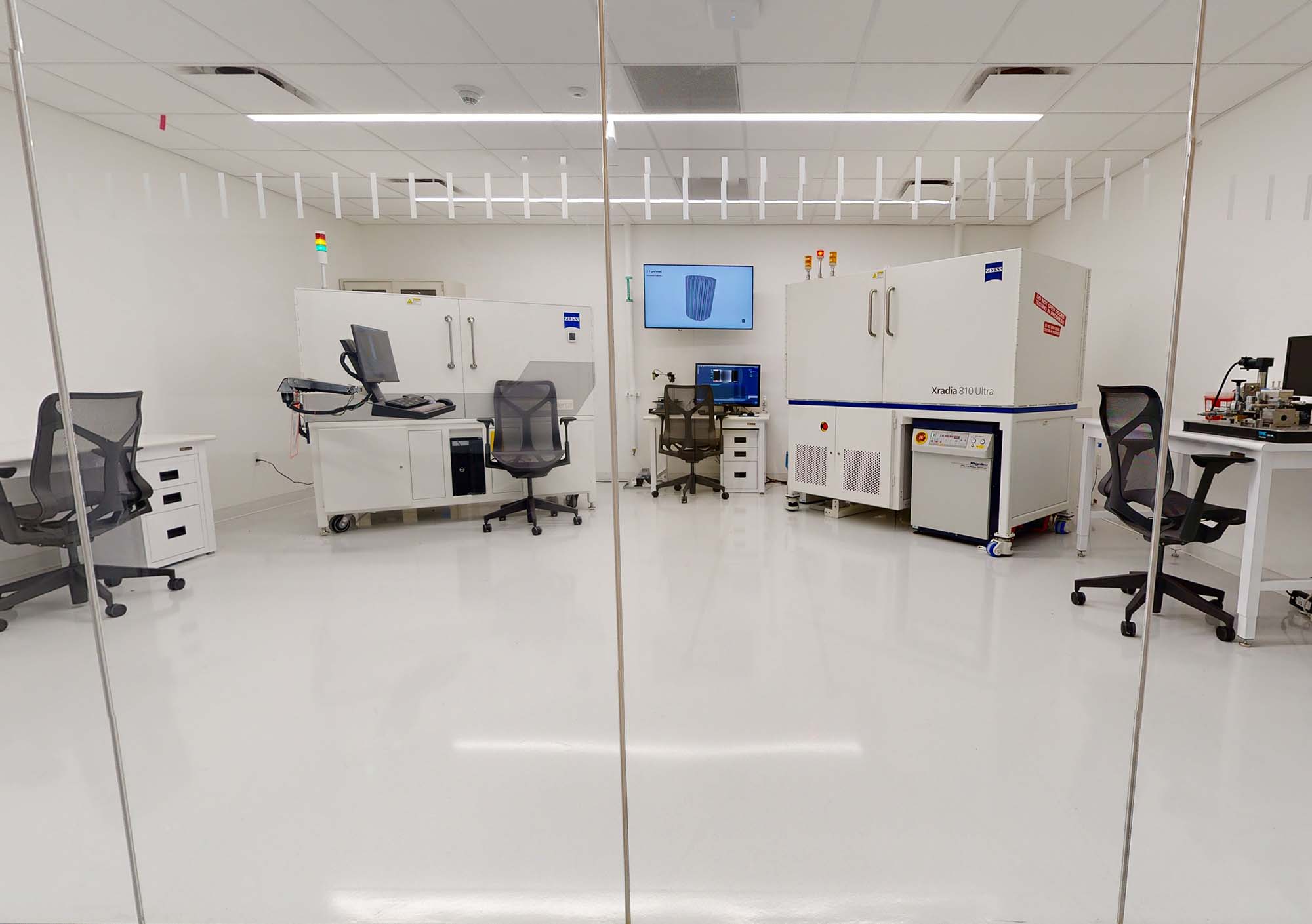
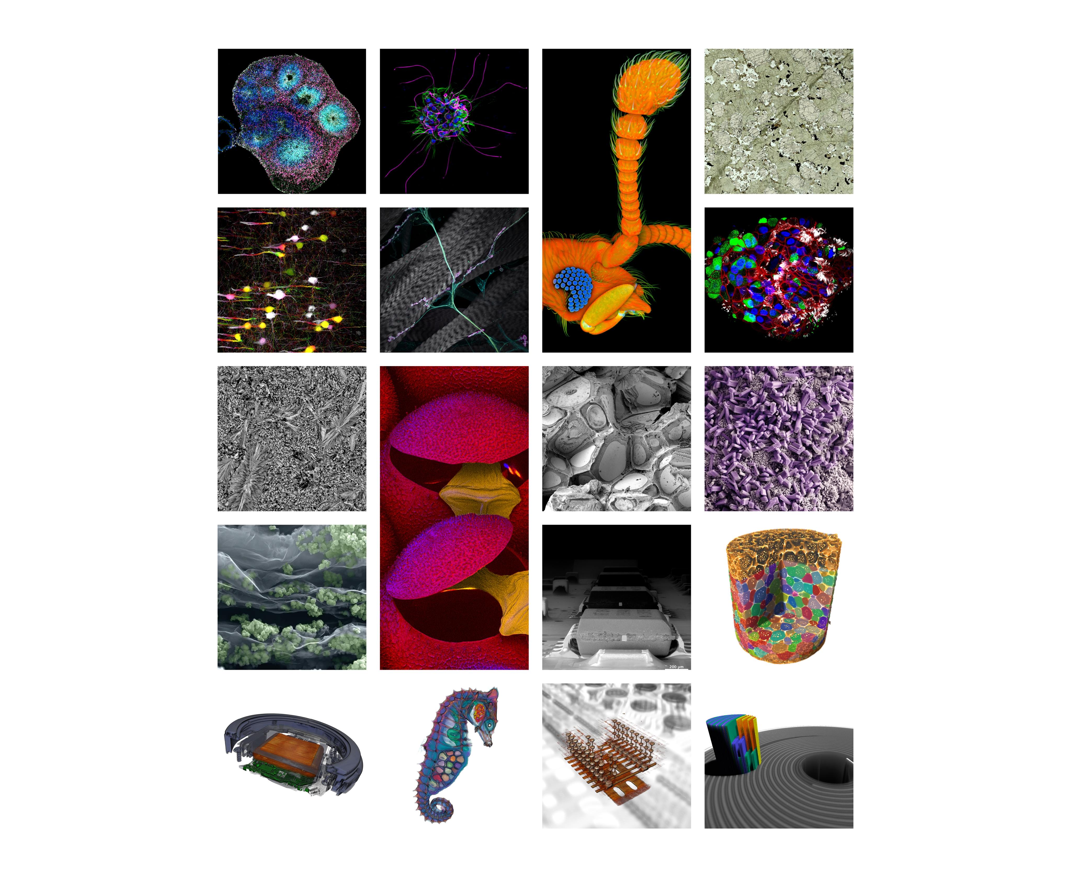
Among the latest ZEISS microscopy solutions on display in the Customer Center are the new Axio Observer with AI Sample Finder, Lattice Lightsheet 7, Gemini SEM 460, Gemini SEM 560, Crossbeam Laser FIB, Cryo correlative workflow, and Crystal CT. The Customer Center is fully equipped with the latest technology for customers to access these and other ZEISS microscopes through remote channels.
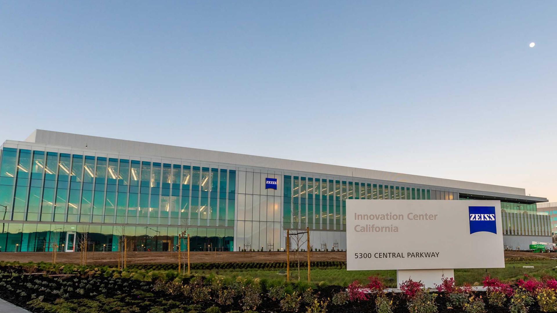
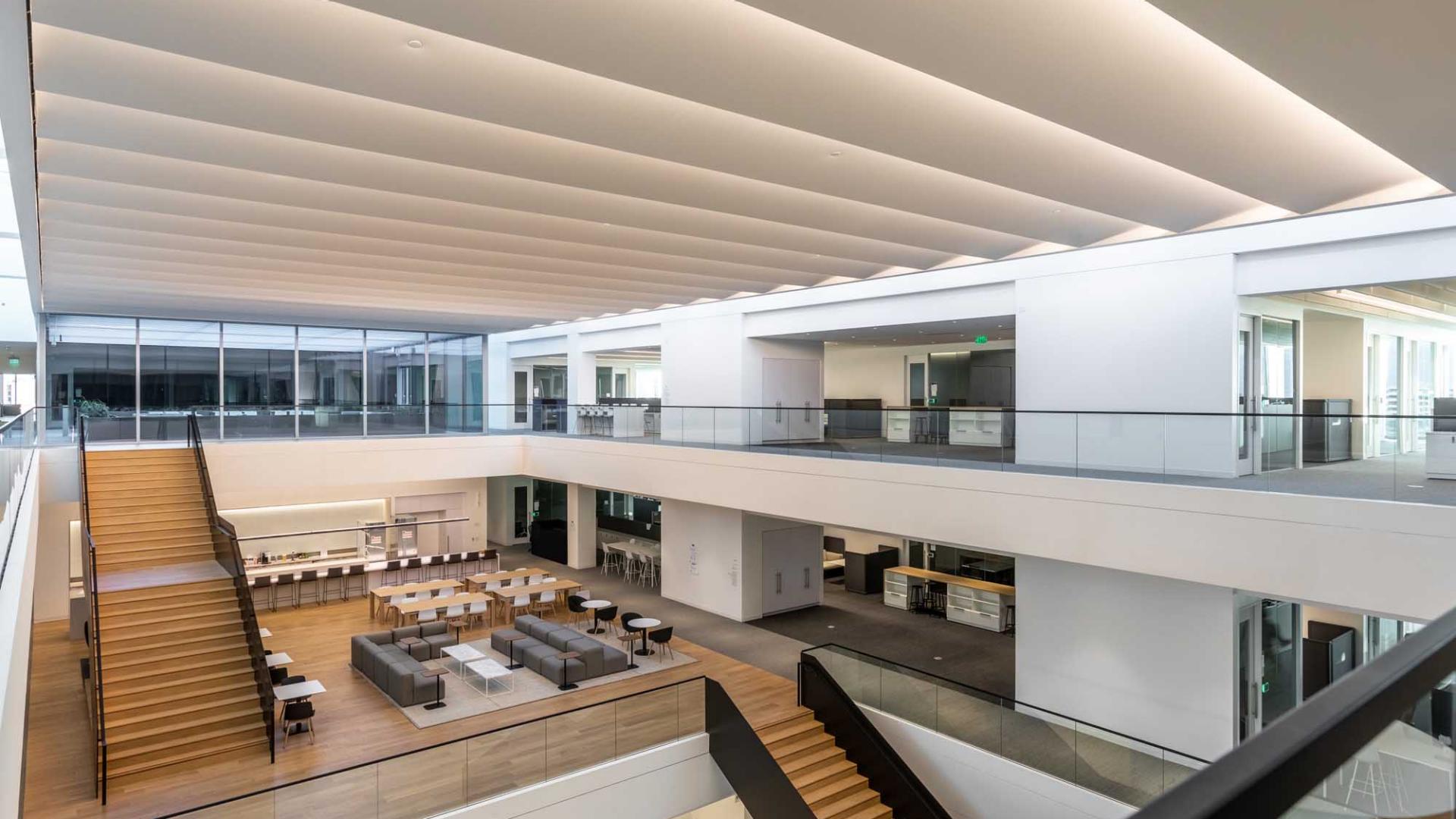
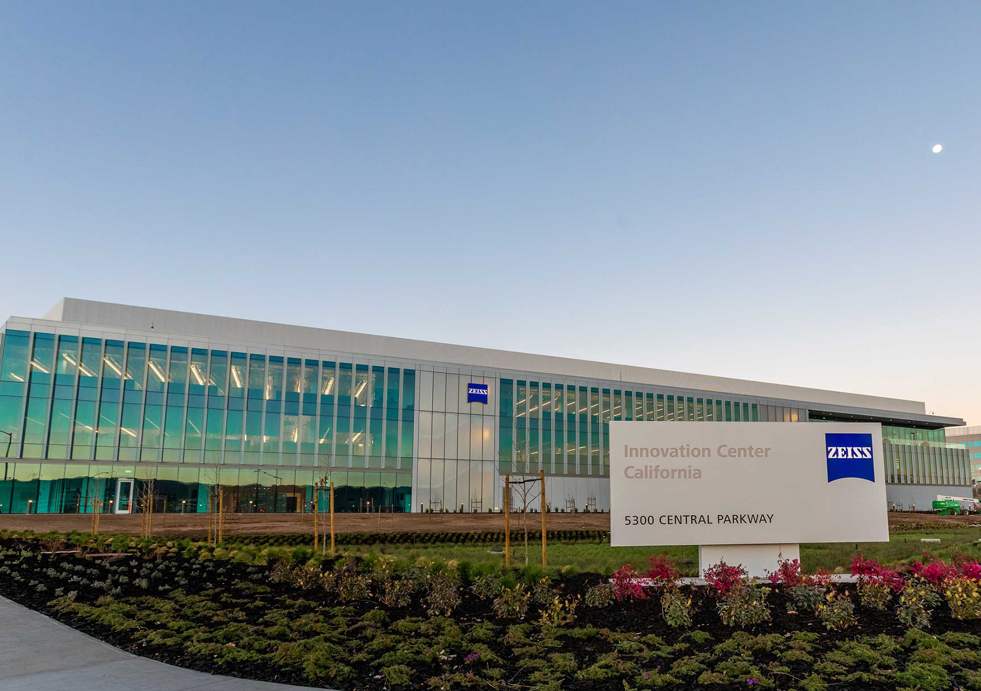
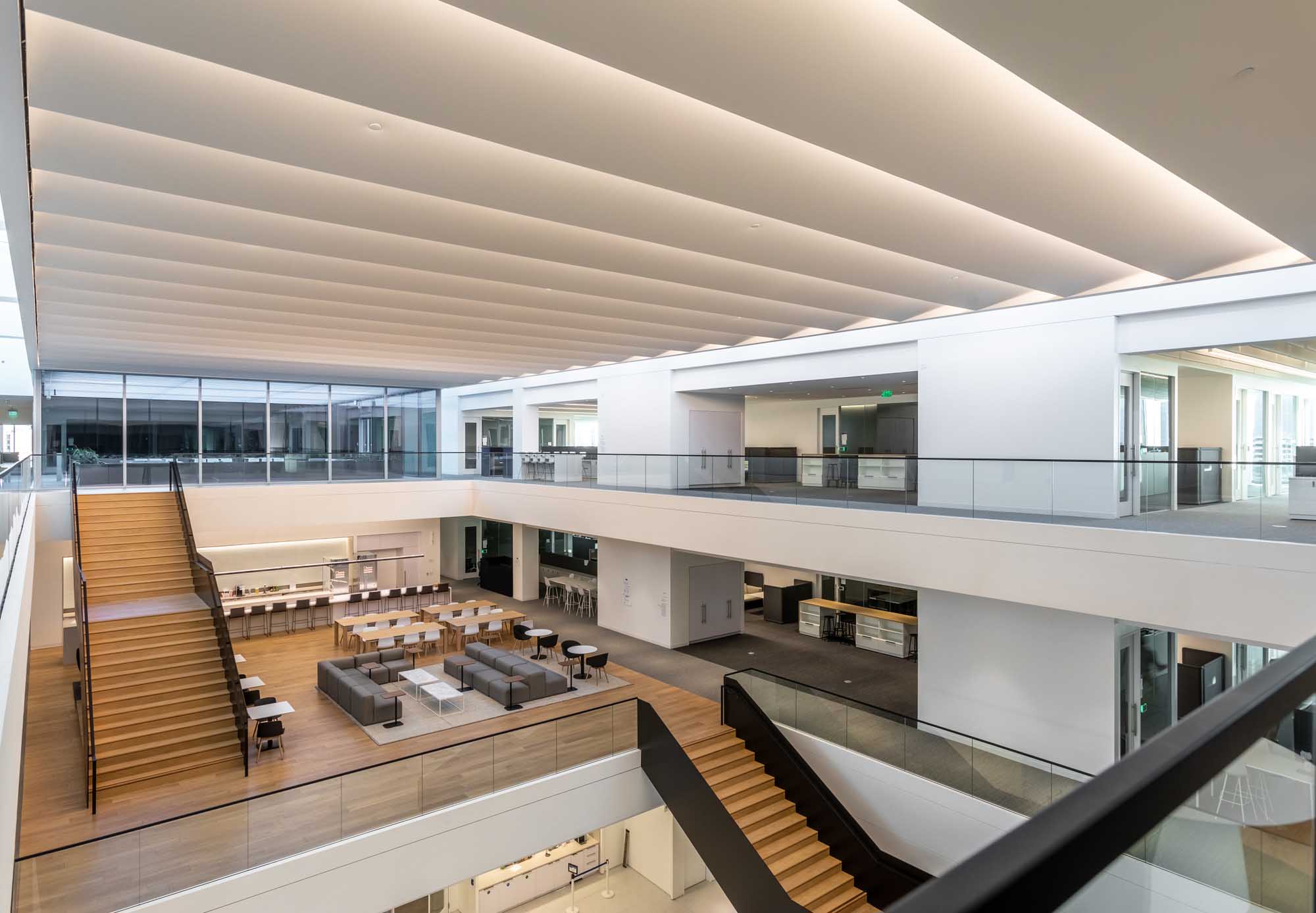
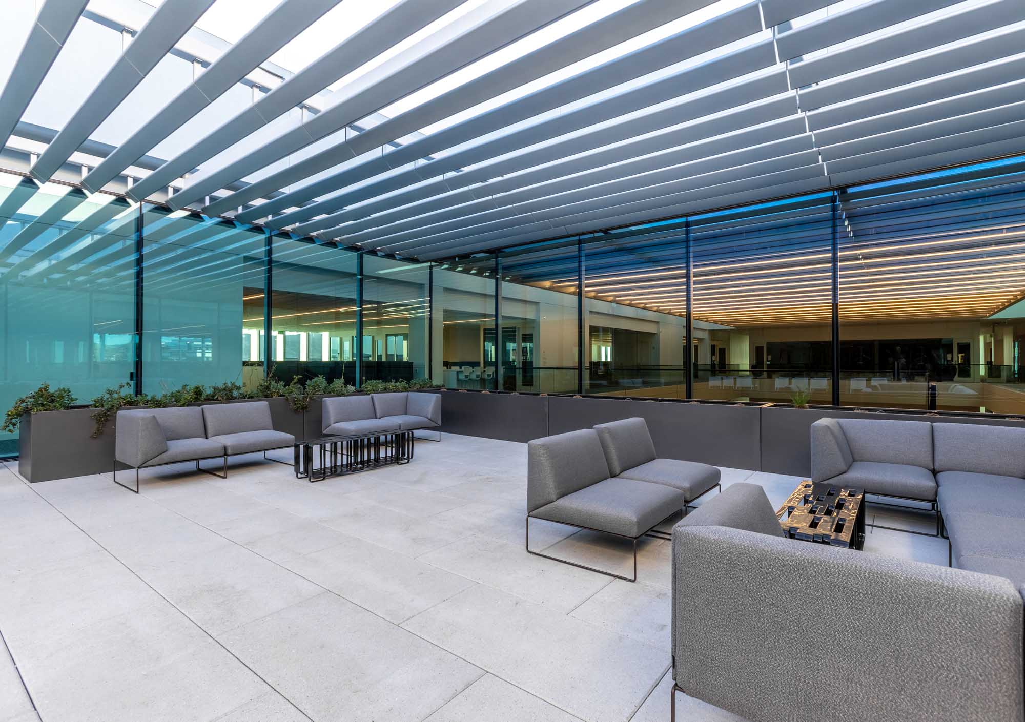
Direct customer interaction with ZEISS technology is an integral part of the commitment by ZEISS to continually drive innovation and shape new markets. The location of the Customer Center in the ZEISS Innovation Center therefore serves as an ideal location to promote the intersection of ideas between various ZEISS businesses and experts, and to serve as a networking hub for academia and industry.
Browse through the tabs below to learn about upcoming events and training options as well as available equipment in our facilities.
Events & Education
Hone your microscope skills and share your experiences
We offer training courses for you to get the best results and highest benefits from your instruments and systems. We also invite you to participate in our user meetings, both so we can learn of your successes and challenges in your day-to-day work, and provide a forum for you to network with other microscopists to exchange best practices.
Upcoming Skillbuilder Workshops
|
September 10 |
Virtual | Register Now |
|
|
October 14-15 |
In-Person | Register Now |
|
Read about our workshop registration, payment and cancellation policy
2026 Skillbuilder Workshops
|
January 14, 2026 |
Conquer the Confocal |
Closed | Contact Us |
|
February 10, 2026 |
FESEM Academy: Establishing Workflows in SmartSEM |
Closed | Contact Us |
|
March 4, 2026 |
ZEN Blue Software: Mastering Modules |
Closed | Contact Us |
|
March 5, 2026 |
Mastering FIB-SEM: Introduction to ATLAS 3D |
Closed | Contact Us |
|
March 6, 2026 |
Conquer the Confocal: Basics |
Closed | Contact Us |
|
March 10, 2026 |
Lights, Camera, Apotome! |
Closed | Contact Us |
|
March 12, 2026 |
Conquer the Confocal: Airyscan |
Closed | Contact Us |
|
April 9-10, 2026 |
Suceed On Your Sigma Bootcamp |
Closed | Contact Us |
|
May 13-14, 2026 |
Mastering FIB-SEM: Comprehensive Intro to Crossbeam |
Closed | Contact Us |
|
May 19-20, 2026 |
XRM University - Versa XRM Crash Course |
Closed | Contact Us |
|
May 21, 2026 |
XRM University - Context MicroCT Operation |
Closed | Contact Us |
|
June 3-5, 2026 |
Conquer the Confocal |
Closed | Contact Us |
|
June 10, 2026 |
FESEM Academy: Imaging Non-conductive Samples at Low kV |
Closed | Contact Us |
Read about our workshop registration, payment and cancellation policy
Electron/Ion Microscopy


|
|
|
|---|---|
|
Product Name |
Product Type |
|
ZEISS Crossbeam 550 with Laser |
FIB-SEM for High Throughput 3D Analysis and Sample Preparation. Equipped with femtosecond laser for site-specific, ultra-fast sample preparation |
|
Your Highest Demands in Sub-nanometer Imaging, Analytics and Sample Flexibility |
|
|
For Your Highest Demands in Sub-nanometer Imaging, Analytics and Sample Flexibility |
|
|
Your ZEISS SIGMA FE-SEMs for High Quality Imaging and Advanced Analytical Microscopy |
|
Light Microscopy


|
|
|
|---|---|
|
Product Name |
Product Type |
|
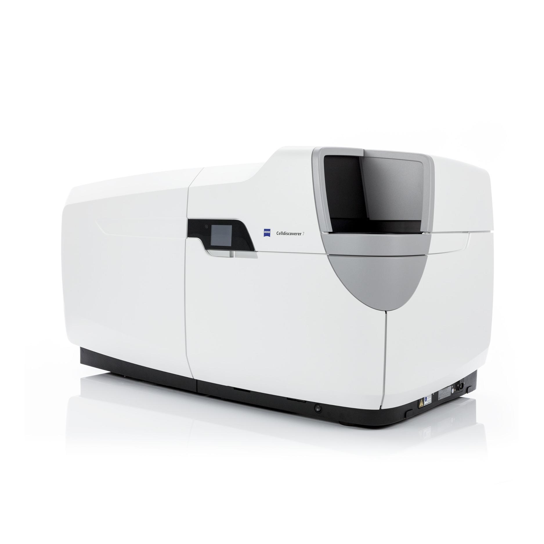
Open and flexible inverted microscope platform with AI assisted experiment startup |
|
|
High-performance Digital Slide Scanner for Fluorescence, Brightfield and Polarization |
|
|
High-performance Digital Slide Scanner Specialized for Multiplexing Worflows |
|
|
Fluorescence Stereo Zoom Microscope for Large Fields |
|
|
Adding Efficiency to Your Fluorescence Imaging |
|
|
Live Imaging System with Unprecedented Resolution |
|
|
Live Imaging System with Super Resolution Capabilities for tissues and organoids |
|
|
Long-term volumetric imaging of living cells and organisms |
|
|
Multi-view imaging of living and cleared specimens |
|
|
Versatile Confocal Microscope for Advanced Imaging and Surface Topography |
|
|
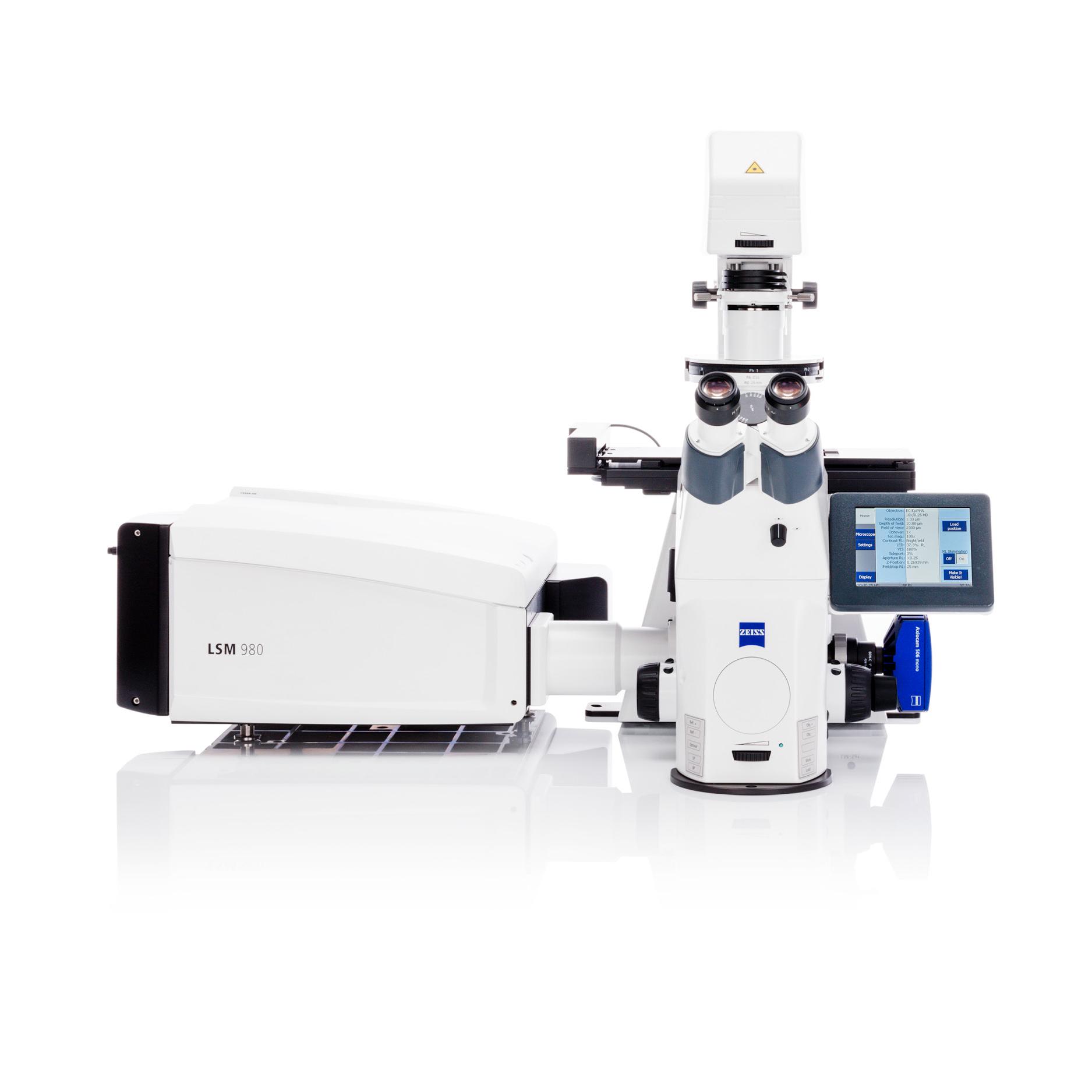
Next Generation Confocal Microscope with Spectral and NIR Detectors, Airyscan, and Lightfield 4D |
|
|
Automated Digital Microscope for Routine and Failure Analysis |
|
|
Crisp and Brilliant Images throughout the Whole 8:1 Manual Zoom Range |
|
X-ray Microscopy


|
|
|
|---|---|
|
Product Name |
Product Type |
|
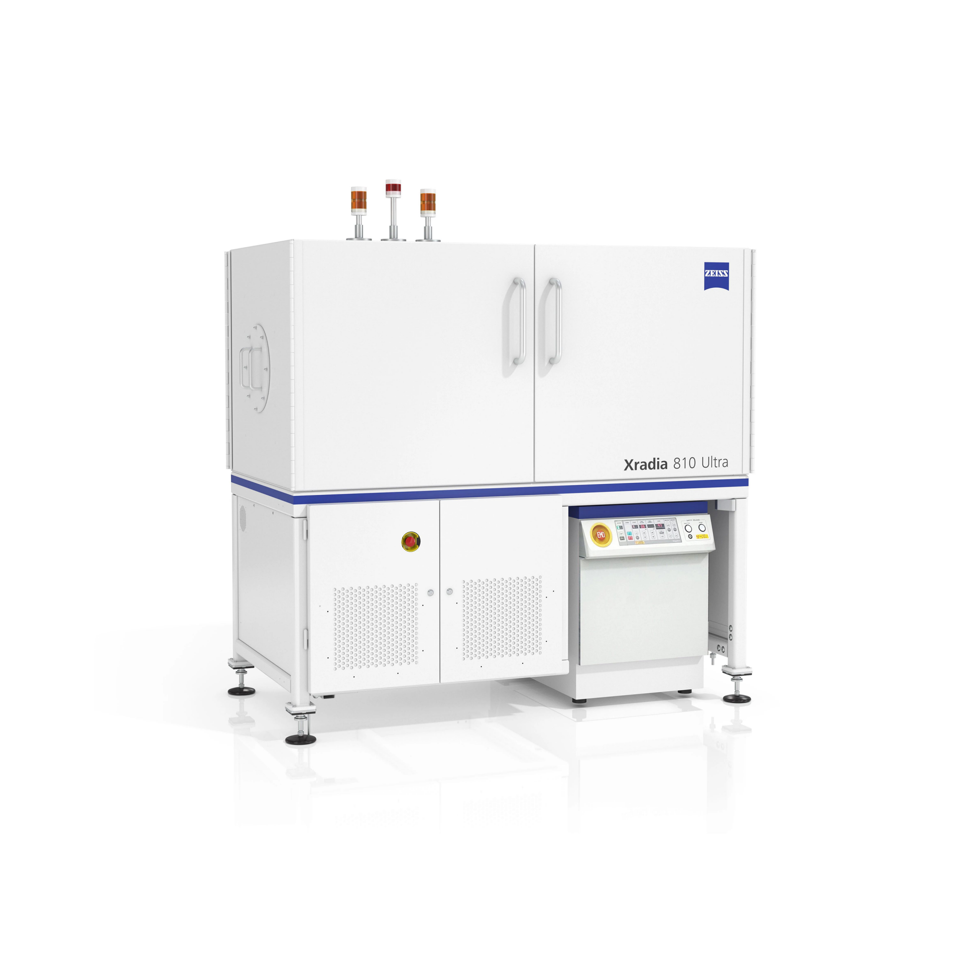
Synchrotron-quality Nanoscale 3D X-ray Imaging |
|
|
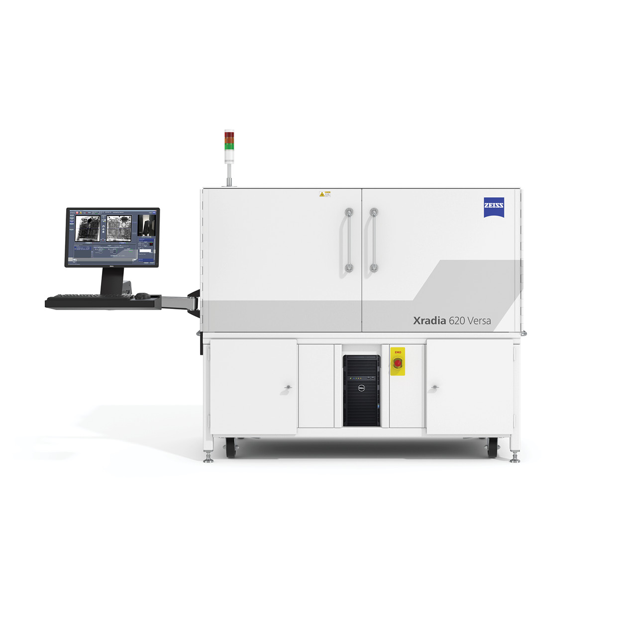
3D X-ray Microscopy for Faster Sub-Micron Imaging of Intact Samples with FAST mode and crystallography capabilities |
|
|
Ground-breaking microCT for unlocking the crystallographic and microstructural secrets of your samples |
|


