You are on our international English website. This site features our entire product portfolio worldwide. The products featured may not be available in the US. If you are a citizen from the US, please visit your country website for local information and contacts.
You are on our international English website. This site features our entire product portfolio worldwide. The products featured may not be available in the US. If you are a citizen from the US, please visit your country website for local information and contacts.
Optimizing retina workflows with OCT and widefield imaging - Dr. Jesse Jung
25 August 202110 minreadbyZEISS
With rapid innovation and advancement in ophthalmic imaging technologies like spectral-domain or swept-source optical coherence tomography (OCT), OCT Angiography (OCT-A) and ultra-widefield cameras, there has never been a more exciting time for eye care professionals to diagnose, monitor and treat retinal disease. Not only has ophthalmic imaging improved clinic workflow and optimized patient care, it has also served as an invaluable tool for telemedicine during the Covid-19 pandemic.
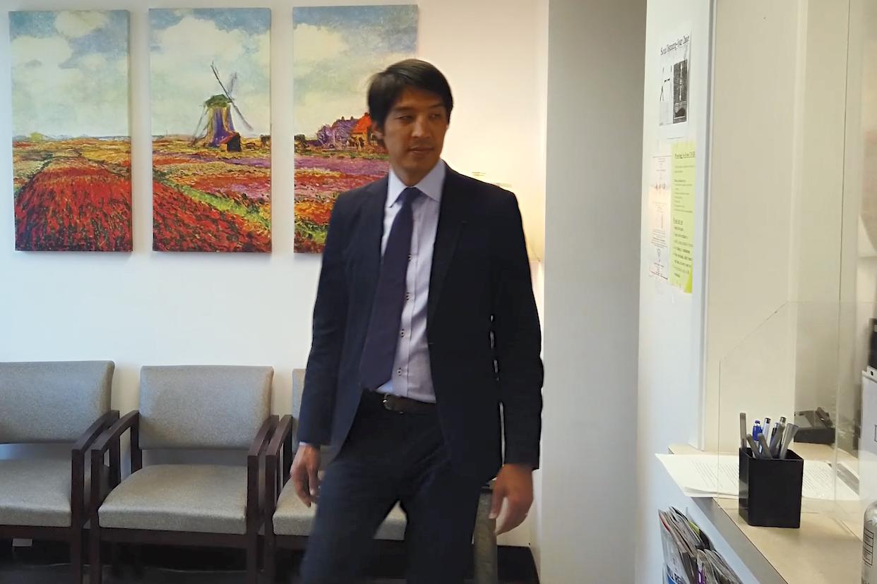
Dr. Jung arrives to his Pleasanton, CA office.
Optimizing a lean practice with ophthalmic imaging
Founded in 1967 by Dr. George F. Hilton, one of the original founders of pneumatic retinopexy, East Bay Retina Consultants, Inc. has been serving the East Bay community for over 50 years. Based on the principle of compassionate service, our board-certified vitreoretinal specialists are committed to each individual patient and the referral doctors in the community.
As the first retinal practice in the East Bay, we continue to dedicate ourselves towards providing the utmost best care, and the continued development of state-of-the art treatments for medical and surgical retinal disease. With 5 locations in the East Bay, we rely heavily on ophthalmic imaging technologies like spectral-domain OCT and ultra-widefield imaging to optimize retinal disease diagnosis and management and improve patient care.
Dr. Jesse Jung explains his practice's retina workflow:
Third-party Content Blocked
The video player is blocked due to your cookie preferences. To change the settings and play the video, please click the button below and consent to use of "Functional" tracking technologies.
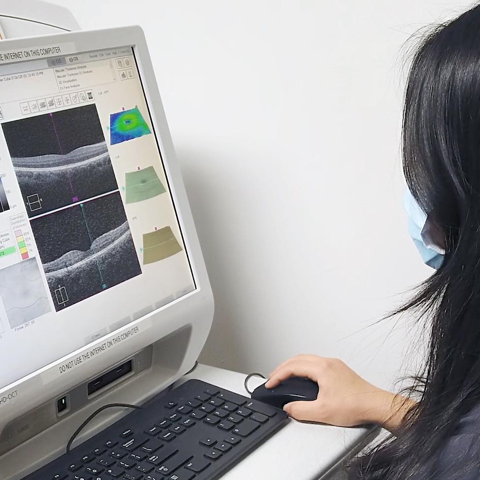
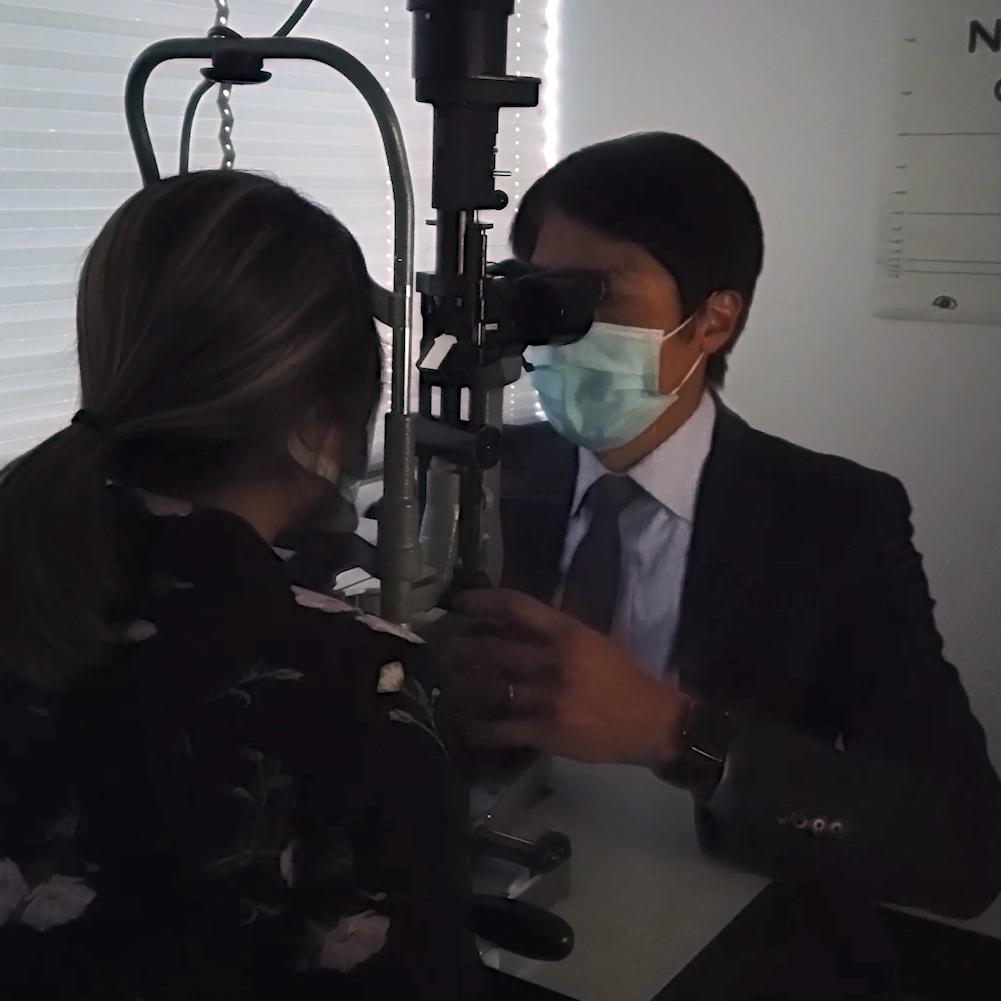
The ZEISS CIRRUS is used regularly in Dr. Jung's retinal practice.
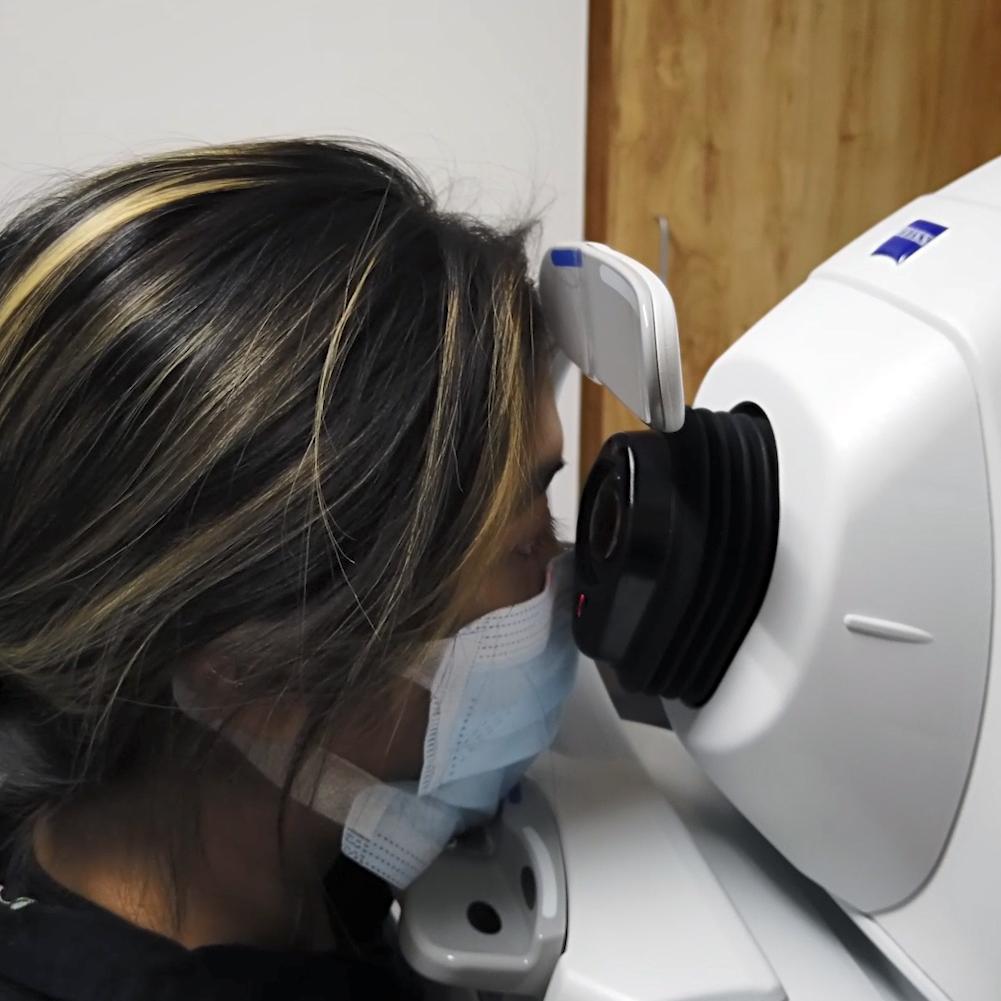
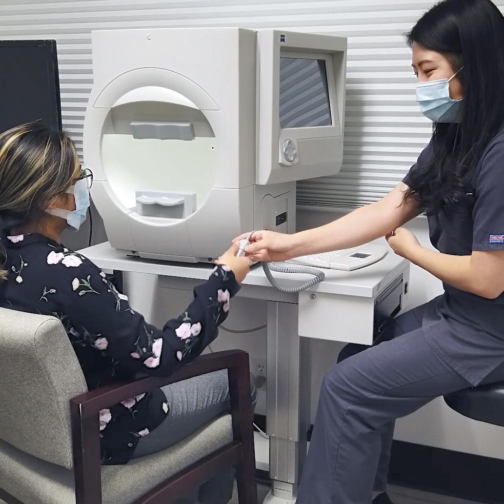
A patient during an OCT exam.
Ophthalmic imaging and improving the standard of care
OCT is considered standard-of-care for the management of most retinal diseases. Despite the myriad of imaging technologies available to retinologists—including fundus photography, fundus autofluorescence (FAF), and fluorescein (FA) and indocyanine angiography (ICGA)—spectral-domain OCT may be the most heavily relied on, non-invasive imaging tool in clinical practice. While once considered a luxury item, OCT has become a mainstay in ophthalmic practice, and an indispensable tool in our practice.
East Bay Retina Consultants relies heavily on integrated imaging as part of the workflow for every patient, and we are actively participating in various clinical trials and imaging research. OCT imaging has enabled our offices to better manage a greater number of patients with more diagnostic accuracy and efficiency. Among the additional imaging technologies utilized in our practice, advancements in ultra-widefield imaging such as the ZEISS CLARUS 700 have made it possible to capture high quality, true-to-color images while combining multiple imaging modalities like FAF, anterior segment external photography and FA all in one device.
Third-party Content Blocked
The video player is blocked due to your cookie preferences. To change the settings and play the video, please click the button below and consent to use of "Functional" tracking technologies.
Watch Dr. Jesse Jung discuss in detail the benefits of CLARUS 700, including some of the the advanced features eye care specialists can look forward to using.
Ultra-widefield imaging allows for imaging of the far periphery outside of the standard 7-field Early Treatment Diabetic Retinopathy Study (ETDRS) field of view, elucidating the extent of disease of peripheral pathology such as predominant peripheral lesions or ischemia in diabetic retinopathy. In our practice, diagnostic tools like the spectral-domain CIRRUS OCT and OCTA have enabled us to improve our clinical decision-making—particularly in determining the level of macular ischemia in diabetes or retinal vein occlusion that may be affecting visual acuity and when to, or not to, treat in cases of subtle pigment epithelial detachment suspicious for indolent quiescent macular neovascularization.
As retinal specialists, we regularly utilize imaging: these tools are essential for a retina practice for diagnosis, education, and treatment planning. During the pandemic, we found that ultra-widefield imaging was an extremely useful supplement to the clinical exam, allowing us to diagnostically evaluate our stable patients while minimizing the amount of exposure . Most imaging devices including the CLARUS 700 today are non-mydriatic and are able to capture high quality images in a non-dilated pupil.
However, while the latest advancements in ophthalmic and ultra-widefield imaging technologies continue to push the limit on what is possible, none should be considered a direct substitute for a thorough dilated fundus evaluation with scleral depression. This is especially true when assessing the peripheral retina for symptomatic patients with suspected holes, tears or detachments. Yet for retinal physicians, this technology provides crucial supplements to exam findings, where we gain a better understanding of those elements of the retina that cannot always be seen with the naked eye.
Among the additional imaging technologies utilized in our practice, advancements in ultra-widefield imaging such as the ZEISS CLARUS 700 have made it possible to capture high quality, true-to-color images while combining multiple imaging modalities like FAF, anterior segment external photography and FA, all in one device.
Optimizing patient workflow with ophthalmic imaging
Coordinating patient flow to prioritize imaging before the patient enters the exam room helps prevent excessive, unnecessary movement throughout the office and can eliminate bottlenecks in clinic flow. This is especially important when offices are seeing a high volume of patients, some of whom may have mobility issues and now even more important during the Covid-19 pandemic to minimize exposure and maximize social distancing.
Unfortunately, many patients were lost to follow up during the Covid-19 pandemic and did not receive the necessary care they needed. Despite temporary office closures, ophthalmic imaging played an integral role in launching the widespread use of telemedicine in ophthalmology. While most clinicians were unable to see their typical patient volume at the height of the pandemic, ophthalmic imaging enabled the remote monitoring of patients’ retinal pathologies and helped supplement telemedicine calls between doctors and patients.
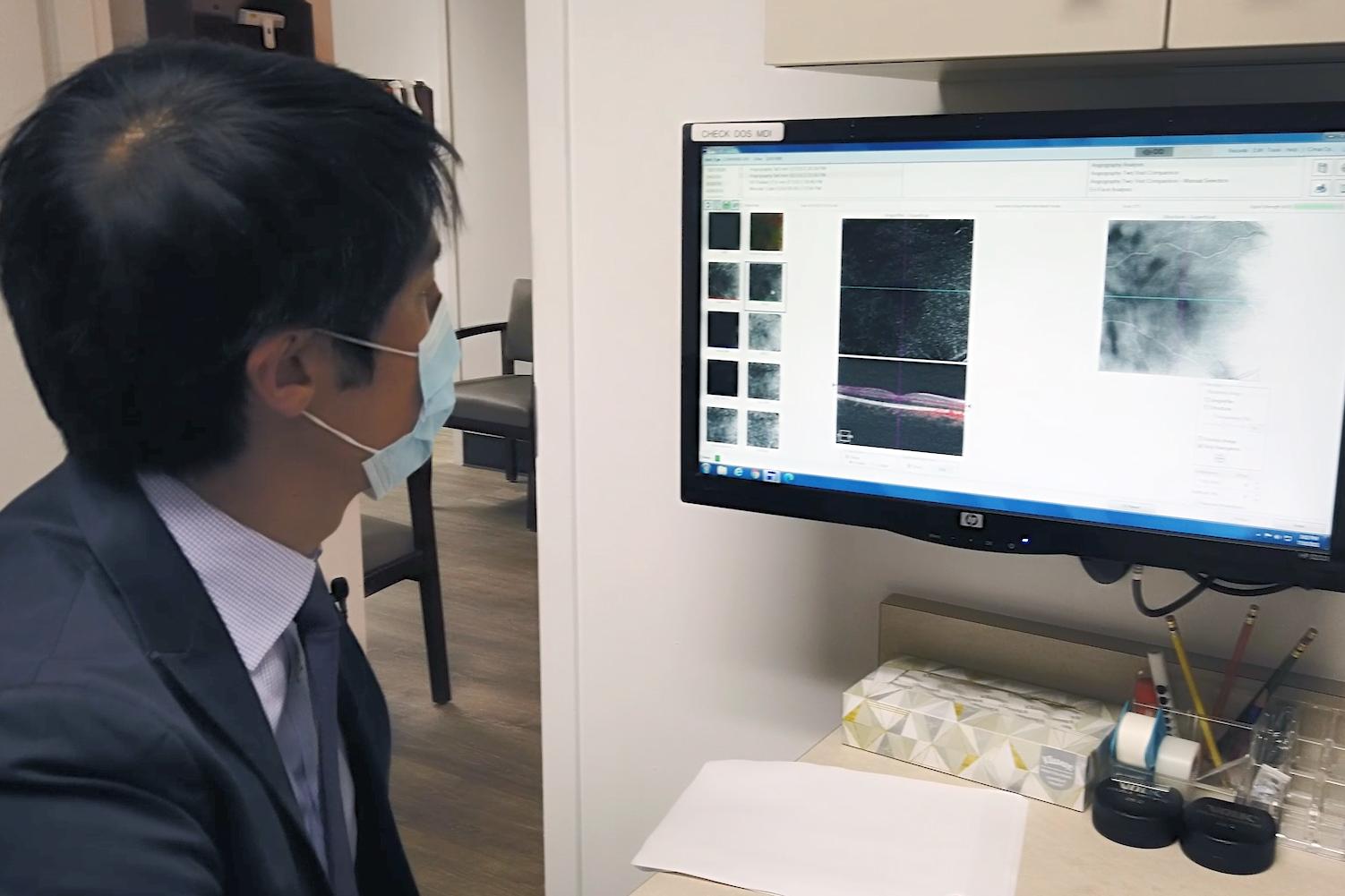
Dr. Jung looks over patient data
When doctors are managing stable retinal diseases like age-related macular degeneration (AMD) or diabetic retinopathy (DR), it is important to have access to many longitudinal data points over time. For this reason, imaging technologies like OCT have become the mainstay in determining treatment algorithms, and serve as a powerful tool in management and prognosis for patients with retinal disease. Moving forward, patients with stable retinal disease like dry AMD or mild nonproliferative diabetic retinopathy may be good candidates for telehealth imaging/telemedicine follow up, as we saw during the pandemic.
Regardless of practice modality, those managing patients with retinal disease will benefit from having fundus photography and OCT on site. In high volume practices like ours, the return on investment (ROI) of acquiring high quality imaging technology such as an ultra-widefield camera or high-definition OCT machine can pay for itself in as little as one year.
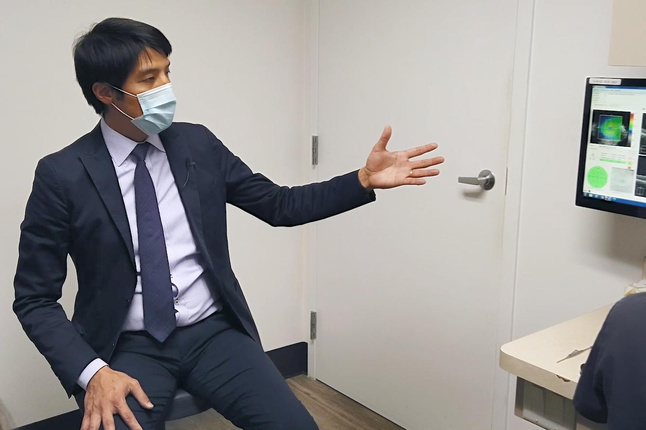
Dr. Jung says patients benefit from seeing pictures of their own eyes when explaining treatment plans.
The uses of imaging beyond clinical decision-making
In our patient workflow, we rely on ophthalmic imaging as an excellent tool for educating patients on their particular retinal disease. The imaging is done before a patient is settled in the exam room, and our doctors utilize a separate monitor with remote reviewing software to walk patients through their images to help them understand their disease diagnosis, progression, and status. OCT, in particular, helps us not only determine the as-needed treatments for our patients, but gives us visual aids for explaining their prognosis and what to expect in terms of future treatments or progression.
For example, when a patient with AMD or a retinal tear views a photo or image of their own eye, they are better able to understand the disease process as well as the required management and treatment they are undergoing. Without a visual depiction of the ocular anatomy, patients may not understand or fully comply with their treatment. Retinal ischemia, assessed via FA or OCT-A, can also help explain decreased best-corrected visual acuity to patients with otherwise unremarkable clinical exams.
At East Bay Retina Consultants, we have also been involved in numerous clinical trials that use imaging to assess treatment outcomes for various retinal diseases. Ophthalmic imaging is now a mainstay in clinical trials. Whether its OCT to assess central foveal thickness or FAF to measure growth of geographic atrophy, imaging serves to determine an objective primary endpoint. Unlike clinical trials in years past, subjective outcomes such as visual acuity alone are no longer always the primary end point that researchers use. By enabling the objective assessment of structural changes to the retina, clinicians and researchers are better able to determine best practices for treating retinal pathology. Imaging research has played a significant role in improving how clinicians diagnose and treat a wide variety of retinal diseases.
One final advice I have for physicians looking to improve their efficiency is to look at their own process. Take time to actually watch the flow of a patient through your practice from check in to check out.
Dr. Jesse Jung
The future of ophthalmic imaging
At the end of the day, the ultimate goal of every ophthalmology practice is to deliver the best possible care to their patients. It is hard to refute the workflow optimization and efficiency that ophthalmic imaging can offer. Future innovations like portable equipment with home device monitoring, teleconsulting, and artificial intelligence will only further improve the patient experience.