Comparison of loupes versus microscope-enhanced CAD-CAM crown preparations: A microcomputed tomography analysis of marginal gaps
Original title:
Comparison of loupes versus microscope-enhanced CAD-CAM crown preparations: A microcomputed tomography analysis of marginal gaps
Source:
Alan M. Atlas DMD, Sridhar Janyavula DMD, MS, Rami Elsabee DMD, Emily Alper DMD, Wael F. Isleem DMD, Michael Bergler CDT, MDT, Frank C. Setzer DMD, PhD, MS. Comparison of loupes versus microscope-enhanced CAD-CAM crown preparations: A microcomputed tomography analysis of marginal gaps. The Journal of Prosthodontic Dentistry, 26 May 2022
Statement of problem
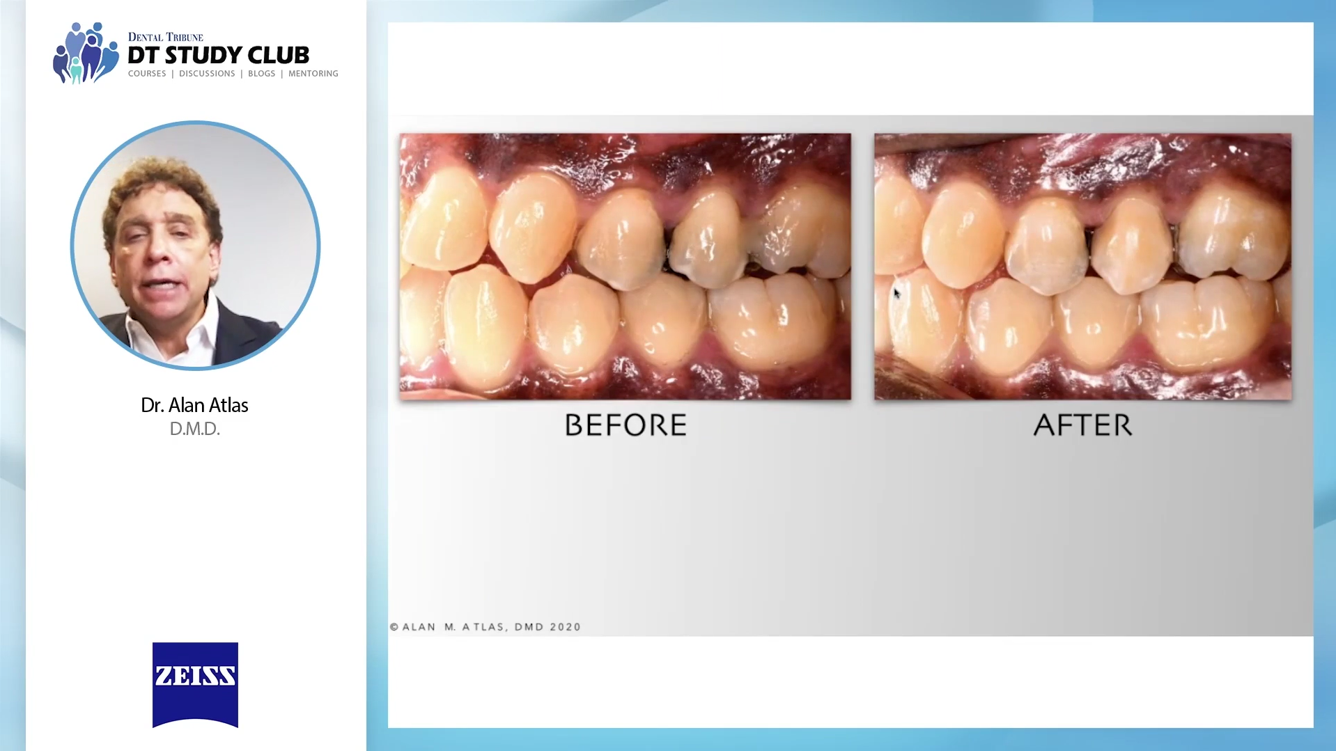
Long-term restoration success depends on a precision marginal fit to prevent marginal leakage and caries. The successful fit of a computer-aided design and computer-aided manufactured (CAD-CAM) crown may be affected by different workflow variables, including preparation, scanning, crown design, milling, sintering, and cementation. Discrepancies in any of these steps may result in poor marginal and internal fit. Evidence suggests that tooth preparation may be the most important step in the workflow for a successful outcome. Compared with the traditional means of crown preparation using the naked eye or loupes, the dental operating microscope provides higher magnification and more direct illumination. However, the impact of high magnification during preparation on the marginal quality of CAD-CAM crowns is unclear.
Purpose
The purpose of this in vitro study was to compare marginal fits of CAD-CAM crowns fabricated after initial preparation with loupes and subsequent preparation refinement with either loupes or a microscope. The null hypothesis was that no significant difference would be found in the marginal gap between the preparations with loupes and those with a microscope.
Material and methods
Mounted extracted molars (N=18) received initial crown preparations with a coarse grit, rounded shoulder, diamond rotary instrument with loupes of ×3.0 magnification. The teeth were then randomly divided into 2 groups and refined for an additional 2 minutes with fine grit, rounded shoulder, diamond rotary instruments with either loupes (LOUP) or a microscope up to ×10.0 magnification (DOM). The prepared teeth were scanned with an intraoral scanner to fabricate zirconia-reinforced lithium silicate crowns manufactured with a 4-axis milling machine, sintered in a dental furnace in accordance with the manufacturer’s instructions, and cemented with self-adhesive resin cement. All teeth with crowns were mounted and scanned with a microcomputed tomography (µCT) system at 21-mm nominal voxel size. The resulting Digital Imaging and Communications in Medicine (DICOM) images were imported into a semiautomatic segmentation software program. Marginal and absolute gaps were measured at 24 consistent circumferential points per specimen. Absolute gaps were labeled, and the total volume was calculated. Paired and unpaired t tests were used for statistical analysis (alpha=.05).
Results
The mean marginal gap was 145.0 ±259.6 mm for LOUP and 35.6 ±110.6 mm for DOM, with a statistically significant difference (P<.001). The mean gap volume for LOUP was 0.975 ±0.811 mm3, and 0.250 ±0.477 mm3 for DOM, also statistically significantly different (P=.023). A significant difference was found between the absolute and marginal gaps for LOUP (P=.007), but for DOM, the difference was not significant (P=.063).
Conclusions
This study demonstrated that the higher magnification used during tooth preparation played a significant role in the size of marginal gaps present around CAD-CAM crowns. Crown preparations finished by using fine grit diamond rotary instruments with a microscope at higher magnification than loupes resulted in a more precise marginal fit with smaller gaps. (J Prosthet Dent 2022)