Indispensable OCT-AMD and Geographic Atrophy Tool Kit
Indispensable OCT-AMD and Geographic Atrophy Tool Kit
Age-related macular degeneration (AMD) seems so ubiquitous that it is often underappreciated or even worse, unseen. It is estimated that 196 million people had AMD in 2020, with that number rising to 288 million by 2040, yet 25% of AMD in the primary eyecare setting goes undiagnosed.1,2 Though the neovascular form of AMD gets most of the spotlight, the non-neovascular or dry form of AMD makes up the majority of all AMD. The Advanced RPE Analysis, available on the ZEISS CIRRUS OCT, was specifically created to help clinicians manage non-neovascular AMD in all its forms.
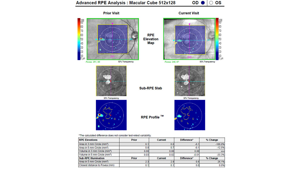
The Advanced RPE Analysis is a combination of two algorithms: RPE Elevation Map to measure drusen burden and Sub-RPE Slab to measure geographic atrophy (GA). The Advanced RPE Analysis reprocesses the standard macular cube scan (512x128 or 200x200) data, to provide reproducible, quantifiable OCT-based measurements of drusen and GA burden. Either algorithm can be applied to any macular cube scan, past or present, and then exported as its own unique report.


RPE Elevation Map and Drusen Detection
The RPE Elevation Map can be considered as an OCT-based surrogate to detect drusen, measure drusen area, and measure drusen volume. Any RPE elevation greater than 19.4μm will be automatically detected and included in the analysis. RPE elevations (OCT surrogate for drusen) can be evaluated with a qualitative approach using the color coded RPE Elevation Map or using the calculated quantitative metrics. The color coded RPE Elevation Map is visualized as a transparent image overlaying the fundus image, to help with correlation with clinical examination. RPE elevation area and volume can be measured within a 3mm or 5mm diameter circle centered on the fovea.
OCT-based detection of drusen is complementary but not equivalent to ophthalmoscopic or color fundus photographic detection of drusen. Whereas ophthalmoscopy and color fundus photography identify pigmentary changes corresponding to drusen, the Advanced RPE Analysis detects RPE elevations corresponding to drusen.
As with many chronic conditions, the detection of non-neovascular AMD initiates a lifelong protocol of monitoring for disease progression. The RPE Elevation Map provides an automatic, reproducible, and objective method with which to monitor for drusen progression or regression. RPE elevation changes are quantified within 3mm and 5mm circles centered on the fovea. Metrics are automatically calculated between the present examination and a previous examination for both RPE elevation area difference and RPE elevation volume difference.
Sub-RPE Slab and Geographic Atrophy Detection
The Sub-RPE Slab is a proprietary ZEISS CIRRUS algorithm for the detection of absent or attenuated RPE. As the RPE thins and is eventually lost in GA, the underlying choroid is hyper-illuminated and more easily visualized on OCT. The Sub-RPE Slab takes advantage of this OCT phenomenon of RPE atrophy to quantify areas of RPE atrophy, which can then be used as an OCT-based surrogate for GA. The Sub-RPE Slab can be visualized qualitatively as an enface overlay or quantitatively as a summation within a 5mm circle centered on the fovea. The Advanced RPE Analysis will also automatically identify the fovea and the shortest distance between any area of sub-RPE illumination and the fovea.
Though short wavelength fundus autofluorescence (FAF) has been the legacy imaging modality of choice for the management of GA, OCT is quickly becoming the modern imaging technology for the diagnosis of GA. Unlike FAF, the Advanced RPE Analysis can automatically and objectively compare an image against a baseline scan to detect GA progression. The progression analytics can be used to detect any increase in GA and encroachment onto the fovea.
A New Era in Geographic Atrophy
GA is a funduscopic or color fundus photography term used to denote areas of retinal and RPE atrophy. The Classification of Atrophy Meetings (CAM) group has created a new international consensus classification system for atrophy secondary to AMD. Complete RPE and retinal atrophy (cRORA) is an OCT based definition (see table 1) of atrophic AMD lesions loosely synonymous to clinical GA in older classification systems.3 When visualized with OCT, the RPE will be attenuated, disrupted, or absent with degeneration of the overlying outer retina. The choroid will be hyper-reflective and seen in greater detail due to the loss of the RPE’s masking effect, which absorbs much of the OCT signal. Incomplete RPE and retinal atrophy (iRORA) is a precursor to cRORA and may become an important entity as treatments for GA are approved.4
|
AMD Finding |
Description |
|---|---|
|
Geographic Atrophy |
Clinical term used to denote areas of retinal and RE atrophy without the presence of present/past CNV |
|
iRORA |
Vertically aligned photoreceptor/outer retinal degeneration, RE attenuation or disruption, and increased signal transmission into the choroid |
|
cRORA |
Vertically aligned zone of hypertransmission of ≥250 um, zone of attenuation or disruption of RPE band of ≥250Mm, and evidence of overlying photoreceptor degeneration whose features include ONL thinning, ELM loss, and EZ or IZ loss |
RPE: retinal pigment epithelium; iRORA: incomplete RPE and retinal atrophy; cRORA: complete RPE and retinal atrophy; ONL: outer nuclear layer; ELM: external limiting membrane; EZ: ellipsoid zone; IZ: interdigitation zone; CV: choroidal neovascularization
The approved use of complement inhibition to treat GA is set to completely alter how clinicians manage GA. The management of GA will transition from a diagnose and loosely monitor approach to a diagnose, carefully monitor, and potentially treat approach. This new paradigm of GA will require careful and accurate diagnosis based on OCT findings as per the CAM group followed by careful and accurate monitoring of the disease course with OCT.
Progression analytics, like those found within the Advanced RPE Analysis, will be key to determine candidacy for treatment and for disease monitoring upon initiation of treatment. These same progression analytics may be used as an educational tool to teach patients about the progressive nature of GA. Advanced RPE Analysis will be able to illustrate the natural progression before treatment and hopefully the slower progression after treatment. This simple illustration of the altered course of disease will hopefully keep patients adherent to treatment and hopefully maintain their vision.
Clinical Implementation of Advanced RPE Analysis
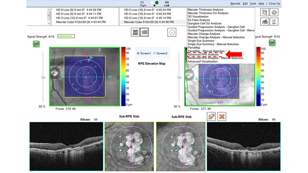
The Advanced RPE Analysis is not a scan protocol but an algorithm that can be applied to any macular cube scan. After completing a macular cube scan, either 512x128 or 200x200, you can choose the Advanced RPE Analysis in the top right-hand corner as seen below (Figure 1). Because it can be used in any form of dry AMD, the Advanced RPE Analysis should be applied to any macular cube scan with signs of dry AMD. For early or intermediate dry AMD, the RPE Elevation Map can be used to detect and monitor macular drusen burden. For late dry AMD, the Sub-RPE Slab can detect areas of GA and their proximity to the fovea, progression, or encroachment onto the fovea. Once the Advanced RPE Analysis algorithm has been applied to a macular cube scan, the analysis can be saved as a stand alone report and exported to the patient chart.


The ZEISS CIRRUS OCT has been a workhorse for the detection, evaluation, and management of neovascular AMD for years but the often-overlooked Advanced RPE Analysis is a comprehensive approach to drusen and GA OCT-based management. By combining macular cube scans, high-definition raster scans, progression analyses, and the Advanced RPE Analysis into the CIRRUS OCT, ZEISS has created the ultimate suite of tools to properly manage all forms of dry AMD.
Get in touch with us!
Receive more information about the product and availability in your country!-
1
Wong, Wan Ling, et al. "Global prevalence of age-related macular degeneration and disease burden projection for 2020 and 2040: a systematic review and meta-analysis." The Lancet Global Health 2.2 (2014): e106-e116
-
2
Neely DC, Bray KJ, Huisingh CE, et al. Prevalence of undiagnosed age-related macular degeneration in primary eye care. JAMA Ophthalmol. 2017;135(6):570-5
-
3
Sadda, Srinivas R., et al. "Consensus definition for atrophy associated with age-related macular degeneration on OCT: classification of atrophy report 3." Ophthalmology 125.4 (2018): 537-548.
-
4
Guymer, Robyn H., et al. "Incomplete retinal pigment epithelial and outer retinal atrophy in age-related macular degeneration: classification of atrophy meeting report 4." Ophthalmology 127.3 (2020): 394-409.