ZEISS provides high-performance imaging solutions designed to enhance diagnostics and education in veterinary medicine. From routine tests such as fecal exams, cytology, urinalysis, and blood smears to histopathology and parasitology, ZEISS microscopes deliver reliable results that veterinarians can trust. Beyond routine diagnostics, ZEISS solutions also support veterinary education and specialized research, making them a versatile choice for a wide range of applications.
Veterinary diagnostics rely on identifying subtle details in fecals, blood smears, or cytology slides. ZEISS microscopes deliver high-contrast, standardized imaging so veterinarians can make confident diagnoses every time.
Time Is Critical in Clinical Workflows
From routine check-ups to urgent cases, speed matters. ZEISS systems enable rapid image capture, efficient documentation, and streamlined digital workflows—saving time while improving accuracy in daily practice.
Scalability for Teaching and Beyond
Veterinary schools and labs must serve many users and applications. ZEISS solutions combine durability with digital connectivity, making it easy to teach microscopy, archive cases, and scale up to specialized research when needed.
Dr. Michelle Oakley, Yukon Vet
Explore Dr. Oakley's Configuration
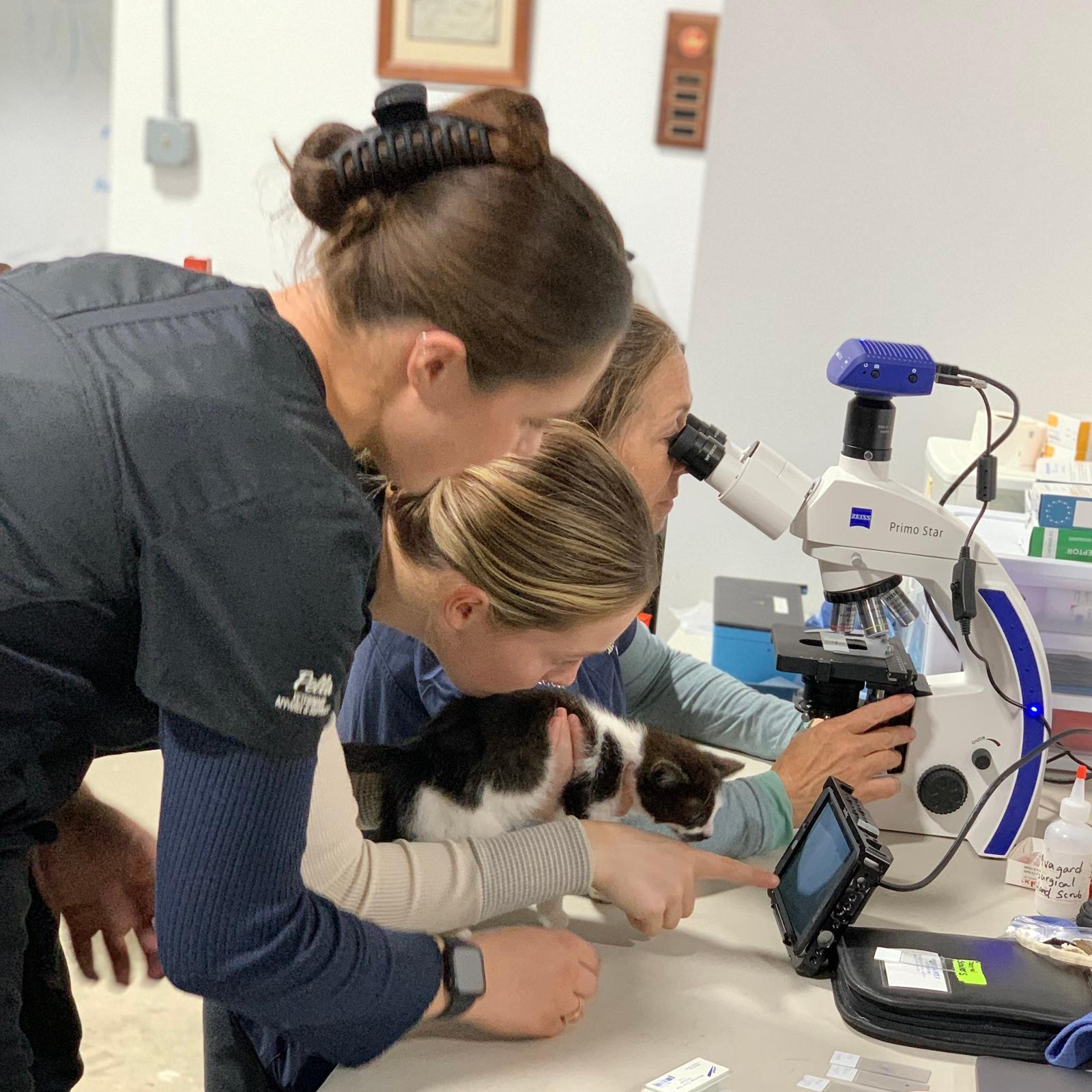
See how the Primostar 3 is enabling veterinary medicine
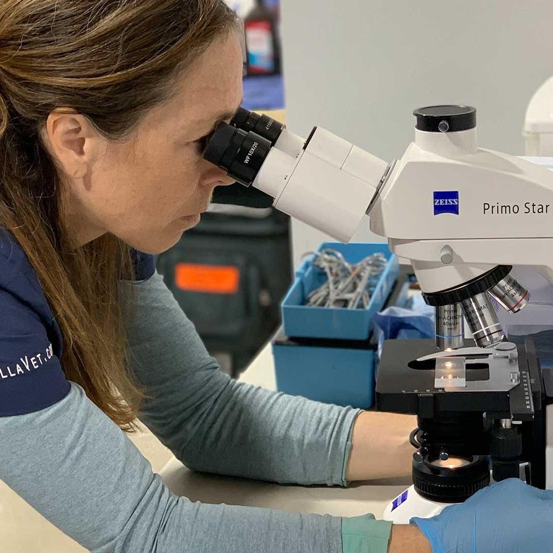
For years, Dr. Oakley has trusted the ZEISS Primostar 3 microscope both in her veterinary clinic and during her global wildlife rescue and research missions. With its reliable performance, she can quickly process blood smears, cytology samples, and tissue biopsies — providing a clear foundation for accurate diagnoses and tailored treatment plans. The Primostar 3 not only strengthens her confidence in every case but also helps her share findings more effectively with clients, colleagues, and conservation partners.
The microscope opens up whole new worlds to them. They become engaged and excited by the opportunity to participate in my work with animals. I am happy to be able to pass on this wonderful gift to them.
See Dr. Oakley’s Scientific Ally
Seek clarity in your subjects
Unlocking new levels of understanding and success in your own education.
Dr. Alejandro Rojas-Fernandez with one of his nanobody generating alpacas
From alpacas to animal health
Leveraging ZEISS imaging platforms to drive diagnostics, therapies, and prevention in veterinary medicine
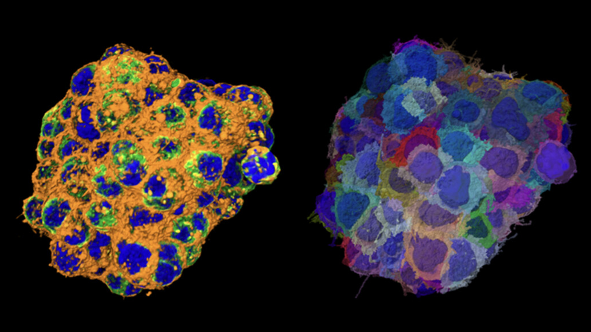
ZEISS microscopy isn’t just for human health — algorithms and imaging tools are helping veterinary science too. In one recent project, researchers used ZEISS Celldiscoverer 7 to rapidly screen alpaca-derived nanobodies, generating potent antiviral agents in response to SARS-CoV-2. Similar technologies can be applied in veterinary settings to identify pathogens, study immune responses, or develop novel treatments for animals. From parasites to viruses, imaging adds precision and speed, improving outcomes for patients across species. By integrating high content microscopy, veterinarians and researchers can accelerate both diagnostics and therapeutic development in animal health care.
Using ZEISS Celldiscoverer 7, we were able to screen 100x 384-well plates in 7 channels, fully automated.
Recommended Products for Veterinary Research
Your Microscope for Digital Teaching and Routine Lab
ZEISS Primostar 3
Primostar 3 is the easy-to-use, compact and long-lasting instrument that turns your investment into the right choice for long working hours even in the most space-limited environments.
Your Smart Microscope for Biomedical Routine and Research
ZEISS Axioscope
The ZEISS Axioscope 5 is a flexible upright microscope optimized for routine and basic research applications. It features excellent optics for brightfield, brilliant LED illumination for transmitted light and optional multi-channel fluorescence.
Inverted Microscope Platform with AI Assisted Experiment Startup
ZEISS Axio Observer
ZEISS Axio Observer is your inverse platform for demanding multimodal imaging of living and fixed specimens. Combine Axio Observer with a wealth of technologies and refine it to support your experiments precisely.
Your Solution for Advanced Image Analysis and Visualization
ZEISS arivis Pro
Create seamless analysis pipelines with just a few clicks. Effortlessly process massive datasets on compatible workstations. “Walk into your sample” using our VR toolkit for a new perspective.
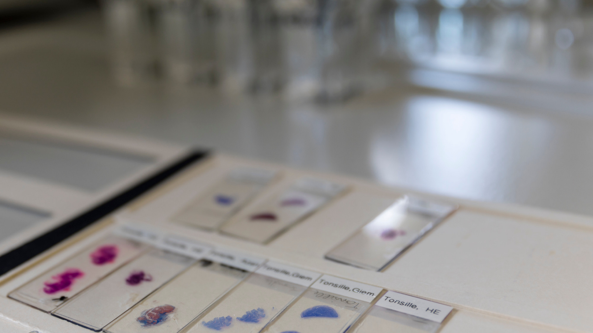
For routine workflows such as fecal exams, blood smears, and urine sediment analysis, ZEISS Axioscope and Primostar microscopes deliver reliable optical clarity and ergonomic design. These systems are durable for daily use in clinics while providing the precision needed for confident diagnoses.
High-contrast ZEISS optics allow subtle details in parasite eggs, cysts, and urinary crystals to stand out clearly. Optional digital cameras and imaging software enable easy documentation, teaching, and consultation with colleagues, improving diagnostic confidence.
Yes. ZEISS digital platforms, including arivis Pro, support automated image analysis pipelines for parasite detection, slide screening, and quantification. These tools reduce manual effort, standardize results, and save time in high-volume diagnostic or teaching labs.
ZEISS microscopes are modular and scalable, meaning you can start with a clinical diagnostic setup and later add digital imaging, automation, or advanced research capabilities. This ensures long-term value whether you’re a private practice, teaching hospital, or veterinary research lab.
Integrated digital cameras and software capture, annotate, and share images quickly, supporting efficient patient records, client communication, and teaching material creation.
Classroom and digital teaching solutions allow multiple users to view the same sample simultaneously, both in person and remotely, strengthening microscopy education and hands-on training.