A Digital Teaching Laboratory for Microscopy-Focused Education
One university's success at creating a modern, microscopy-based, teaching facility
In the university setting, the resources for microscopy-based education are often eclipsed by research-centric goals for equipment allocation and core resources. However, university graduates require advanced microscopy training to be competitive, productive and successful in their STEM career paths.
Dr. Glen Marrs, the microscopy facility director at Wake Forest University (WFU), USA, and his colleagues took on this challenge and created the WFU Microscopy Training Center, an extensive teaching laboratory designed to promote microscopy education which is equipped with ZEISS Axiolab and ZEISS Stemi 305 microscopes. The facility now supports eight courses ranging from Parasitology, Developmental Biology, Developmental Neurobiology, Mycology and – of course – Biological Microscopy.
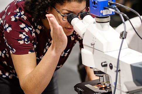
Students learning about digital lab books while working with ZEISS Axiolab and ZEISS Stemi 305 microscopes in Wake Forest University's Microscopy Teaching Center.
Microscopy Training Center Design
Up to sixteen students have access to ZEISS Axiolab compound microscopes and ZEISS Stemi 305 stereo microscopes. These microscopes have real-time camera displays on iPads that can be wirelessly transmitted to any monitor. Wall-mounted monitors display live camera output for the entire class.
The WFU Microscopy Training Facility has succeeded in:
Utilizing active microscopy-based learning as a dynamic lecture/laboratory tool
Providing access to modern imaging modalities, including fluorescence
Maximizing digital image sharing and evaluation capabilities
Creating a unified teaching setting for an entire group of students
We were able to meet our goals economically, in particular, with the acquisition of the cost-effective scientific ZEISS ERc 5s cameras that utilize a variety of sharing mechanisms and can be controlled wirelessly.
Example Application Images
Collected by Students in the Biological Microscopy Course at Wake Forest University
Insect Specimen - Honey Bee Leg
Darkfield stereo microscopy
Plant Sample - Lycopodium strobilus
Brightfield microscopy
Plant Sample - Pumpkin Stem
Brightfield microscopy
Mixed Pollen Specimen
Brightfield microscopy
I am able to use the excellent technology and personnel of the Wake Forest Microscopy Teaching Center for my Parasitology course.
Dr. Cordy works with the ZEISS Axiolab classroom microscope.
Dirofilaria immitis (dog heart worm) parasites, imaged in a blood smear from a canine using brightfield microscopy.
Dirofilaria immitis (dog heart worm) parasites, imaged in a blood smear from a canine using brightfield microscopy.
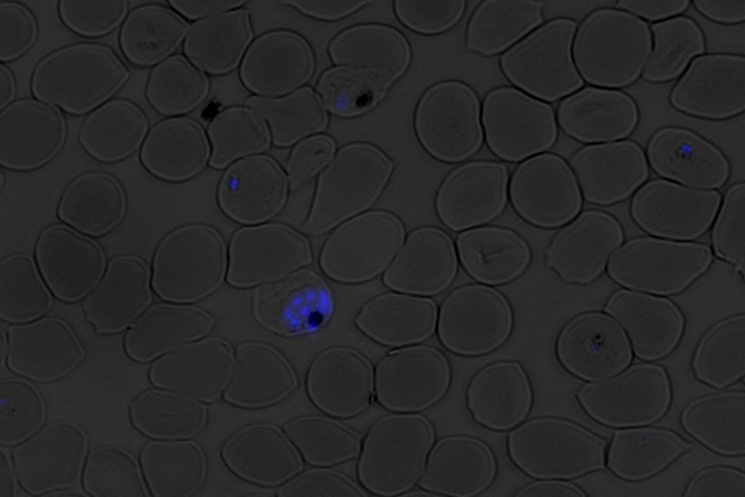
DAPI-labeled nuclei of the malaria parasite Plasmodium falciparum inside of human red blood cells imaged using fluorescence microscopy.
DAPI-labeled nuclei of the malaria parasite Plasmodium falciparum inside of human red blood cells imaged using fluorescence microscopy.
Parasitology with Dr. Cordy
Hands-on Learning with Real Specimens
In my Parasitology course, students learn the basics of stereo and compound light microscopy.
They are introduced to various applications including isolating live nematodes from a petri dish, mounting and staining samples, using an oil immersion lens, doing live cell microscopy, performing white balance and taking clear images, and performing post-image processing.
With a smaller group of undergraduate students who take research for credit, we are also able to venture into fluorescence microscopy using fixed cells and nuclear dyes.
Dr. Cordy Explains the Benefits for Teachers and Students
I really love the fact that the microscope images can be displayed to a large screen so easily; I can quickly glance around the room and see how the students are progressing with the laboratory exercises. I can offer instant feedback from across the room. I can also quickly show them something from my microscope using my own screen, providing fast and effective guidance.
I believe that the students enjoy the digital format of our classroom. They digitally photograph what they see, edit images on an iPad, airdrop their photos to their laptop, finalize their assignments using a digital lab notebook, and then submit them via a course website. There is no pen and paper. I feel that this fully digital format suits this particular generation quite nicely.


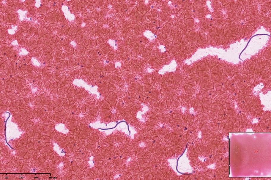


 parasites, imaged in a blood smear from a canine using brightfield microscopy.")
 parasites, imaged in a blood smear from a canine using brightfield microscopy.")
 parasites, imaged in a blood smear from a canine using brightfield microscopy.")
 parasites, imaged in a blood smear from a canine using brightfield microscopy.")


