The Role of OCTA in the Management of AMD and Geographic Atrophy
The Role of OCTA in the Management of AMD and Geographic Atrophy
The neovascular form of age-related macular degeneration (AMD) makes up 10% of all AMD but is responsible for up to 90% of all cases of AMD-related legal blindness.1 This disproportionate burden of vision loss is the reason why all clinicians must stay hyper-vigilant when managing AMD patients, identifying neovascularization early and initiating treatment promptly. OCT has revolutionized how we detect and manage neovascular AMD but OCT angiography (OCTA) systems such as the ZEISS CIRRUS AngioPlex®, continue to push the boundaries of ocular imaging so that clinicians can better care for these patients.
Advantages of OCTA in Age-related Macular Degeneration
Unlike dye-based angiography, such as fluorescein angiography, which uses an injectable dye and excitatory signals to image retinal vasculature, OCTA is a non-invasive modality for imaging ocular vasculature. OCTA takes advantage of motion-based variance of erythrocytes to generate depth encoded images of retinal and choroidal vasculature. OCTA is unaffected by dye accumulation or signal blunting from macular pigments, which makes it particularly useful for detailed macular vascular imaging. For these reasons, OCTA is an incredibly effective imaging modality for detecting and following macular neovascularization (MNV). [DE1] Unlike structural retinal OCT scans which are analyzed in cross-section, OCTA scans are evaluated as enface slabs. OCT angiograms provide structural OCT data and vascular OCTA data that can be analyzed simultaneously. This combination of angiographic and structural data allows for visualization of MNV, along with the resultant structural OCT changes that we are accustomed to seeing.


Dye-based fluorescein angiography is often considered the gold standard for detecting MNV in AMD, but due to its invasive nature, it is often reserved for patients in which there is a relatively high suspicion for MNV. But some equivocal or high-risk cases might require MNV screening even though they do not reach the threshold for performing fluorescein angiography; for these cases we have OCTA.
Intermediate AMD eyes with intraretinal hyper-reflective foci, both large drusen and pigmentary changes, or neovascular AMD in the fellow eye are at especially high risk for conversion to neovascular AMD. These high-risk intermediate AMD eyes should be monitored carefully and considered for MNV screening to aid in the early detection of neovascular AMD.
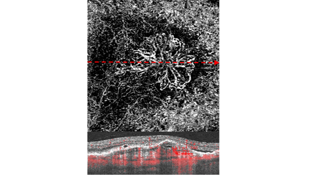
Non-exudative Macular Neovascularization
With the advent of OCTA, a new form of MNV has been described: nonexudative MNV. Nonexudative MNV is an asymptomatic form of MNV with minimal changes to retinal architecture and no fluid, hemorrhage, or exudation. Nonexudative MNV is considered by some to be a precursor to exudative MNV (the standard type of MNV with fluid, hemorrhage, and/or exudation) because up to 80% of nonexudative MNV patients will convert to exudative MNV within 2 years.2 OCTA is key to diagnosing and properly managing these cases because they can be easily missed with funduscopy, color fundus photography, and even fluorescein angiography but are readily visualized with OCTA.


The Importance of Detecting Macular Neovascularization Early
Screening with OCTA is particularly important in high-risk intermediate AMD and nonexudative MNV patients because MNV can grow at a rate of 26μm per day and cause devastating vision loss.3 Real world data has shown that the average VA upon initiation of neovascular AMD therapy is 20/83.4 Considering that the average patient may have exudative neovascular AMD for up to a year before treatment initiation, it is no surprise that intermediate AMD patients will lose 3-5 lines of visual acuity before their neovascular AMD diagnosis.5 Studies have demonstrated that exudative neovascular AMD patients with better initial acuity at the time of treatment initiation, tend to have a better final acuity.4,6 With the ability to screen for MNV, hopefully we can improved these trends and keep more patients from losing vision.
OCTA in the Era of Geographic Atrophy Treatment
The era of complement inhibition for the treatment of geographic atrophy (GA) has opened up a brand-new clinical arena. For the first time, clinicians will be able to treat GA instead of watching patients slowly lose vision. Complement inhibition has been shown to slow down the progression of GA but at the cost of an increased risk of MNV. Patients undergoing GA treatment will require frequent screening in order to promptly detect MNV and initiate anti-VEGF treatment. Using serial OCTA imaging, neovascularization will ideally be detected in its nonexudative form, before exudation and vision loss occur. With new protocols such as this, clinicians will be able to ensure that GA patients attain all the benefits of complement inhibition without suffering the potential adverse events from exudative neovascularization.
For years, choroidal alterations have been implicated in GA pathogenesis but recent OCTA studies have proven that choroidal alterations precede GA.7 Reductions of choriocapillaris flow, as visualized with OCTA, tend to be larger than the clinical GA lesions. When monitored longitudinally, the GA lesions were more likely to expand into the areas with the greatest reductions in extra-marginal choriocapillaris flow. These unique OCTA features of visualizing the choriocapillaris provide important differential and prognostic indicators, that can help tailor GA treatment.
Much the same way that structural OCT has revolutionized retinal and choroidal structural imaging, OCTA has revolutionized how we evaluate retinal and choroidal vasculature. As the need for MNV screening becomes more vital in the era of GA, clinicians will further rely on OCTA systems such as ZEISS CIRRUS with AngioPlex® to render high-quality care to our patients and keep our AMD patients from losing vision.
-
1
Ferris, Frederick L., Stuart L. Fine, and Leslie Hyman. "Age-related macular degeneration and blindness due to neovascular maculopathy." Archives of ophthalmology 102.11 (1984): 1640-1642.
-
2
Laiginhas, Rita, et al. "Nonexudative macular neovascularization–a systematic review of prevalence, natural history, and recent insights from OCT angiography." Ophthalmology Retina 4.7 (2020): 651-661.
-
3
Liu, Tin Yan A., Ankoor R. Shah, and Lucian V. Del Priore. "Progression of lesion size in untreated eyes with exudative age-related macular degeneration: a meta-analysis using Lineweaver-Burk plots." JAMA ophthalmology 131.3 (2013): 335-340.
-
4
Ho, Allen C., et al. "Baseline Visual Acuity at Wet AMD Diagnosis Predicts Long-Term Vision Outcomes: An Analysis of the IRIS Registry." Ophthalmic Surgery, Lasers and Imaging Retina 51.11 (2020): 633-639.
-
5
Ho, Allen C., et al. "The potential importance of detection of neovascular age-related macular degeneration when visual acuity is relatively good." JAMA ophthalmology 135.3 (2017): 268-273.
-
6
Comparison of Age-related Macular Degeneration Treatments Trials (CATT) Research Group. "5-year outcomes with anti-VEGF treatment of neovascular age-related macular degeneration (AMD): the comparison of AMD treatments trials." Ophthalmology 123.8 (2016): 1751.
-
7
Müller, Philipp L., et al. "Optical Coherence Tomography-Angiography in Geographic Atrophy." Ophthalmologica 244.1 (2021): 42-50.