
ZEISS CALLISTO eye Computer assisted cataract surgery
With ZEISS CALLISTO eye® markerless alignment, manual marking steps can be skipped altogether for an efficient1 and precise2 toric IOL alignment to reduce residual astigmatism. It helps you meet patient expectations with assistance functions projected directly into your surgical field.

Cataract assistance functions for every step of the surgery
The assistance functions of ZEISS CALLISTO eye are completely surgeon-controlled – with either the foot control panel or handgrips.
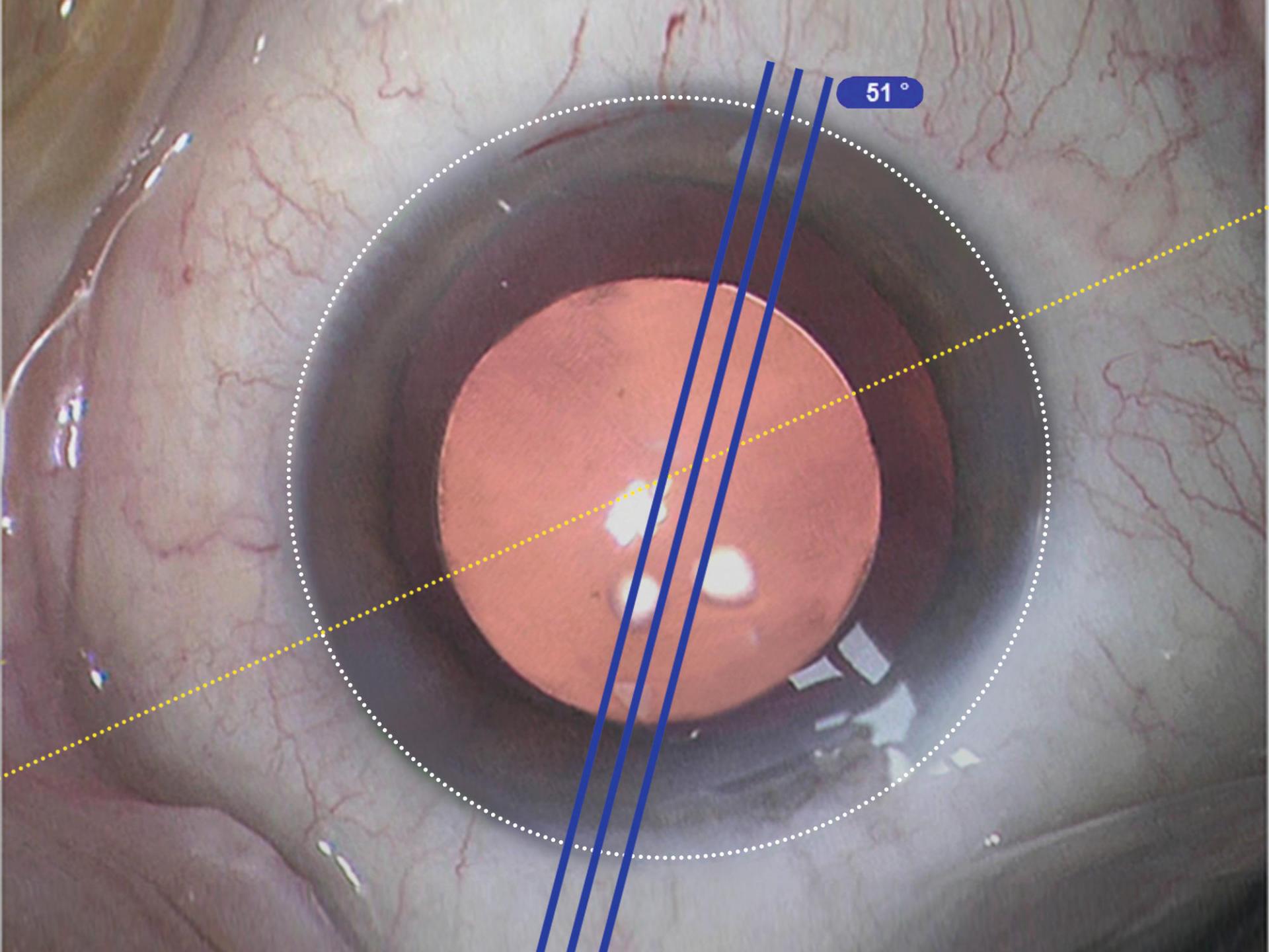

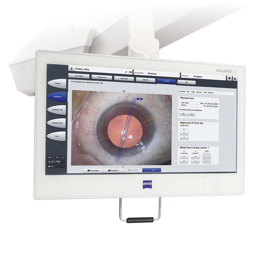
Z ALIGN®
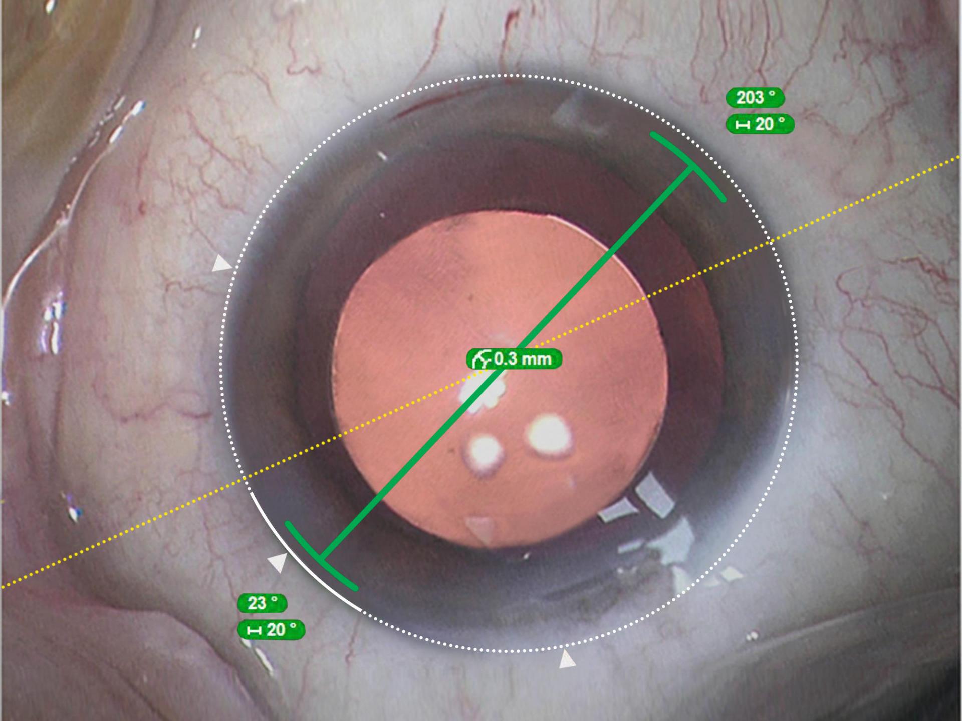
Perform toric IOL centration on the visual axis provided by the IOLMaster and perform rotational alignment.
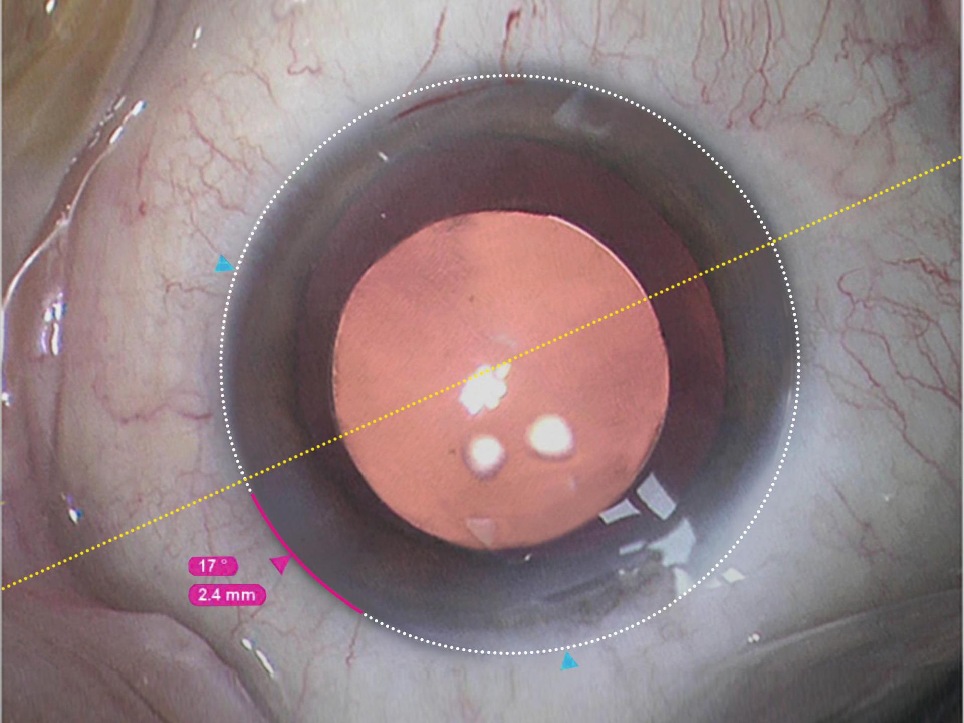

Incision
Position incisions, optionally on the steep axis; add opposite clear cornea incision and paracenteses.

Rhexis
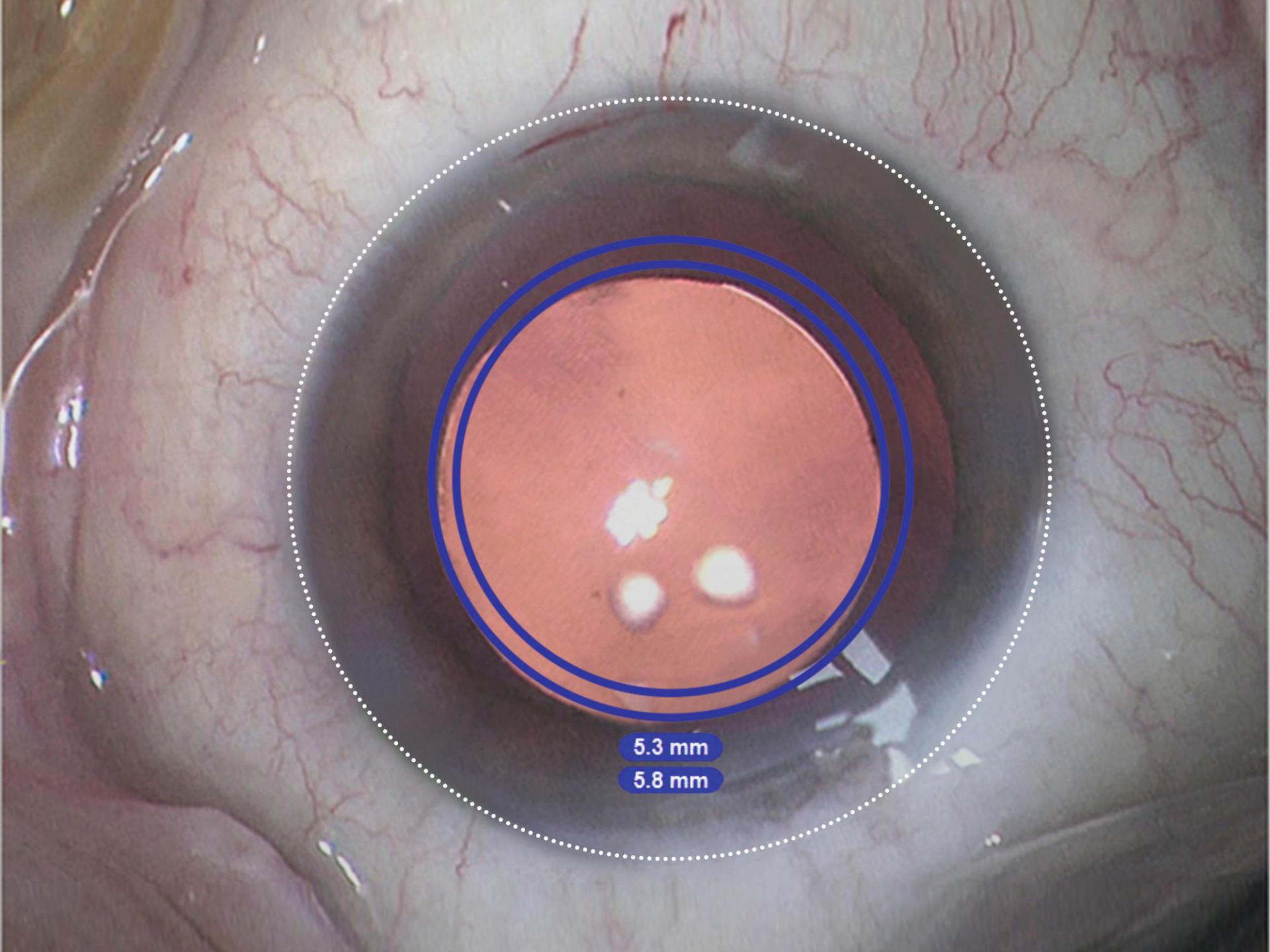
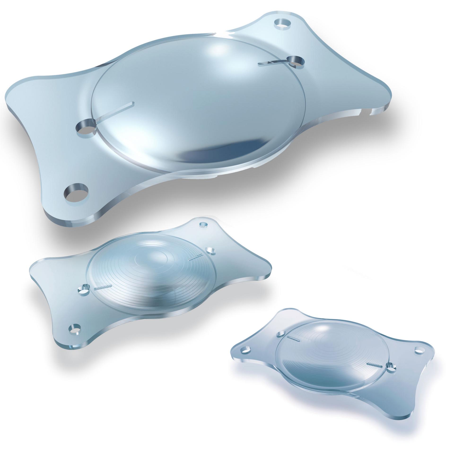
Precisely2 size and shape capsulorhexis and align the IOL on the visual axis provided by the IOLMaster.

LRI
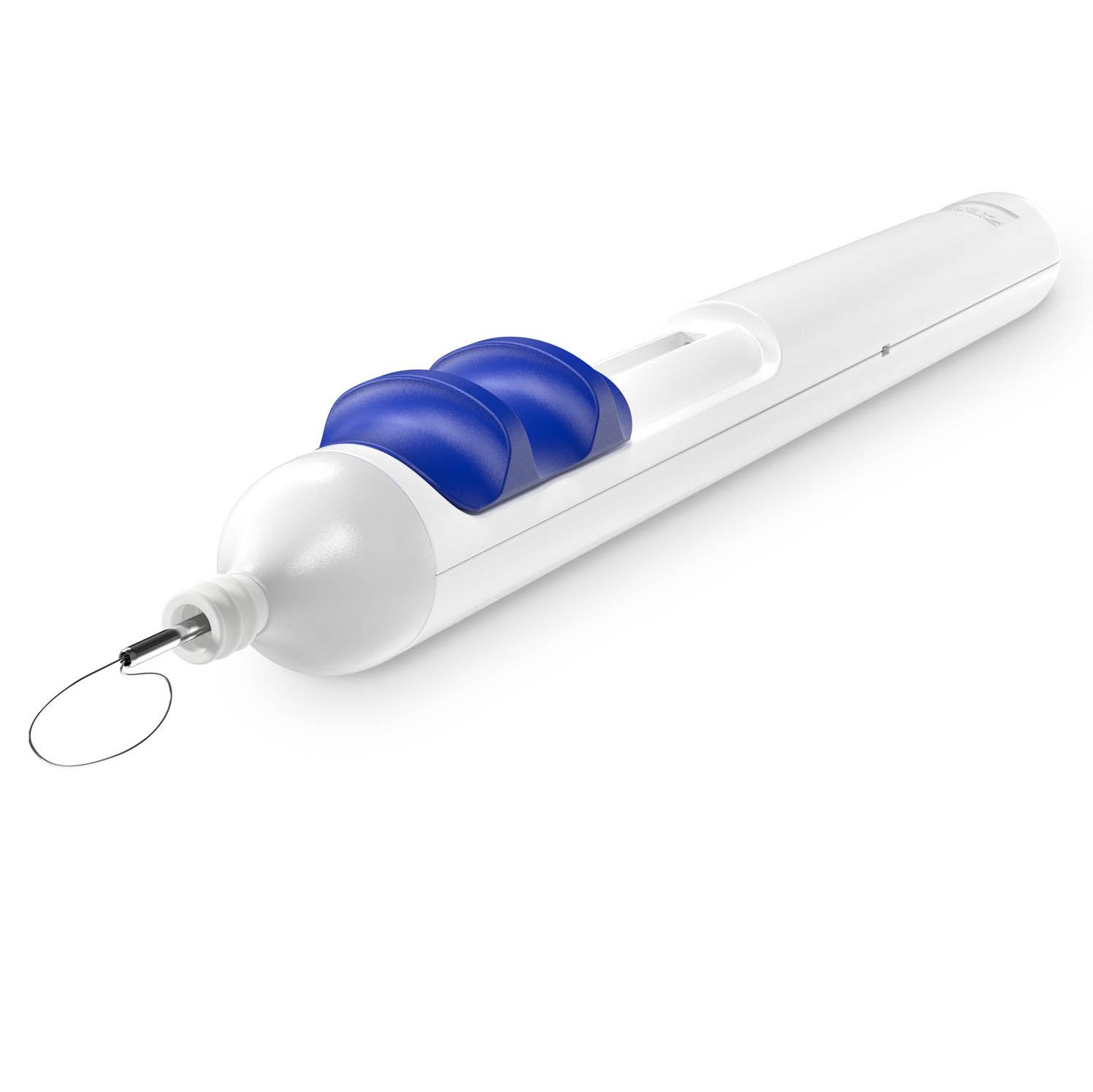
Perform limbal relaxing incisions.

K TRACK®
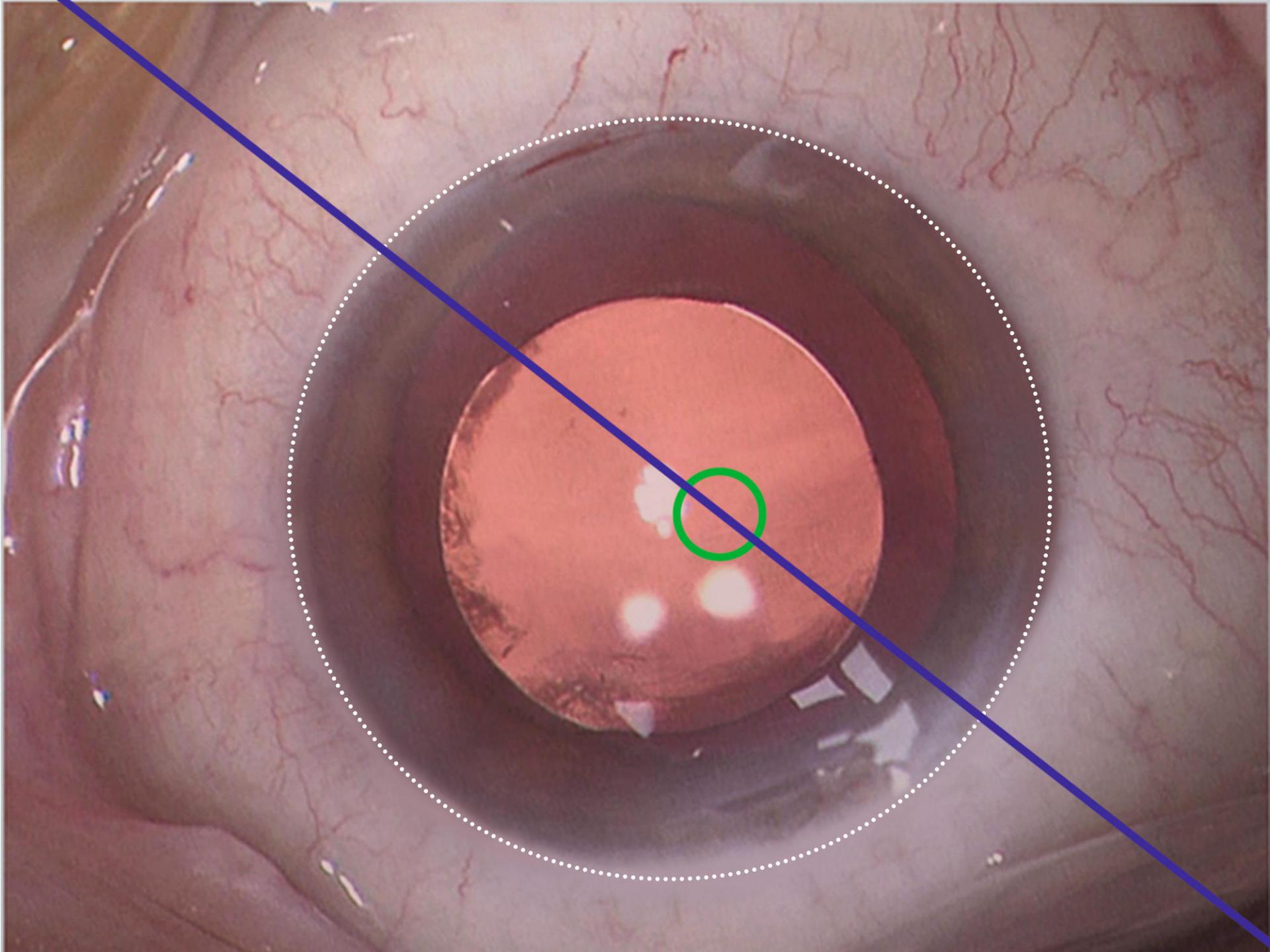
Estimate the local corneal curvature in combination with a keratoscope.

Markerless toric IOL alignment
Starting with a biometry reference image from the IOLMaster® from ZEISS, data is transferred smoothly to ZEISS CALLISTO eye. This data is used to create overlays in the eyepiece. Save time, increase efficiency and reduce residual astigmatism when you:
- skip manual preoperative marking
- skip manual data transfer
- skip manual intraoperative marking


Convenient data management
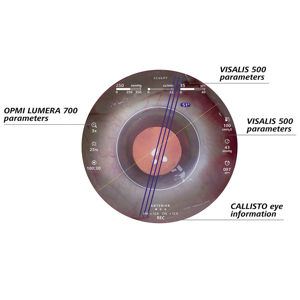
HD video recording and photos that include assistance functions meet demanding requirements in quality management, teaching and for presentations. Import patient lists via a network connection and DICOM modality worklist or USB stick. Export videos and photos via a DICOM network connection or USB stick. Your OR team can easily follow the surgery thanks to the full-screen live video.
Downloads


Specifications
ZEISS CALLISTO eyeCompatibility Overview
Which product works with which CALLISTO eye feature?|
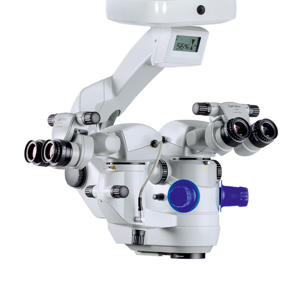
ZEISS OPMI LUMERA 700 with IDIS
|
ZEISS OPMI LUMERA family3 with EDIS
|
|
|---|---|---|
|
CALLISTO eye BASIC
|
||
|
High quality video and photo documentation
|
✓
|
✓
|
|
Mounting options: |
|
|
|
- Floor stand of the ZEISS surgical microscope |
✓ |
- |
|
- Ceiling mount |
✓ |
- |
|
- Wheeled stand |
✓ |
✓ |
|
- Table stand |
✓ |
✓ |
|
Full remote control of surgical microscope |
✓ |
- |
Get in touch with us!
Receive more information about the product and availability in your country!Related products


-
1
Clinical data of Dr. Mayer: "Toric IOL implantation was significantly faster using digital marking" published in J Cataract Refract Surg 2017; 43:1281–1286.
-
2
VIROS research team of Prof. Findl: Clinical data of Dr. Varsits "Deviation between the postoperative (at the end of surgery in the operating room) and aimed IOL axes was 0.52 degrees± 0.56 (SD)" published in J Cataract Refract Surg 2019; 45:1234–1238 and Clinical data of Dr. Hirnschall presented at ESCRS 2013.
-
3
S88/OPMI Lumera T, OPMI Lumera i, S7/OPMI Lumera