

ZEISS OPMI LUMERA 700 Seeing to succeed with the microscope for every ophthalmic specialty
Whether preserving or restoring a patient’s sight, the OPMI LUMERA® 700 from ZEISS is the surgical microscope for every ophthalmic specialty. Experience markerless IOL alignment and integrated intraoperative OCT3 imaging – all in one device – from the ophthalmic microscope market leader.
ZEISS OPMI LUMERA 700 is part of our commitment to helping you succeed in your OR. It’s also part of the ZEISS Cataract Suite, which includes leading products designed to work together for markerless toric IOL alignment.


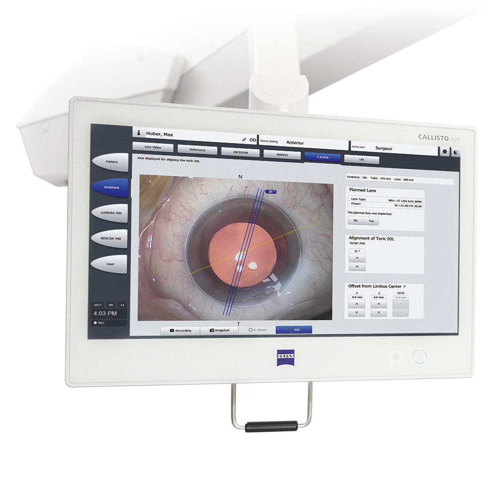
Markerless toric IOL alignment
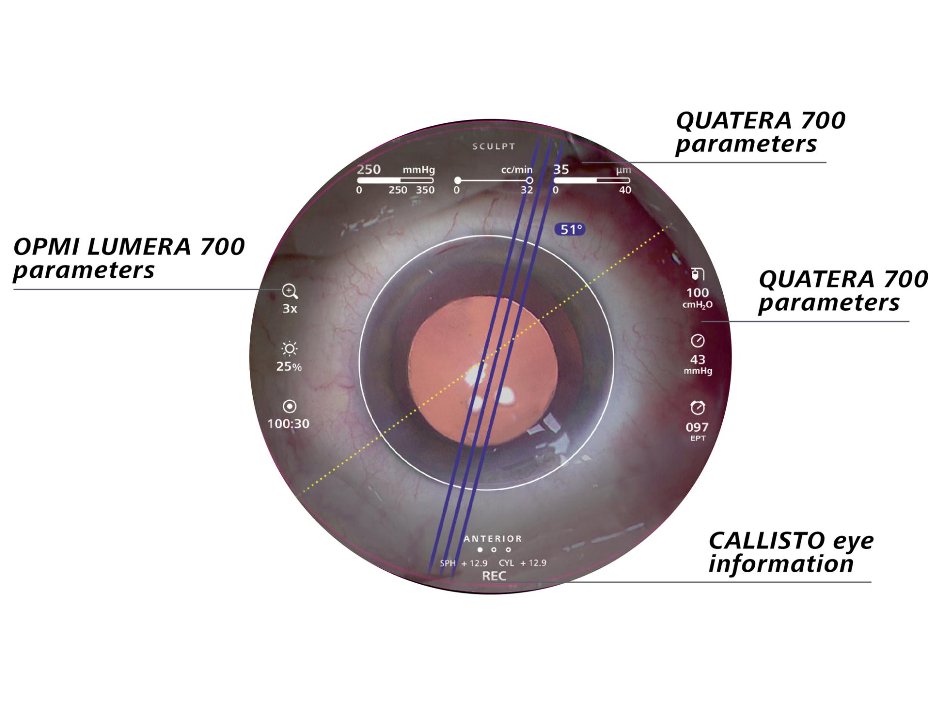
With ZEISS CALLISTO eye® markerless alignment, manual marking steps can be skipped altogether for an efficient2 and precise1 toric IOL alignment to reduce residual astigmatism. For all cataract surgeries, ZEISS OPMI LUMERA 700 provides outstanding anterior views and precise1 assistance functions, thanks to its patented SCI illumination, ZEISS optics and ZEISS CALLISTO eye.

Seeing to succeed in cataract surgery
- Get cataract assistance: The assistance functions of ZEISS CALLISTO eye are completely surgeon-controlled with either the foot control panel or handgrips.
- Achieve efficient2 markerless IOL alignment: Data is transferred smoothly to CALLISTO eye from the IOLMaster® from ZEISS to create overlays in the eyepiece.
- Enhance efficiency: Save time and reduce residual astigmatism3 by skipping manual preoperative marking, manual data transfer and manual intraoperative marking.
- Improve surgery setup: Optimize light intensity, magnification and centration of the live image with the guided image quality check.
- Get exceptional details: The well established Stereo Coaxial Illumination (SCI) delivers highly stable and high contrast red reflex, even with advanced cataracts.
I save 6 minutes per patient and improve alignment precision by 40% compared to manual marking.


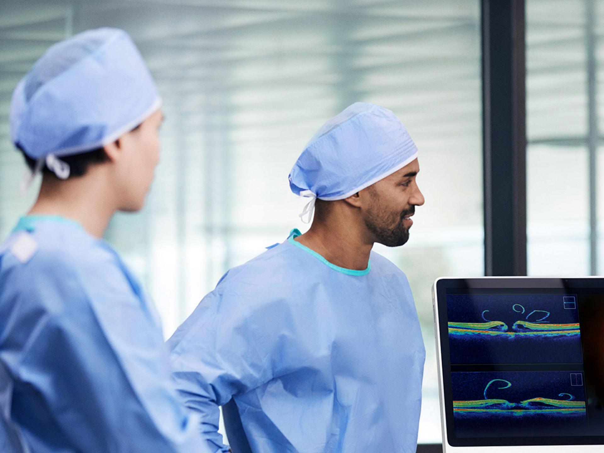
Improved visualization
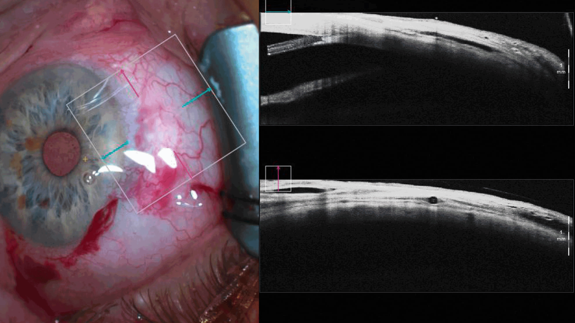
The integrated intraoperative OCT4 images of the ZEISS OPMI LUMERA 700 enable a clear visualization of each surgery, helping to achieve excellent outcomes. Experience superb clarity and critical insights during procedures. Studies have revealed that intraoperative OCT4 from ZEISS can lead to quicker decisions4.

Seeing to succeed in glaucoma surgery
- Make well-informed decisions: The distortion-free computer-enhanced intraoperative OCT4 images enable you to visualize detailed structures in the correct physiological shape.
- Stay focused: The automatic XY tracker maintains the selected intraoperative OCT4 scan location and compensates for movements of the eye or microscope.
- Protect the retina: The integrated retina protection filter shields the retina from excessive light exposure.
- Maintain flexibility: Tilt the microscope head as needed to better observe the iridocorneal angle.
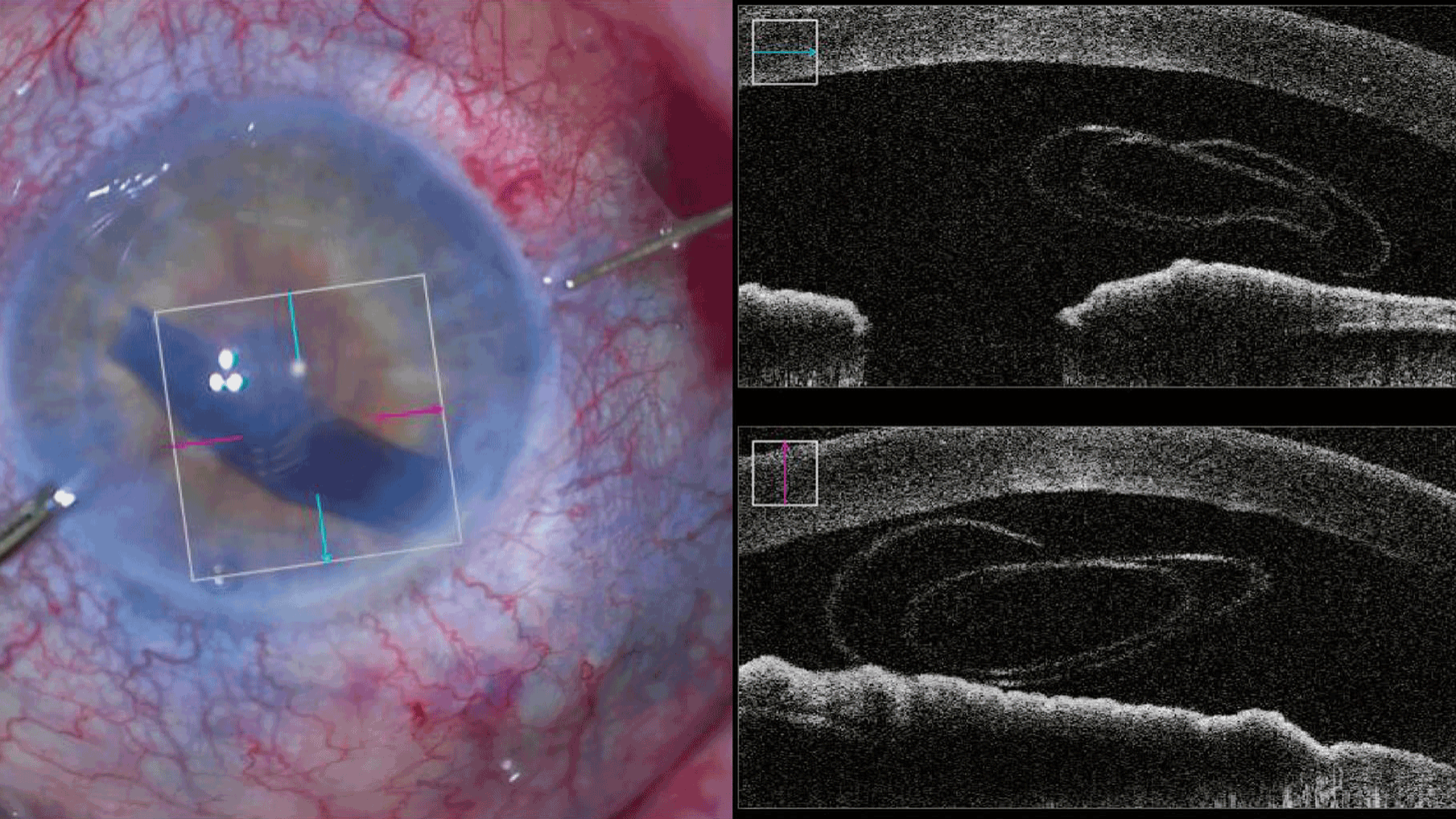

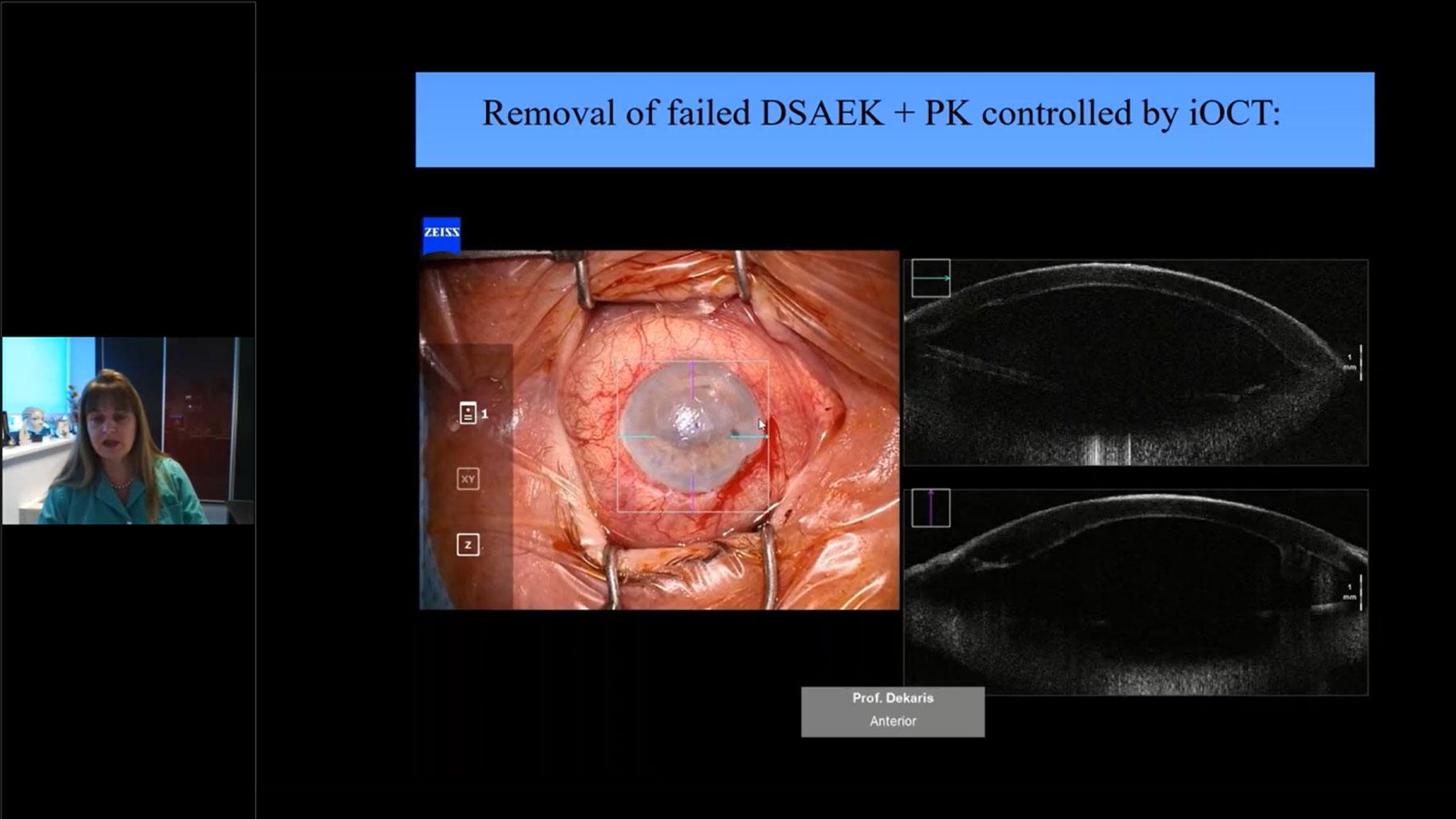
Seeing to succeed in cornea surgery
- Make faster decisions: Quickly change between high-resolution OCT scans (2.9 mm scan depth in tissue) and large overview images (5.8 mm scan depth in tissue) to visualize and assess graft orientation.
- Save time with easy graft monitoring: In DMEK procedures, monitor the graft orientation and assess the interface with the patient’s cornea, as well as verify proper graft positioning.
- Secure big-bubble procedure: Better evaluate DALK dissection depth using OCT imaging and reduce perforation risk.
- Get full integration: The integrated slit illuminator6 provides four slit widths with left-right slit movement for easy observation of the cornea and anterior chamber.
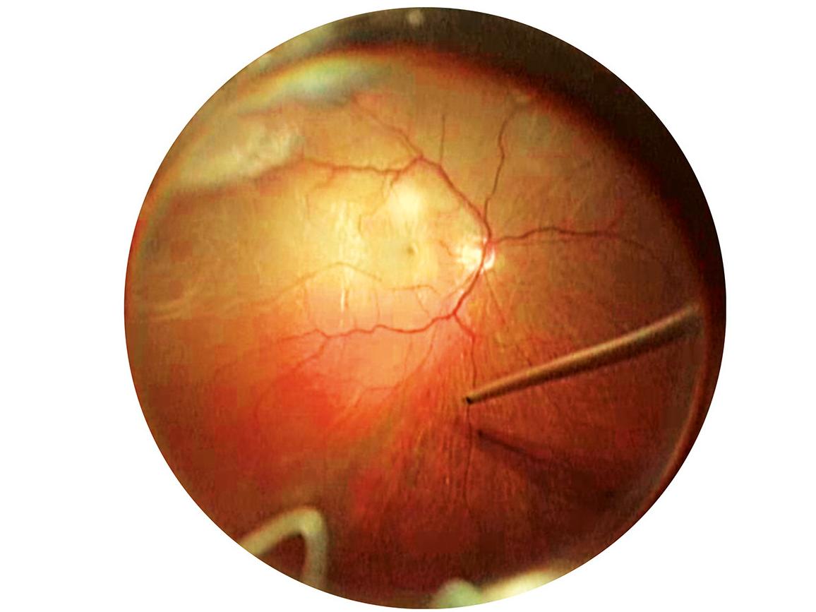
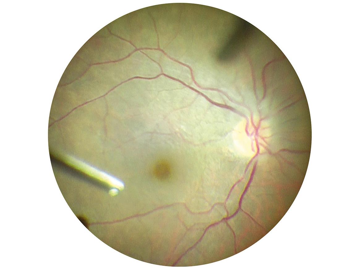
Seeing to succeed in retina surgery
- Get superb OCT images: The integrated intraoperative OCT4 adds a real-time third dimension to visualization capabilities for viewing transparent structures of the eye.
- Stay focused: The automatic XY tracker maintains the selected intraoperative OCT4 scan location and compensates for movements of the eye or microscope.
- Complete your surgery with confidence: Verify that all necessary membrane residue has been completely removed following ILM peelings with OCT4 imaging, plus detect macular holes.
- See more details: ZEISS RESIGHT 700 provides detailed views of the retina, enabling surgeons to switch magnification quickly and stay focused on the area of interest.


-
1
VIROS research team of Prof. Findl: Clinical data of Dr. Varsits "Deviation between the postoperative (at the end of surgery in the operating room) and aimed IOL axes was 0.52 degrees± 0.56 (SD)" published in J Cataract Refract Surg 2019; 45:1234–1238 and Clinical data of Dr. Hirnschall presented at ESCRS 2013.
-
2
Clinical data of Dr. Mayer: "Toric IOL implantation was significantly faster using digital marking" published in J Cataract Refract Surg 2017; 43:1281–1286.
-
3
Clinical data of Dr. Black presented at ESCRS 2014 – 99% of patients had a postoperative refractive cylinder within +/- 0.50 D.
-
4
ZEISS RESCAN 700
-
5
Clinical data of Cost B, Goshe JM, Srivastava S, Ehlers JP published in Am J Ophthalmol.2015 Sep; Intraoperative optical coherence tomography-assisted descemet membrane endothelial keratoplasty in the DISCOVER study.
-
6
Not available in combination with intraoperative OCT.


Teaching possibilities
ZEISS OPMI LUMERA 700 features excellent tools for enhancing the learning experience by providing students a clear understanding of the surgical process. Whether during surgery, viewing through the assistant scope or reviewing post-surgery, ZEISS delivers images with excellent contrast, color and high resolution.

Share what you see during surgery
- Record everything: use a USB device to document the cockpit view, assistance functions and intraoperative OCT1 images, while ZEISS CALLISTO eye, together with a data management system such as FORUM from ZEISS, records live images on multiple drives.
- Share all the details: The new ZEISS CALLISTO eye cockpit allows doctors and students to see data in the eyepiece, from all connected devices.
-
1
VIROS research team of Prof. Findl: Clinical data of Dr. Varsits "Deviation between the postoperative (at the end of surgery in the operating room) and aimed IOL axes was 0.52 degrees± 0.56 (SD)" published in J Cataract Refract Surg 2019; 45:1234–1238 and Clinical data of Dr. Hirnschall presented at ESCRS 2013.
-
2
Clinical data of Dr. Mayer: "Toric IOL implantation was significantly faster using digital marking" published in J Cataract Refract Surg 2017; 43:1281–1286.
-
3
Clinical data of Dr. Black presented at ESCRS 2014 – 99% of patients had a postoperative refractive cylinder within +/- 0.50 D.
-
4
ZEISS RESCAN 700
-
5
Clinical data of Cost B, Goshe JM, Srivastava S, Ehlers JP published in Am J Ophthalmol.2015 Sep; Intraoperative optical coherence tomography-assisted descemet membrane endothelial keratoplasty in the DISCOVER study.
-
6
Not available in combination with intraoperative OCT.


Downloads


Specifications
OPMI LUMERA 700 from ZEISSGet in touch with us!
Receive more information about the product and availability in your country!Related products


-
1
VIROS research team of Prof. Findl: Clinical data of Dr. Varsits "Deviation between the postoperative (at the end of surgery in the operating room) and aimed IOL axes was 0.52 degrees± 0.56 (SD)" published in J Cataract Refract Surg 2019; 45:1234–1238 and Clinical data of Dr. Hirnschall presented at ESCRS 2013.
-
2
Clinical data of Dr. Mayer: "Toric IOL implantation was significantly faster using digital marking" published in J Cataract Refract Surg 2017; 43:1281–1286.
-
3
ZEISS RESCAN 700
-
4
Clinical data of Cost B, Goshe JM, Srivastava S, Ehlers JP published in Am J Ophthalmol.2015 Sep; Intraoperative optical coherence tomography-assisted descemet membrane endothelial keratoplasty in the DISCOVER study.
-
5
Not available in combination with intraoperative OCT.