
A Microscopy Journey Through Time
ZEISS has been producing high-precision microscopes since the middle of the 19th century. From 1857 onwards, the simple models were followed by compound microscopes. Thanks to the work of scientist Ernst Abbe, microscopes have been based on theoretical calculations since 1872. This enabled the production of large numbers of microscopes to the same exceptional quality.
In addition to scientific applications, the microscopes came to be used for routine tasks in clinics, for checking materials, and for educational purposes. The development of microscopes kept advancing, resulting in new models with new technologies.
A Visionary Entrepreneur


Carl Zeiss
(1816 – 1888)
Milestones of ZEISS Microscopy
A Microscopy Journey Through Time
-
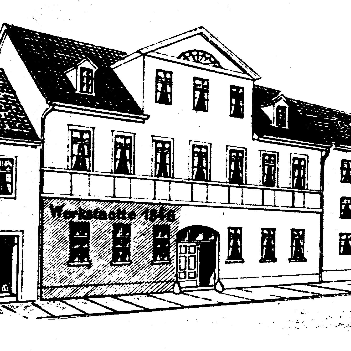
1846
Carl Zeiss opens a workshop for precision mechanics and optics in Jena.

-
1847
Simple microscope with doublet and triplet optics. Production of simple microscopes begins.

-
1857
Carl Zeiss sells his first compound microscope.

-
1866
Begin of partnership with Ernst Abbe.

-
1869
Illumination apparatus with focusable condenser: Ernst Abbe

-
1872
Ernst Abbe's research results allow microscope optics to be produced on the basis of mathematical calculations for the first time.

-
1884
Partnership between Zeiss, Abbe and Schott. Optical glass from Otto Schott enables more effectively corrected microscope systems. © Carl Bräunlich, ZEISS Archives.

-
1886
First apochromatic microscope lens, a color-corrected objective lens for three wavelengths based on the calculations of Ernst Abbe.

-
1893
Illumination device with separate control of the luminous field and condenser aperture: August Köhler (1866-1948).
.")
-
1896
ZEISS manufactures the first Greenough-type stereomicroscope.

-
1903
Invention of the ultramicroscope by Henry Siedentopf and Richard A. Zsigmondy.

-
1931
Beginning of TEM development by/at AEG

-
1936
First prototype of a phase-contrast microscope based on Zernike's original design; he wins the Nobel Prize in 1953.

-
1938
Plan Apochromats and Plan Achromats with a flat image field for micro-photography based on calculations by Hans Boegehold (1876-1965).
.")
-
1942
Cooperation for electron microscopy started by AEG and ZEISS.

-
1949
Electrostatic AEG-ZEISS transmission electron microscope EM 8.

-
1950
The Standard microscope becomes one of the most successful models in the history of ZEISS.

-
1962
Beginning of SEM development in association with the Cambridge University. Cambridge Instruments establishes as a scientific instrument company by Horace Darwin.

-
1965
Cambridge Scientific Instruments releases the first commercial SEM, the Stereoscan Mark I.

-
1973
Axiomat, a microscope with unparalleled stability and image quality.

-
1982
The laser scanning microscope, a microscope system with object scanning through an oscillating laser beam and electronic image processing.

-
1984
EM 902 with imaging electron energy filter becomes first system on the market to generate high-resolution element mapping images.

-
1985
ZEISS launches the first fully digital SEM, the DSM 950.

-
1986
ZEISS unveils the "pyramids", a new generation of microscopes. The design includes special features of Axioplan, Axiophot, and Axiothron: ICS (Infinity Color Corrected System) and SI (System Integration).
 and SI (System Integration).")
-
1993
Market launch of DSM 982 GEMINI field emission scanning electron microscope featuring combined electrostatic-magnetic lens (GEMINI technology).
.")
-
1995
Founding of LEO Electron Microscopy 50/50 Cooperation between ZEISS and Leica.

-
1999
PlasDIC by ZEISS allows the use of plastic dishes for microscopic examinations.

-
2004
LEO fully integrated in ZEISS as Nano Technology Systems Division.

-
2005
The LSM 5 LIVE, a light microscope, with which living cells can be examined 20 times faster and in a particularly gentle manner, enters series production in Jena and receives the R&D Award for its performance in real-time research.

-
2007
ZEISS introduces the ORION helium-ion microscope. Samples are scanned with helium ions instead of electrons. This provides markedly better resolution and improved material contrast.

-
2010
The first super-resolution microscope system ELYRA PS.1 from ZEISS includes structured illumination microscopy (SIM) and photoactivated localization microscopy (PALM) modes. It is far surpassing the diffraction barrier and enables the observation of structural details with unprecedented accuracy.

-
2011
Carl Zeiss NTS GmbH and Carl Zeiss MicroImaging GmbH jointly form the new ZEISS Microscopy business group offering light and electron microscopes to customers.

-
2012
ZEISS introduces its first light sheet microscope system: ZEISS Lightsheet Z.1 works with an expanded light beam, the light sheet, that illuminates only a thin section of the sample, thus protecting the rest of the specimen. Biologists can use the microscope system to observe the development of entire organisms over several days or more.

-
2013
With the acquisition of U.S.-based Xradia, Inc. ZEISS Research Microscopy Solutions becomes the only manufacturer of light, electron and X-ray microscopes, with unique solutions for research and routine inspection in materials and life sciences application fields.

-
2014
ZEISS MultiSEM 505, a 61-beam (multi-beam) SEM and fastest SEM in the world, is introduced to the market.
 SEM and fastest SEM in the world, is introduced to the market.")
-
2018
APEER image analysis cloud platform is launched to a small group of people, being rapidly adapted by the academic community.

-
2020
Based on the pioneering research and developments of Ernst H. K. Stelzer and Nobel laureate Eric Betzig, ZEISS Lattice Lightsheet 7 allows researchers to observe cellular processes within cells and small organisms in 3D for hours or days – all at subcellular resolution.

-
2022
ZEISS experts Dr. Thomas Kalkbrenner, Dr. Jörg Siebenmorgen, and Ralf Wolleschensky win the Deutscher Zukunftspreis 2022 for their significant contribution in the development of the microscope system ZEISS Lattice Lightsheet 7.

-
2023
ZEISS is fully acquiring and integrating arivis AG, offering a scientific image analysis ecosystem to researchers and creating a sub-product category for file format-agnostic software called ‘ZEISS arivis’. At the same time, APEER is becoming arivis Cloud.


.")


.")


 and SI (System Integration).")
.")


 SEM and fastest SEM in the world, is introduced to the market.")


Behind the Scenes
Microscopes Made in GermanyIn Jena production, components are mounted and whole light microscope systems are integrated. What has started as a small workshop for precision mechanics and optics in the middle of the nineteenth century, now has become a solid production facility. Today, over 120 employees manage this complexity and ensure the fast and precise production of our microscopes.


A short glance back
ZEISS has been producing high-precision microscopes since the middle of the 19th century. From 1857 onwards, the simple models were followed by compound microscopes (image: compound microscope from Carl Zeiss, Stand I, from 1891).
Thanks to the work of scientist Ernst Abbe, microscopes have been based on theoretical calculations since 1872. This enabled the production of large numbers of microscopes to the same exceptional quality. The development of microscopes kept advancing, resulting in new models with new technologies.


Foreman August Löber with mechanics and apprentices in 1864. Many employees who helped the company grow at the beginning were trained by him.
Fighting for Nanometers
Insights into the production facilityIn Jena production, components are mounted and whole light microscope systems are integrated. What has started as a small workshop for precision mechanics and optics in the middle of the nineteenth century, now has become a solid production facility. Today, over 120 employees manage this complexity and ensure the fast and precise production of our microscopes.
Today, ZEISS microscopes like Axio Imager and Axio Observer, as well as high-end systems like Lightsheet, Lattice Lightsheet, LSMs and the Axioscan slide scanner leave production in Jena to be shipped to research institutes worldwide.
There are two main areas in production: assembly and system integration.


ZEISS Axio Observer is assembled in several steps on the production line.


When calibrating high-resolution microscope systems, accuracy in the nanometer range is essential.


An employee is adjusting the optical components in the scan head of ZEISS LSM 980.


ZEISS Axioscan is set up by loading the 100 sample magazine to start the application test.
Made to order
80% of the production workers only start working with the order in hand. Every system is tailored to the requirements of the customer.
Did you know that?
- … every production employee has its own specification? To do his part of the work he needs a specific certificate to be renewed every three to six months.
- … ZEISS ships microscope systems and components worldwide between production facilities in reusable transport boxes?
- … it takes up to 32 days to put a microscope system into operation?