

ZEISS Axio Observer live
Capture and evaluate dynamic processes in living samplesCollecting and analyzing imaging data from living samples presents added complexity in comparison to fixed samples. ZEISS Axio Observer live provides you with an efficient data collection workflow that comes with stable environmental controls and the highest possible imaging quality, even at different temperatures. Unravel new insights in your research – from reliable image acquisition to complex data analyses.
Speed up your live cell experiments
Incubators typically restrict the access to your sample, making formerly simple tasks like positioning, focusing and generation of an overview image more complicated. AI Sample Finder performs these tasks with the push of a button. Application of immersion media to your water immersion objectives is performed in less than a second without changing focus, stage position, or disturbing the incubation chamber for independent, automated imaging. Environmental parameters are automatically recorded so that you can be confident that the observed dynamics are not caused by fluctuating temperature or atmosphere. Additionally, ZEISS Axio Observer live is equipped with advanced software tools for streamlined analyses of large, multivalued timelapse experiments.
Bundle components
-
Microscope
- Axio Observer 7 (inverted widefield microscope stand)
- Scanning stage 130 × 100
- Motorized condenser NA 0.55
- Definite Focus 3
- AI Sample Finder
- Autoimmersion Module
- Dual filter wheel mot.
Light source / camera
- Viluma 7
- Filter sets LED SBP, 91, 112
- Axiocam 820 mono
Objectives
- EC Plan-Neofluar 5× / 0.16
- Plan-Apochromat 10× / 0.45
- Plan-Apochromat 20× / 0.8
- LD LCI Plan-Apochromat 40× / 1.2 Imm Corr DIC
Environmental control
- Glass Lid CO₂/O₂ heated
- Temperature Controller
- Gas Mixer CO₂
- Heated Plate K with 3 Inserts
Workstation
- Z8 Workstation with 128 GB RAM and nVidia Quadro RTX A4000 16 GB DP
-
Motorized Acquisition
- Acquire images and control motorized components in multi-dimensional experiments.
2D Toolkit
- Create flexible automatic measurement programs for the analysis of your 2D images.
Molecular Quantification Toolkit
- Investigate diverse dynamic and molecular interaction phenomena of diverse specimens.
Deconvolution Toolkit
- Sharpen your images with mathematically exact deconvolution and make previously unknown structures visible by increasing your image contrast and resolution.
Applications
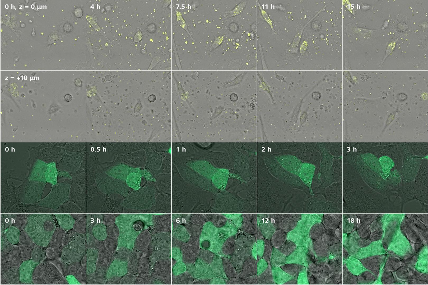


Multi-position, extended time lapse experiment with automated immersion:
When working with living samples, you might not know where your event of interest may occur. To capture the uptake of nanoparticles by macrophages, many locations from a multi-well plate are acquired as well as multiple z-planes over several hours at 37°C using re-immersion. The region shown above is a subset of the much larger dataset that was captured using automated imaging and shows the uptake of nanoparticles inside the cells (top row). The surface of the cells were also imaged to verify that the nanoparticles are inside the cells and not simple sitting on the cell surface (bottom row).
Sample courtesy: F. Páez Larios and C. Eggeling, Institute for Applied Optics and Biophysics, Friedrich-Schiller-Universität Jena, Germany
Live cell experiments over extended periods with automated immersion:
HEK KO PEX5 cells expressing eGFP with a photocaged peroxisomal targeting signal type 1 were reconstituted with the peroxisomal import receptor PEX5. A light induced conformational change of the photocage leads to exposure of the peroxisomal targeting signal. If the WT PEX5 is expressed, accumulation of the eGFP signal in the dotted peroxisomes can be monitored (top row). In case of the mutated PEX5 (bottom row), even after 18 hours, no peroxisomal import could be detected.
Sample courtesy of K. Reglinski, Institute for Applied Optics and Biophysics, Friedrich-Schiller-Universität Jena, Germany


