SK8 K18 Mouse Cells. Vimentin stained with Alexa 488 (green), nuclei stained with DAPI (blue).

Preconfigured widefield microscope bundles

Tailored to your applications. Flexibly expandable as needed.

From prepared specimens to living cells, explore your research with a widefield microscope designed around your workflow. Axio Imager brings high-resolution imaging to tissue, specimens, and materials, while Axio Observer enables powerful imaging of cells, cultures, and dynamic experiments. Choose the platform that fits the way you work — with the imaging performance and flexibility of ZEISS.

Automated acquisition and advanced data analyses

Axio Observer scan

Axio Observer scan

  • Fully automated x/y scanning of slides, dishes, and multi-well plates​
  • LED transmitted and fluorescence illumination​
  • ZEN Bio Apps software​
  • Advanced version equipped with Definite Focus, 2D and AI Toolkits
Axio Imager scan

Axio Imager scan

  • Fully automated x/y slide scanning ​
  • LED transmitted and fluorescence illumination​
  • ZEN Bio Apps software​
  • Advanced version equipped with 2D and AI Toolkits​

High-resolution 3D visualization and evaluation

Axio Observer section

Axio Observer section

  • Fully automated inverted microscope for fluorescence optical sectioning of slides, dishes, and plates​
  • Standard version generates sections with Deconvolution​
  • Advanced version generates sections with Apotome 3 structured illumination​
Axio Imager section

Axio Imager section

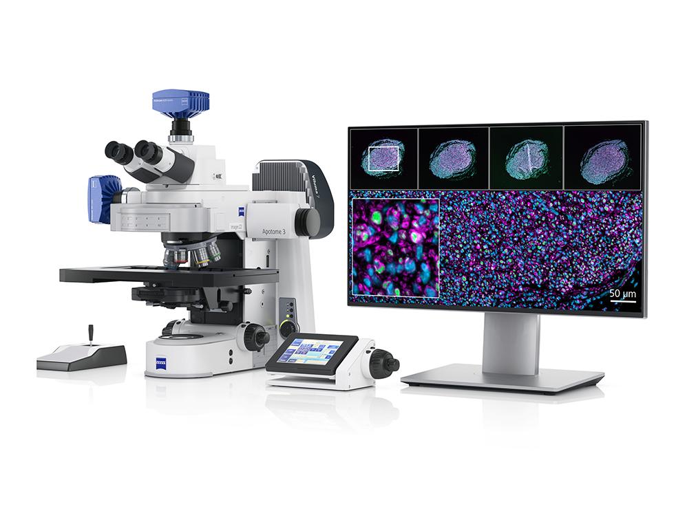
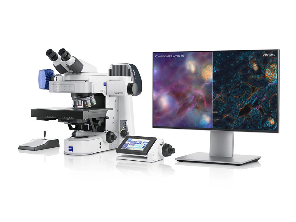
  • Fully automated upright microscope for fluorescence optical sectioning of slides​
  • Standard version generates sections with Deconvolution​
  • Advanced version generates sections with Apotome 3 structured illumination​
     

Capture and evaluate dynamic processes

Axio Observer live

Axio Observer live

  • Automated inverted microscope and stage-top incubation for long-term imaging of living specimens. ​
  • Fluorescence and/or transmitted illumination of slides, dishes or multi well plates.​
  • Standard version generates optical sections with Deconvolution​
  • Advanced version generates optical sections with Apotome 3 structured illumination​