The Link Between Biofilm Infections and Wound Healing Investigated with Whole Slide Imaging
Researchers use automated whole slide imaging to link biofilm properties of bacterial infection to specific pathogenic mechanisms in wound healing.
Bacteria can form biofilms, multicellular communities which are held together by a self-produced extracellular matrix. A growing body of evidence establishes biofilm infection as a major cause of delayed wound healing and non-healing chronic wounds. However, it is unclear how biofilms interfere with wound healing.
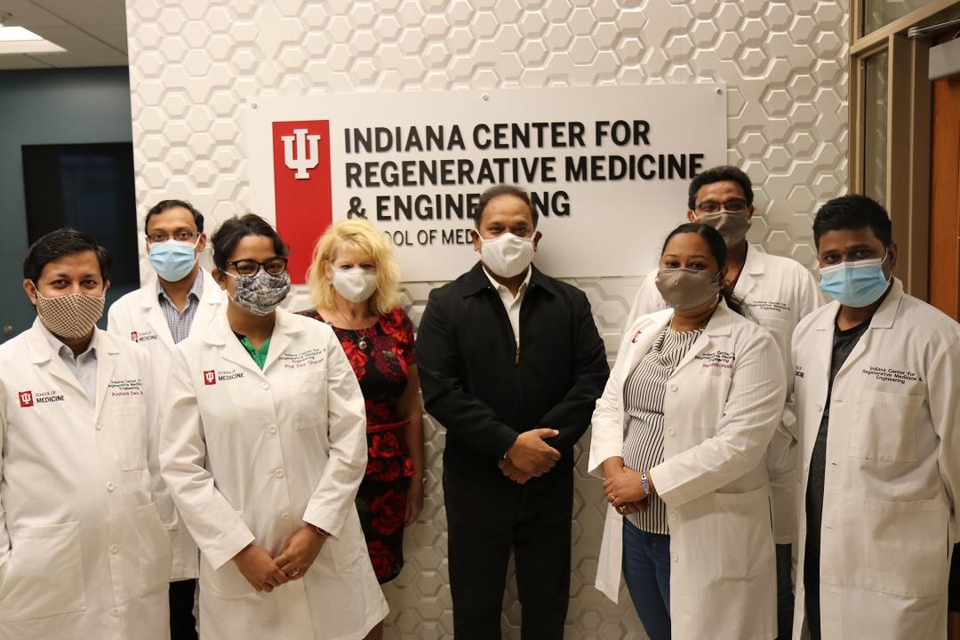
Dr. Chandan K. Sen, Associate Vice President of Military & Applied Research, at the Indiana University School of Medicine, USA, has published work linking biofilm properties of bacterial infection to specific pathogenic mechanisms in wound healing. One of the techniques used in their research is automated whole slide imaging.
Dr. Chandan K. Sen (center in the dark jacket) with some of his team members.
An Infection Wound Model to Study Biofilms
While Dr. Sen's team is actively pursuing discoveries in many areas, his primary interests are tissue injury, repair, regeneration and infection. Previously, his lab has developed a preclinical, porcine biofilm infection wound model. Now, they utilize this wound model with bacterial strains with biofilm-forming abilities to better understand biofilm-dependent mechanisms of action in healing wounds.
Their findings show a biofilm-induced degradation of cutaneous collagen, specifically collagen 1. Collagen 1 is a major structural protein of the extracellular matrix (ECM), and, in wounds, causes poor tensile strength in the repaired skin. This supports the notion that healed wounds with a history of biofilm infection are likely to recur. Automated whole slide imaging with ZEISS Axioscan was instrumental to their findings.
Visualizing Wound Tissue Architecture
Brightfield Imaging with ZEISS Axioscan Digital Slide Scanner
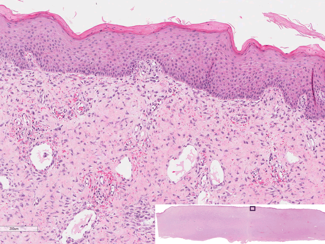
H+E stain
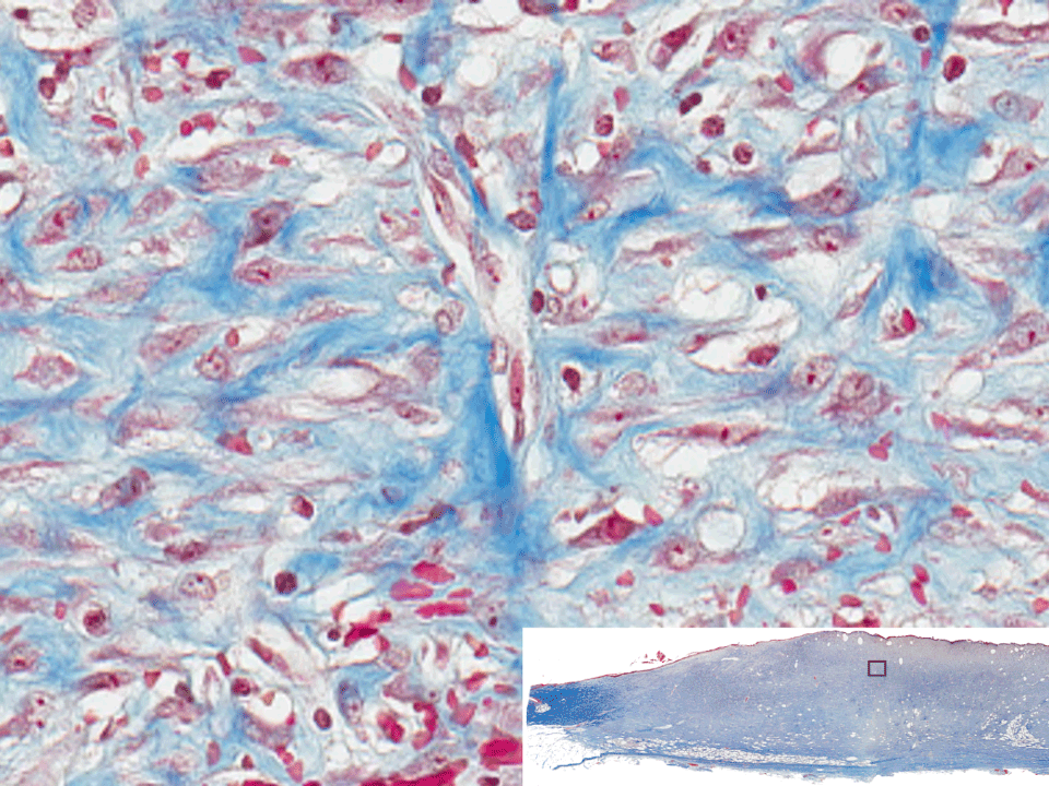
Masson’s trichrome
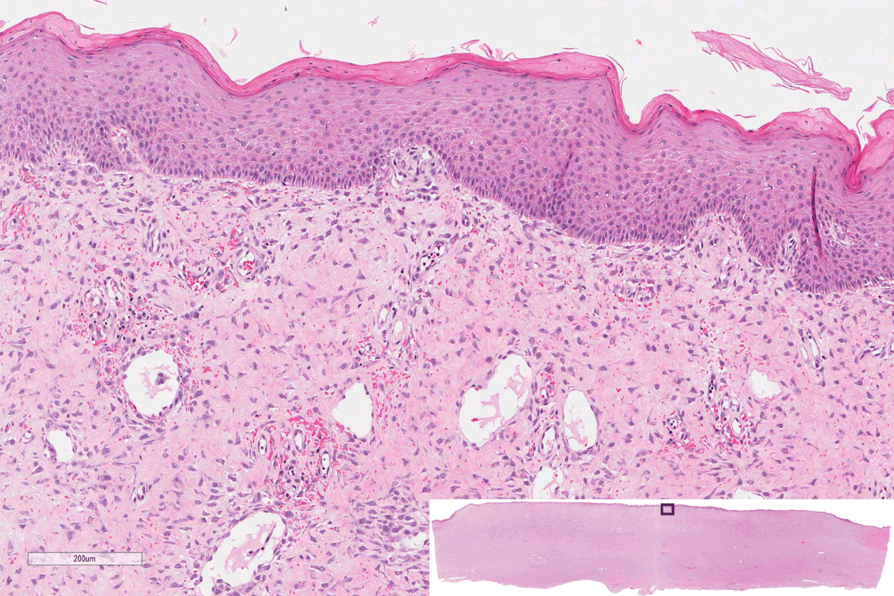
Hematoxylin and eosin-stained whole mount cross sections of infected porcine burn wounds showing re-epithelialization. Overview image is lower right. Imaged with ZEISS Axioscan.
Hematoxylin and eosin-stained whole mount cross sections of infected porcine burn wounds showing re-epithelialization. Overview image is lower right. Imaged with ZEISS Axioscan.
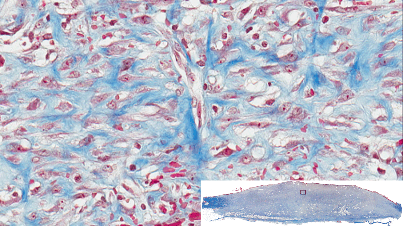
Masson’s trichrome staining of whole mount cross sections of infected porcine burn wounds. Staining results in blue-black nuclei, blue collagen, and light pink/red cytoplasm. Epidermal cells appear reddish. Imaged with ZEISS Axioscan digital slide scanner.
Masson’s trichrome staining of whole mount cross sections of infected porcine burn wounds. Staining results in blue-black nuclei, blue collagen, and light pink/red cytoplasm. Epidermal cells appear reddish. Imaged with ZEISS Axioscan digital slide scanner.
Whole slide imaging allowed us to easily image the entire tissue section and analyze specific regions of the wound. Collagen fiber analysis of the wounded tissue pointed towards our finding of deficient granulation tissue formation.
Specific Proteins in Wound Skin Tissue
Fluorescence Microscopy with ZEISS Axioscan Digital Slide Scanner
Collagen
Actin
Tight junctions
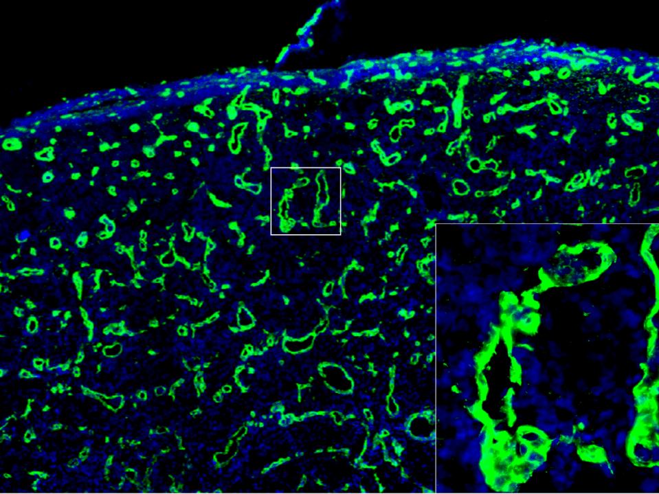
Biofilm infected porcine burn wound stained for collagen (green) and nuclei (DAPI/blue). Images collected with ZEISS Axioscan digital slide scanner.
Biofilm infected porcine burn wound stained for collagen (green) and nuclei (DAPI/blue). Images collected with ZEISS Axioscan digital slide scanner.
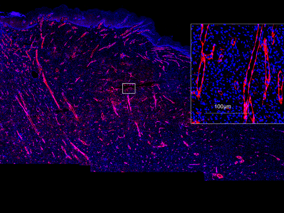
Biofilm infected porcine burn wound stained for smooth muscle actin (red) and nuclei (DAPI/blue). Image acquired with ZEISS Axioscan digital slide scanner.
Biofilm infected porcine burn wound stained for smooth muscle actin (red) and nuclei (DAPI/blue). Image acquired with ZEISS Axioscan digital slide scanner.
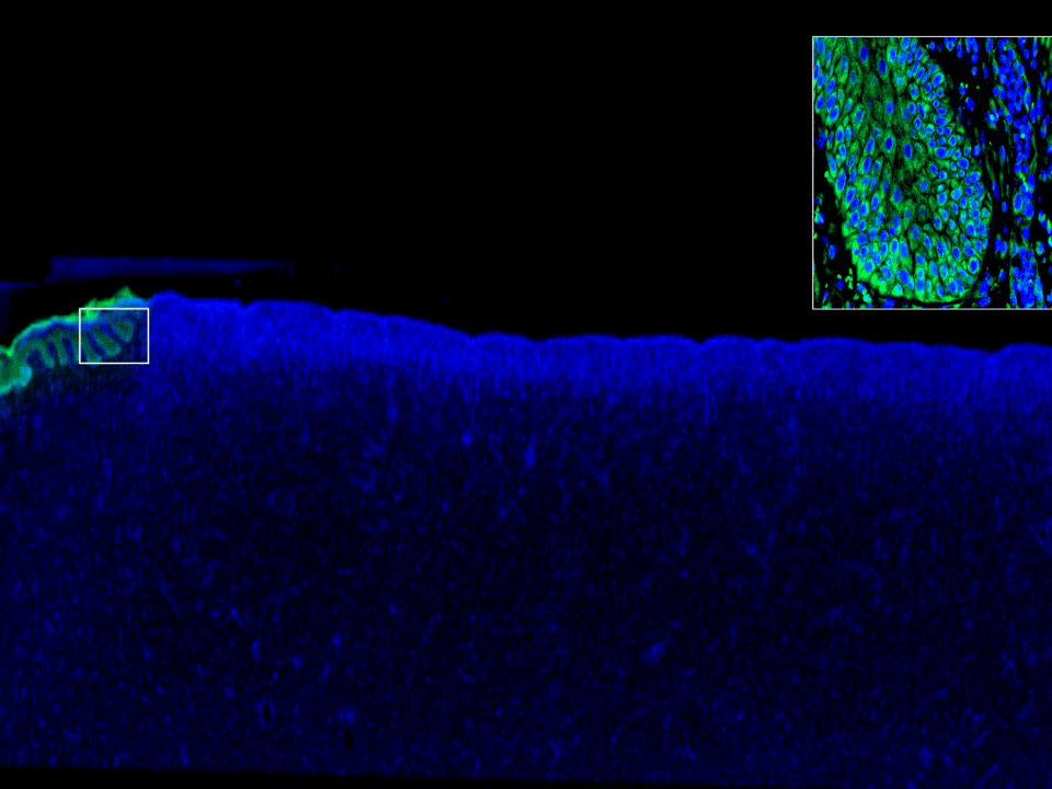
Biofilm infected porcine burn wound stained for tight junctions (green) and nuclei (DAPI/blue). Images collected with ZEISS Axioscan digital slide scanner.
Biofilm infected porcine burn wound stained for tight junctions (green) and nuclei (DAPI/blue). Images collected with ZEISS Axioscan digital slide scanner.
Our finding of deficient collagen in the repaired skin as a direct consequence of biofilm infection was very interesting. Our focus moving forward is to apply the knowledge we have gained to improve diagnosis and management of infected patient wounds.


 and nuclei (DAPI/blue). Images collected with ZEISS Axioscan digital slide scanner.")
 and nuclei (DAPI/blue). Images collected with ZEISS Axioscan digital slide scanner.")
 and nuclei (DAPI/blue). Images collected with ZEISS Axioscan digital slide scanner.")
 and nuclei (DAPI/blue). Images collected with ZEISS Axioscan digital slide scanner.")
 and nuclei (DAPI/blue). Image acquired with ZEISS Axioscan digital slide scanner.")
 and nuclei (DAPI/blue). Image acquired with ZEISS Axioscan digital slide scanner.")
 and nuclei (DAPI/blue). Image acquired with ZEISS Axioscan digital slide scanner.")
 and nuclei (DAPI/blue). Image acquired with ZEISS Axioscan digital slide scanner.")
 and nuclei (DAPI/blue). Images collected with ZEISS Axioscan digital slide scanner.")
 and nuclei (DAPI/blue). Images collected with ZEISS Axioscan digital slide scanner.")
 and nuclei (DAPI/blue). Images collected with ZEISS Axioscan digital slide scanner.")
 and nuclei (DAPI/blue). Images collected with ZEISS Axioscan digital slide scanner.")


