

Imaging Large Brain and Tissue Sections
Whole Slide ImagingWhen imaging large brain and tissue sections, you may find it difficult to set up a workflow that successfully collects your entire sample in high resolution. Or the length of time required to scan your entire sample or all your slides in the resolution you need is unachievable.
ZEISS offers solutions to make imaging brain and tissue sections quick and efficient for high throughput, yielding data that is reproducible and easy to analyze.
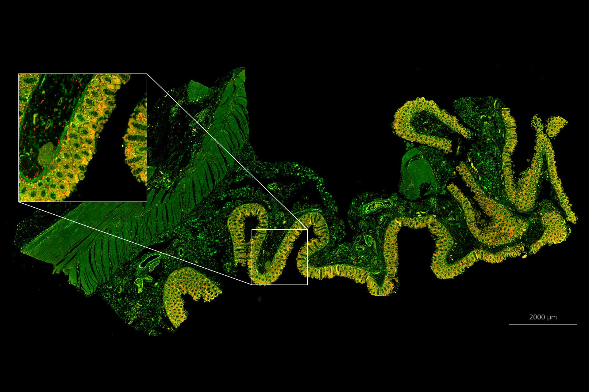

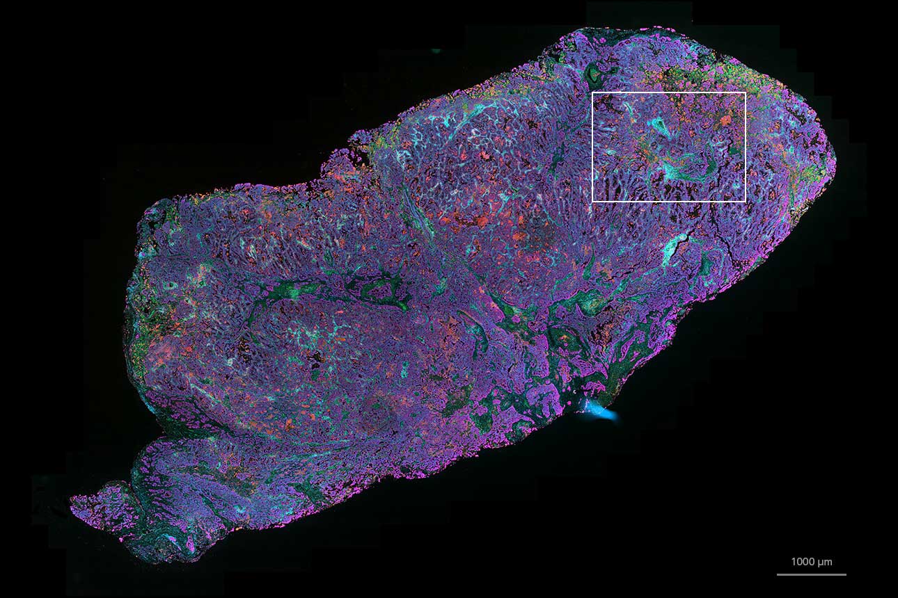

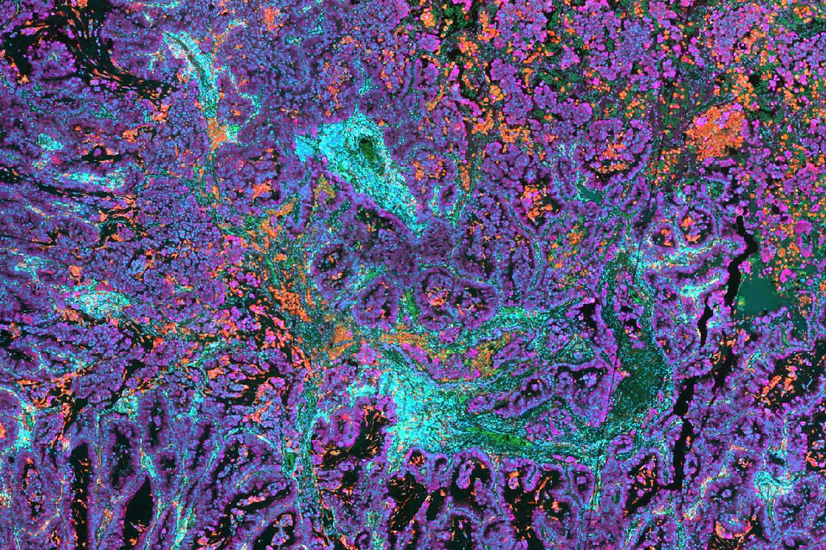


 tissue section imaged using digital slide scanner. Fluorescence staining with UltiMapper I/O PD L1 kit. Sample courtesy of Ultivue, Inc., USA*")
 tissue section imaged using digital slide scanner. Fluorescence staining with UltiMapper I/O PD L1 kit. Sample courtesy of Ultivue, Inc., USA*")


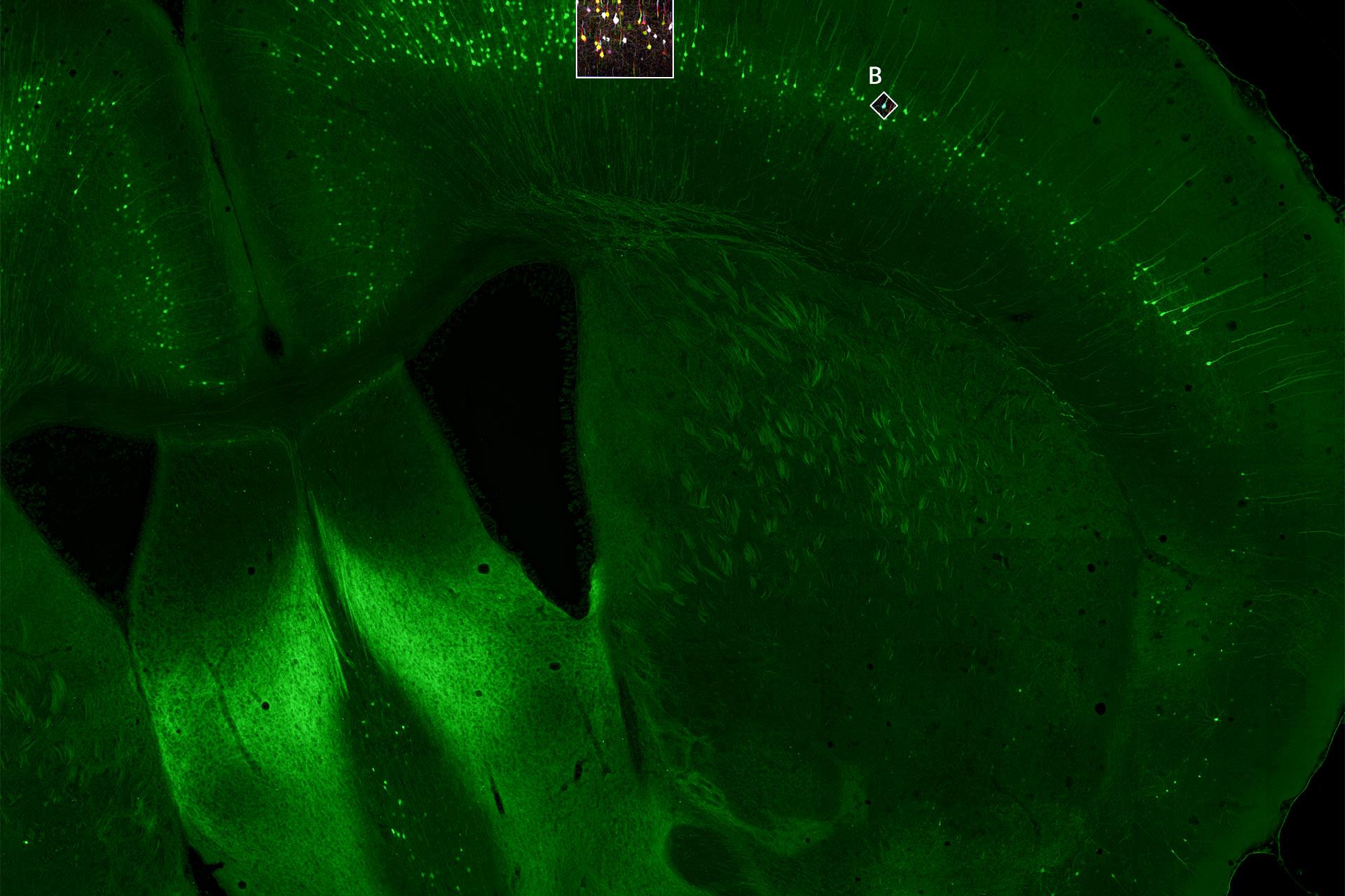
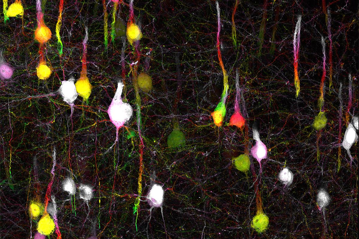
Mouse brain slice, captured using widefield fluorescence microscopy and tiling. Sample courtesy of D. Mi, School of Life Sciences, Tsinghua University, China
Mouse brain slice, captured using widefield fluorescence microscopy and tiling. Sample courtesy of D. Mi, School of Life Sciences, Tsinghua University, China
Easy Workflows for Whole Slide Imaging Using Widefield or Zoom Microscopy
With a motorized stage and ZEISS ZEN software, automated workflows are easy to setup to collect multiple images in brightfield or fluorescence that can be stitched together to generate one seamless image. Widefield microscopes, such as the ZEISS Axio Observer inverted microscope, or the ZEISS Axio Zoom.V16 zoom microscope can support your whole slide imaging needs. Add ZEISS AI Sample Finder to increase the speed and efficiency of set-up and initiation of your imaging acquisition.


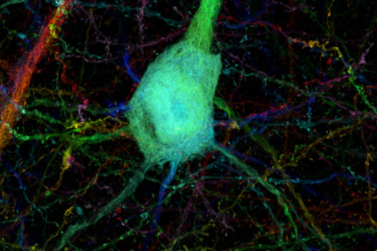


Mouse brain section, area 1 mm². Sample: courtesy of J. Lichtman, Harvard University, USA
Mouse brain section, area 1 mm². Sample: courtesy of J. Lichtman, Harvard University, USA
Large Area Ultra-resolution Imaging of Brain and Tissue Sections
Ultra-resolution imaging with scanning electron microscopes often limits researchers to imaging very small regions of their samples. The ZEISS MultiSEM family features 61 or even 91 electron beams scanning in parallel, resulting in an imaging speed of up to 1820 megapixels per second. Complemented by the advent of automated sample preparation, the promise of imaging larger (1 mm³) tissue volumes at high resolution is now within reach.


Related Products


-
1
* The images shown on this page represent research content. ZEISS explicitly excludes the possibility of making a diagnosis or recommending treatment for possibly affected patients on the basis of the information generated with an Axioscan 7 slide scanner.