

Advanced Microscopy in Life Sciences
Pushing the Boundaries in Cancer Research
Precision tools designed to advance our understanding and treatment of cancer


Interactive Software Demos
Take a tour of our cutting-edge software featuresInterested in learning more?
Contact usScroll animation items

Are you leveraging the latest innovations?
Discover the techniques your peers are using

Explore Deep Brain Activity
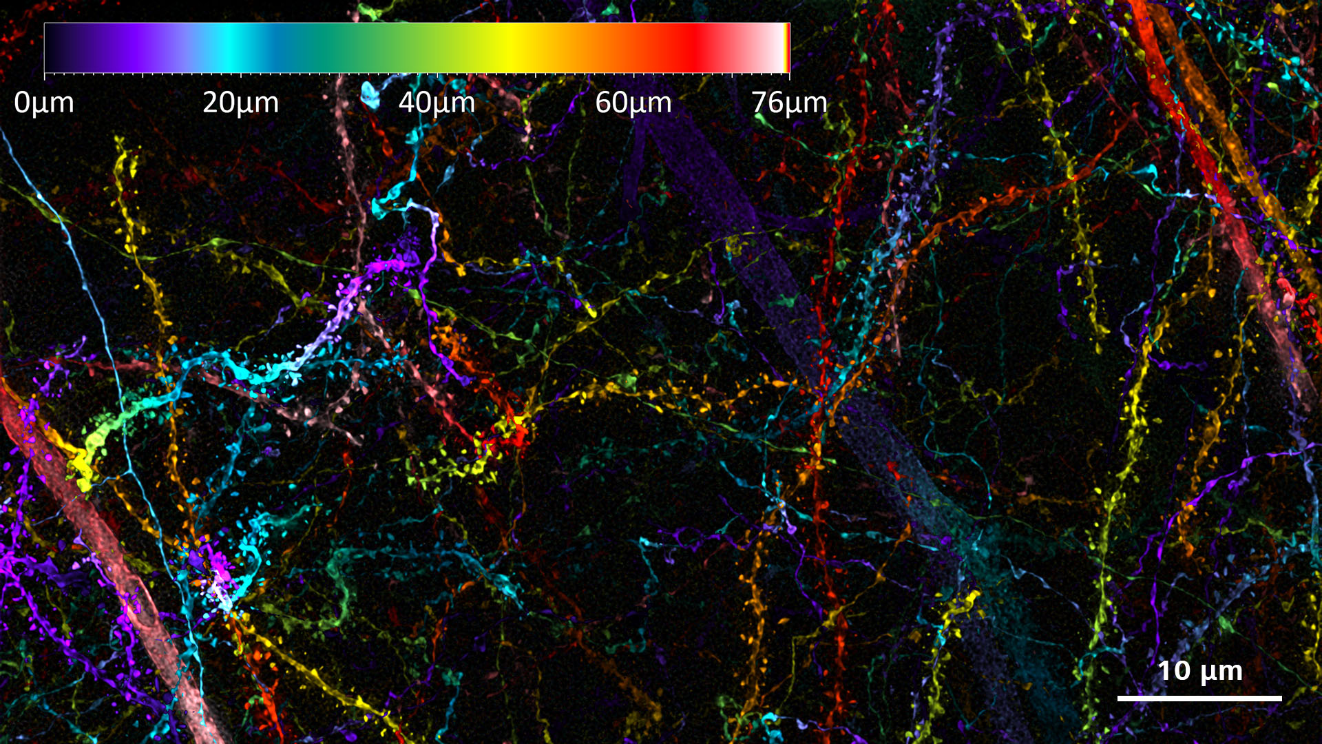
Consider What Can Be Learned from New Structural InsightsDeep brain imaging provides high-resolution images of neural structures, allowing scientists like you to analyze the morphology and organization of neurons, glial cells, and synapses. This detailed visualization aids in understanding the physical layout of neural circuits.
Studying Synaptic Function
Resolving Details Hiding in the DepthZEISS Lattice SIM 3 is specifically designed to meet the imaging requirements of multicellular organisms and tissue sections. This system exploits the full potential of the SIM Apotome technology: fast optical sectioning at superior quality, large fields of view with access to smaller regions of interest, near-isotropic resolution, and the gentlest super-resolution imaging possible.Update


Image Large Specimens
In your Preferred Clearing SolutionLightsheet 7 is designed to match all these conditions. Image specimens at up to 2 cm in size at any refractive index between 1.33 and 1.58, and in almost all clearing solutions. Acquire overview images and data with subcellular resolution – whether you work with optically cleared organoids, spheroids, organs, brains or other specimens.

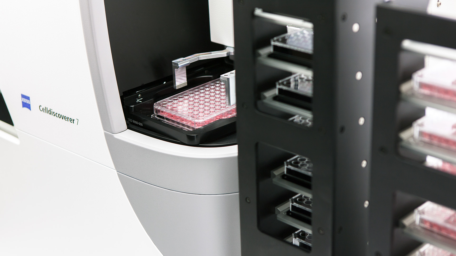
Top Quality Data from Your Samples
Reproducible ResultsObserving live samples over a number of days or imaging lots of multiwell plates really puts your microscope through its paces. To get reproducible, unbiased data, you must control environmental conditions such as light, temperature, CO2 etc. That’s why Celldiscoverer 7 brings you a unique combination of a stable box, darkroom and integrated inverted research microscope with optional incubation. It simplifies your laboratory setup and makes work more comfortable
Optical Sectioning in Widefield Fluorescence Microscopy
Confocal-like Image Quality with ZEISS New UpgradesOptical sectioning with ZEISS Apotome 3 allows you to efficiently minimize out-of-focus light. Create crisp images and 3D renderings, even of thicker specimen, while your microscope remains just as easy to operate as always. Go one step further with Apotome Plus and achieve confocal-like image quality with your widefield microscope.
Scroll animation items

ORIGINAL SCROLL MODULE (VISION)
Discover techniques for your research

Explore Deep Brain Activity
Consider What Can Be Learned from New Structural InsightsDeep brain imaging provides high-resolution images of neural structures, allowing scientists like you to analyze the morphology and organization of neurons, glial cells, and synapses. This detailed visualization aids in understanding the physical layout of neural circuits.


Studying Synaptic Function
Overcome critical challenges with resolution limitations, and enable the visualization of synaptic components at the nanoscale through super-resolution methods. These limitations are lifted by adding processing techniques to see more activity than ever before.
Understanding Neural Circuitry
To accurately map the intricate connections between neurons, the technique of multi-view imaging allows for the simultaneous capture of data from multiple angles, thereby enhancing the three-dimensional reconstruction of neural circuits for research.
Neurodegenerative Disease Connections
See more in real-time and acheive the ability to identify critical changes in cell behavior and morphology. Thereby gaining deeper understanding of neurodegenerative diseases and visualizing cellular processes and interactions that contribute to disease progression.


Studying the Role of Glial Cells
Enhanced optical sectioning capabilities can minimize out-of-focus light, allowing for clearer visualization of these cells in their native environment. This precision enables scientists to investigate the complex interactions between glial cells and neurons, leading to a better understanding of their contributions to brain function and pathology.