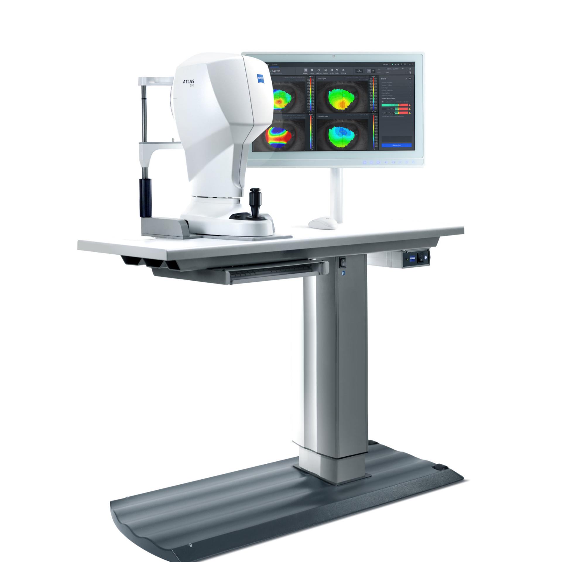

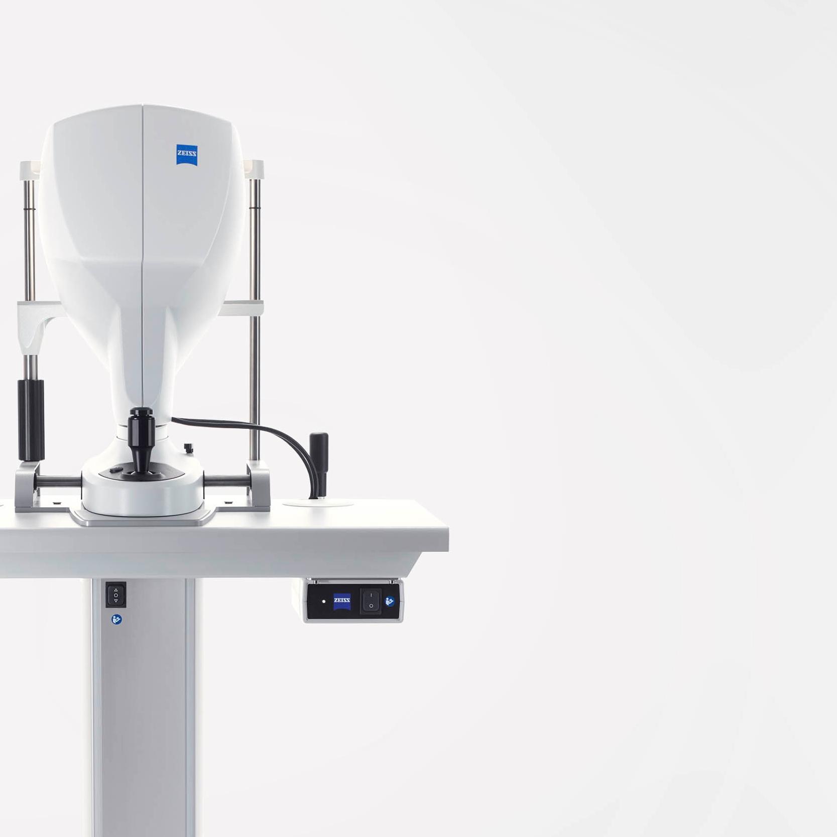
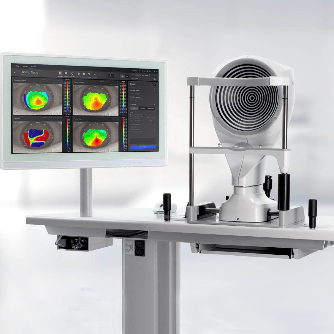
ZEISS ATLAS 500 Multi-modality ocular surface analysis.
A state-of-the-art, multi-modality solution for the anterior eye segment, ATLAS® 500 from ZEISS offers corneal topography. Additionally, the system enables clinicians to carry out dry eye assessments in a single workstation, delivering increased efficiency in a compact design.

Extended insights
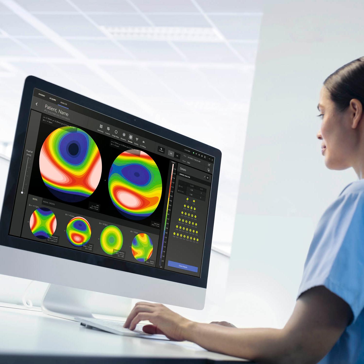
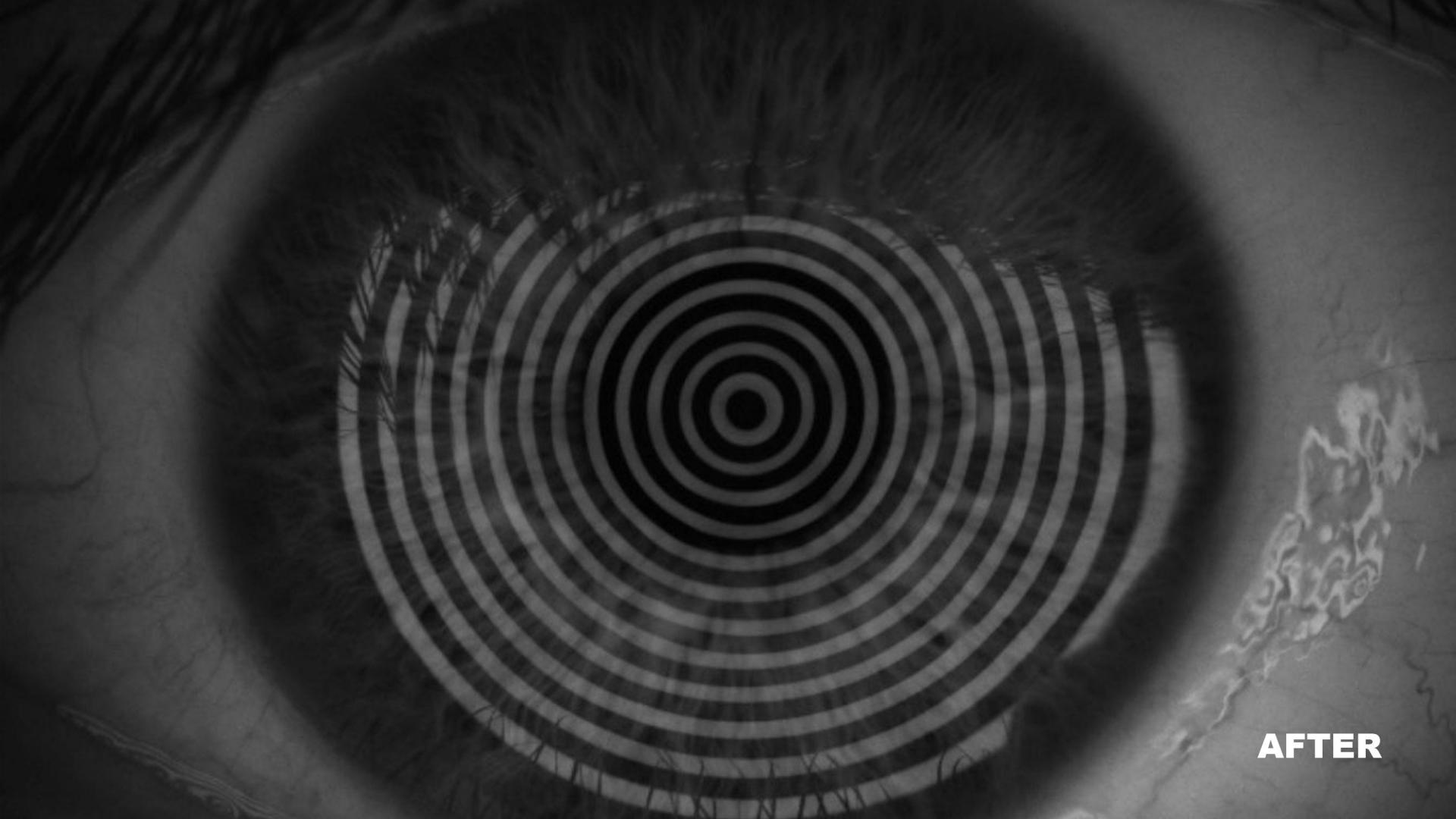
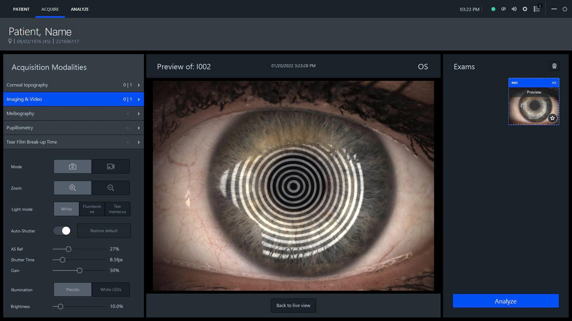
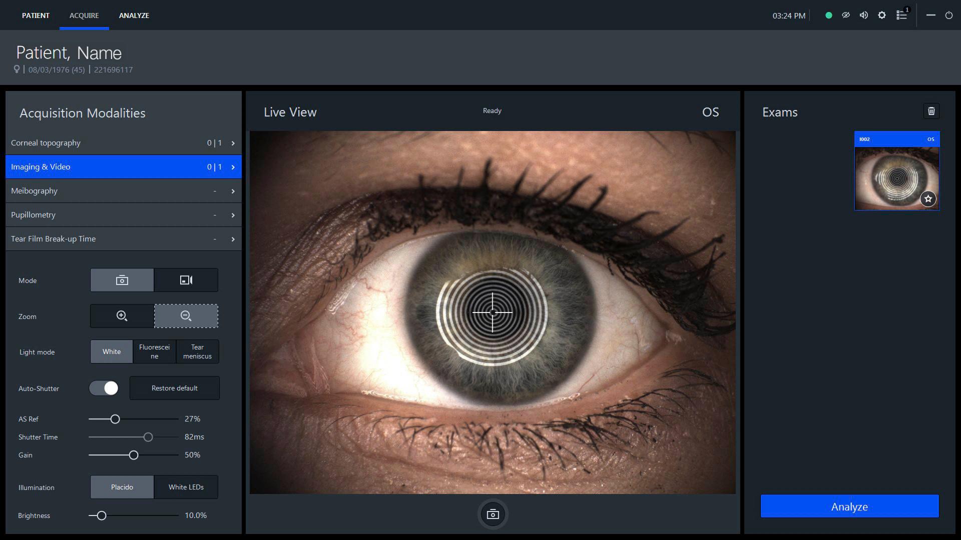
Precise corneal topography and visualizationEquipped with a variety of measurement capabilities, the ZEISS ATLAS 500 quickly captures relevant corneal properties with high resolution images and videos.
The Placido-based measurements provide a variety of corneal topography analysis options:
- Summary/custom view including all common topographic maps
- Elevation (spheric, aspheric and aspheric-toric)
- Corneal wavefront analysis
- Optical quality analysis
- Contact lens fitting
- Keratoconus screening


Keratoconus screening including classification
The summary screen provides details on corneal symmetries and ectasia. Additionally, a keratoconus screening system provides information on the potential risk of keratoconus and classifies each patient’s cornea into one of the following groups: keratoconus, keratoconus suspect, normal or treated.
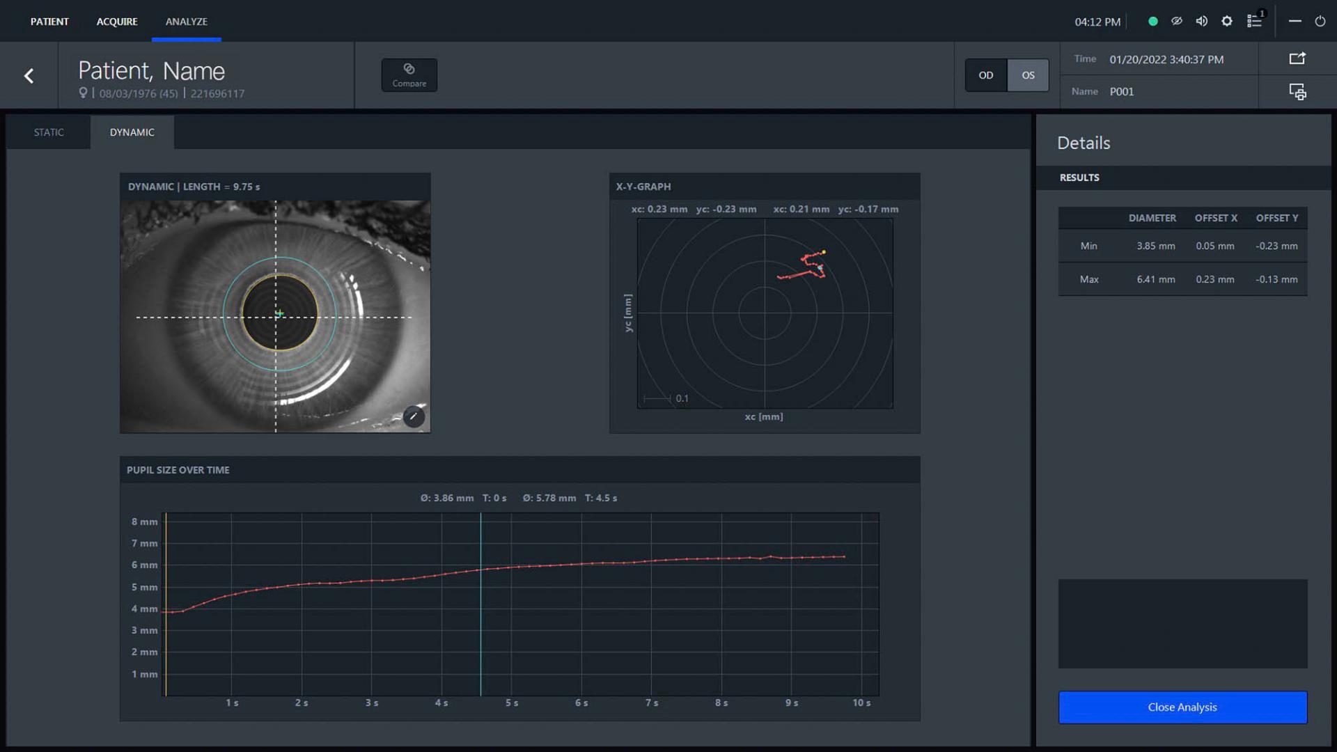

Static and dynamic pupillometry
Analyze pupil size and decentration in scotopic, mesopic and photopic light conditions. Choose between static and dynamic acquisition modes.

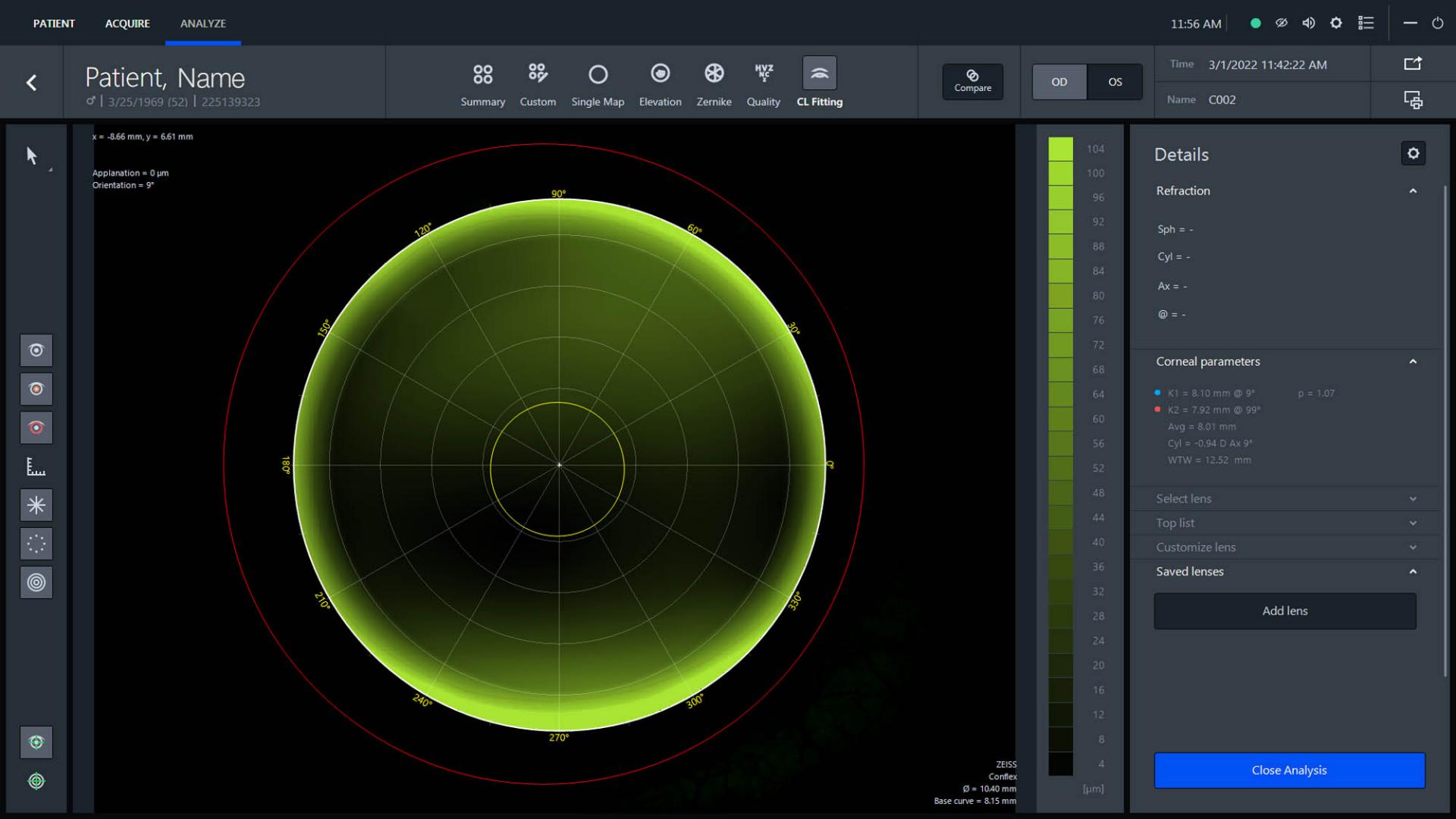

Contact lens fitting
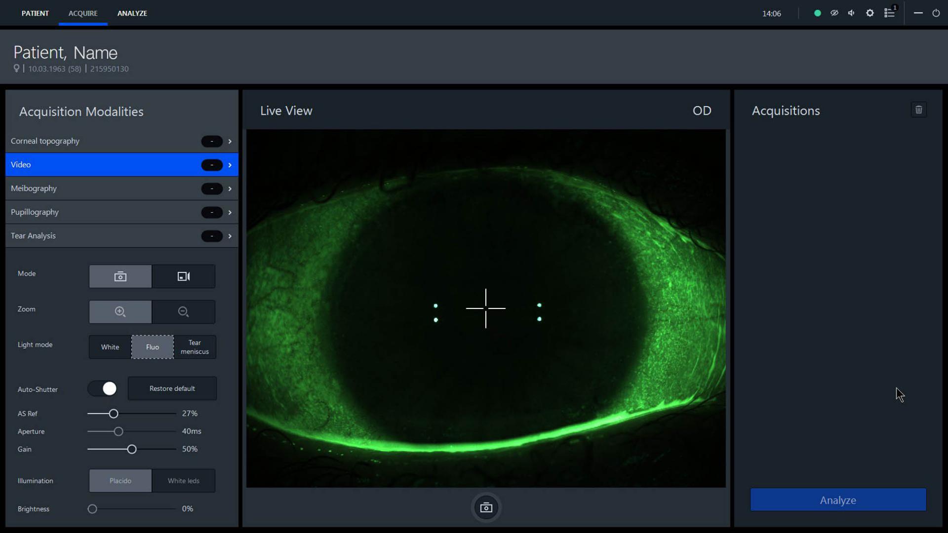
The luminous source with cobalt blue light allows you to analyze the clearance of rigid contact lenses in fluorescein and detects corneal staining and scars. Simulate the fit of rigid contact lenses based on the internal databases of the lens manufacturers.

Profound decision-making
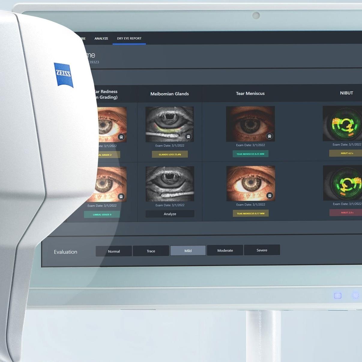
Comprehensive dry eye reportAs a multifactorial disease of the ocular surface, dry eye management requires a collection of measurements and their analysis. The ZEISS ATLAS 500 provides a comprehensive dry eye report, which contains detailed information on a variety of parameters:
- Meibography including calculation of the area of loss
- Ocular redness including grading by Nathan Efron
- Tear meniscus height
- Tear film break-up time
- Ocular Surface Disease Index (OSDI) questionnaire
- Osmolarity (optional data input)
Comprehensive dry eye report


Dry eye report
After you perform single measurements of the dry eye indicators of your patient, you can conveniently pull them into the dry eye report overview.
ATLAS 500 automatically displays the user-graded severity of the single indicators using a traffic-light highlight system.
In the end you can assess all indicators at a glance and select from the following degrees of severity: normal, trace, mild, moderate or severe.
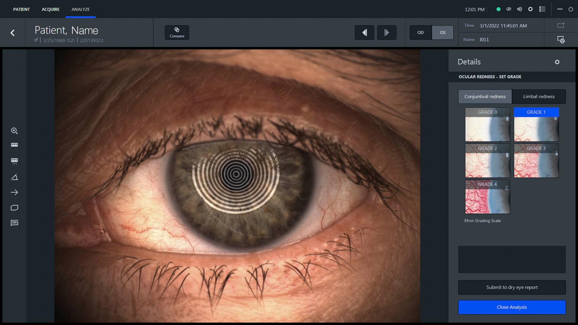

Ocular redness
An overview image of the eye is captured with the imaging and video modality. Afterwards, the operator can assess conjunctival redness or limbal redness visually comparing to the scale published by Nathan Efron.1
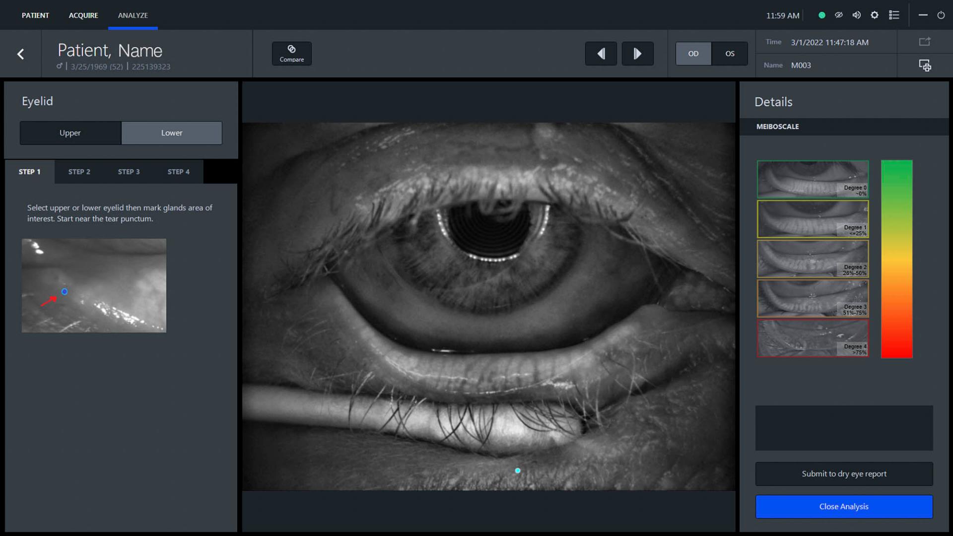

Meibomian glands
Analyze the upper and lower eyelid using a marking wizard that helps identify the meibomian glands loss using the meibomian grading scale by Heiko Pult.2
Non-invasive Tear Film Break-Up Time (NIBUT)
Assess the integrity of the tear film on the patient’s anterior corneal surface over time through dynamic videokeratoscopy.

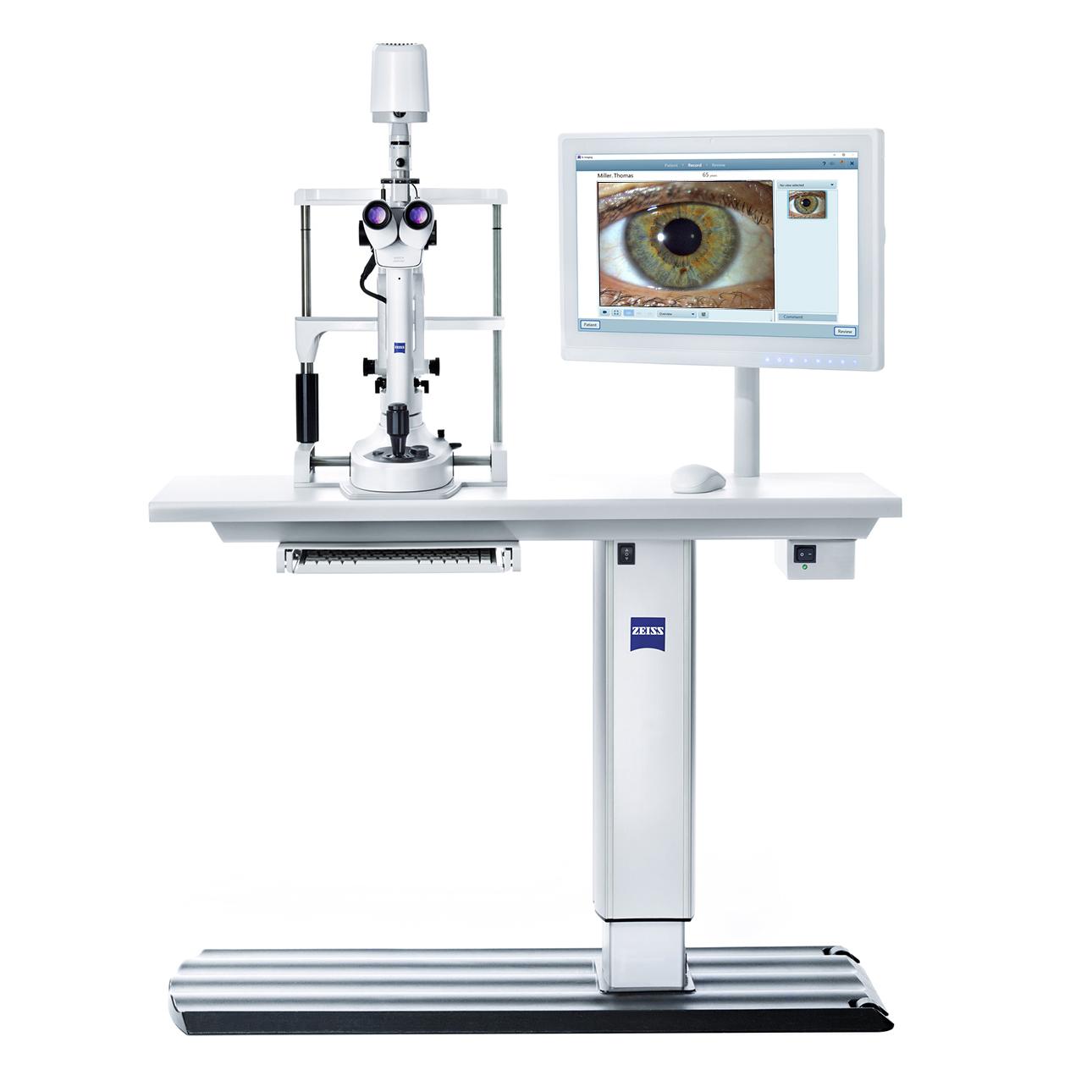
Optimized design
Easy operation and quick data acquisitionWith its proven performance, advanced features and streamlined, contemporary design, the ZEISS ATLAS 500 is designed to fit into any modern clinic or practice environment and provides a positive experience for both operator and patients.
Easy operation and quick data acquisition


Swivel-out measurement head
The unique, swivel-out measurement head helps reduce nasal shadows during data acquisition to increase the captured area of the cornea.


Enhanced working distance
ZEISS ATLAS 500 features two image capture options, which provide both close-up and overview images to evaluate the details of the ocular surface and photograph external structures.
Acquisition support
Alignment instructions on the left guide the operator for correct acquisition.

Digital documentation
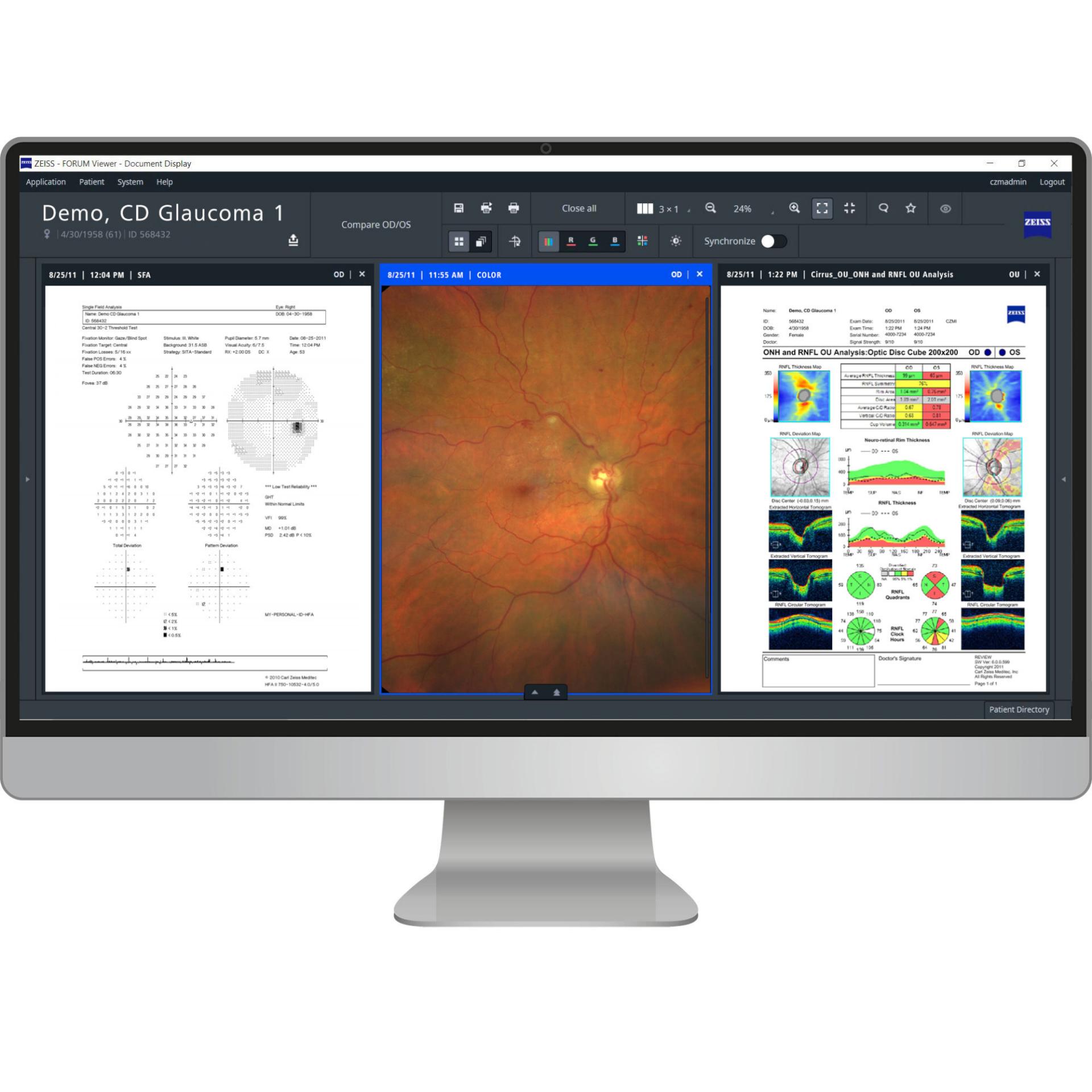
Speed up practice workflow with less manual errorsThe ZEISS ATLAS 500 is optimized for integration into the ZEISS medical ecosystem. The ZEISS FORUM connectivity enables patient data export, including PDF reports, to optimize your workflow.
Speed up practice workflow with less manual errors


Connectivity to FORUM
Link up ZEISS ATLAS 500 with ZEISS FORUM and schedule patients via modality worklists to speed up patient throughput. If required, patient data can also be entered via the ZEISS ATLAS 500 acquisition and analysis suite and transferred to ZEISS FORUM. ATLAS 500 PDF reports are visible in the FORUM viewer


Reports and data export
Store images, videos and reports within the ZEISS ATLAS 500 acquisition and analysis software, or directly send PDF reports to ZEISS FORUM, without the need for manual data transfer.
ATLAS 9000 data import
Simply import ZEISS ATLAS 9000 data and continue to review past recordings and compare to the latest recordings without having to switch between old and new systems. Upgrading and transitioning your ZEISS corneal topographer has never been easier.
Downloads


Technical specifications
ZEISS ATLAS 500 corneal topographer-
Key Parameters
Placido rings
24
Measurement points
6144
Topographic covering (in 42.2D)
9.3 mm
Dioptric range
up to 95D
Accuracy
Type A (ISO 19980 / ANSI Z80.23, ISO 10343)
Measurement head position
Straight, +/- 15° swivel-out
Working distance
74 mm
Adjustment range instrument base (W x H x D)
110 mm x 30 mm x 110 mm
Light sources
Placido
Fluorescence stimulation
Pupillometry and meibographyWhite light LED 450-650 nm
LED 465 nm
LED 950 nmDimensions (W x H x D)
319 mm x 504 mm (+/- 15 mm) x 420 mm
Weight of measurement head
12.2 kg
Power supply
100 V to 240 V AC 50/60 Hz
Data transfer
USB 3.0
Compatibility with standard
DICOM
-
ZEISS PC workstation 22" touch screen monitor including: PC mouse, PC keyboard
Dimensions (W x H x D)
546 mm x 351 mm x 66 mm
Weight
Approx. 8 kg
Monitor resolution
1,920 x 1,080 pixels LCD touch screen
Processor
Intel® Core™ i5/i7 Quad Core Processor
Hard disc
2 TB HDD
RAM
16 GB
Interfaces
4x USB 3.0
2x Isolated Ethernet Port
2x RS-232
1x HDMI and Display Port
Audio (Mic-in/Line-out) -
Processor
Intel® Core™ i7 6th generation or higher
SSD capacity
250 GB
RAM
16 GB
Recommended HDD capacity
2 TB
Interface
1x USB 3.0
3x USB 2.0 or higher
LANMonitor resolution
1,920 x 1,080 pixels (Full-HD)
Scaling 100%Recommended monitor size
22” (15” minimum)
Operating system
Windows 10 x 64 Professional
Data export formats
PDF (report)
Get in touch with us!
Receive more information about the product and availability in your country!Related products


-
1
Efron, “Grading scales for contact lens complications”, Ophthalmic Physiol. Opt., vol. 18, no. 2, pp. 182–186, Mar. 1998, doi: 10.1016/S0275-5408(97)00066-5
-
2
H. Pult and B. Riede-Pult, “Comparison of subjective grading and objective assessment in meibography”, Cont. Lens Anterior Eye, vol. 36, no. 1, pp. 22–7, Feb. 2013, doi: 10.1016//j.clae.2012.10.074