New protocol for efficient generation of seven-color, whole slide imaging with ZEISS Axioscan
Tissue and tumor sections have long been studied using immuno-detection techniques, such as immunohistochemistry (IHC). Depending on the imaging platform available, labeling of these samples was often limited either to one chromogen or three to four fluorophores, restricting researchers and drug developers in their experimental designs. Multiplex immunofluorescence (mIF) has emerged as a process that allows a higher number of fluorescent probes on a single tissue section, permitting in-depth comprehensive studies to identify specific proteins, cell types, spatial biology studies, and more.
Dr. Caroline Bouzin is the facility manager of the 2IP Imaging Platform at the Institute of Experimental and Clinical Research at the UCLouvain, Brussels, Belgium. She has worked with oncologists and pulmonologists to fine-tune a protocol for customized mIF stainings on formalin-fixed paraffin-embedded (FFPE) tissue samples followed by data collection with the ZEISS Axioscan digital slide scanner. A combination of signal amplification (TSA) with carefully selected filtersets matched to a panel of seven fluorophores results in the fast generation of ready-to-analyze digital images without the need for image post-processing, such as spectral unmixing or tissue alignment. This workflow is equipping researchers in her facility for high-throughput, spatial biology analyses, such as the immune microenvironment of tumors.
Using ZEISS Axioscan, we can image whole tissue sections with up to 7 fluorophores. Combined with powerful software, this allows us to assess spatial phenotyping of tumor microenvironments and is now applied to a wide range of projects.
Multiplex Immunofluorescence with Seven Fluorophores
Whole Slide Imaging of Breast Cancer Tissue with ZEISS Axioscan
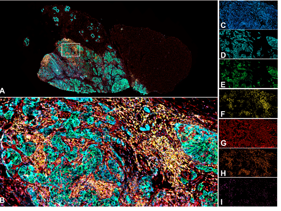
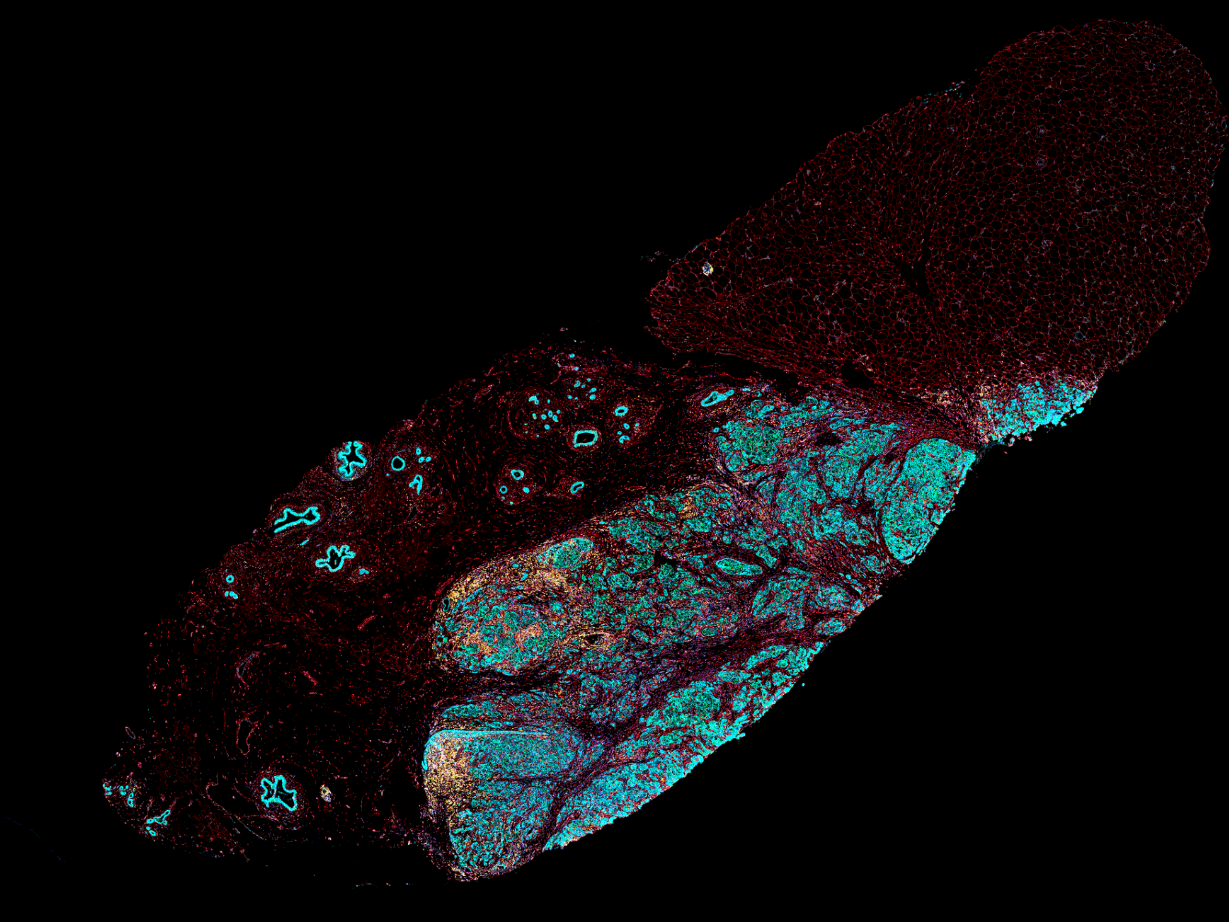
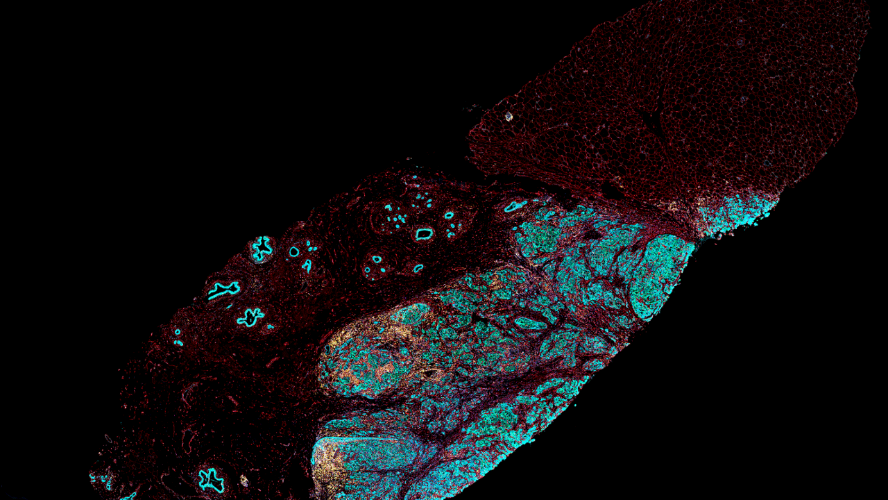
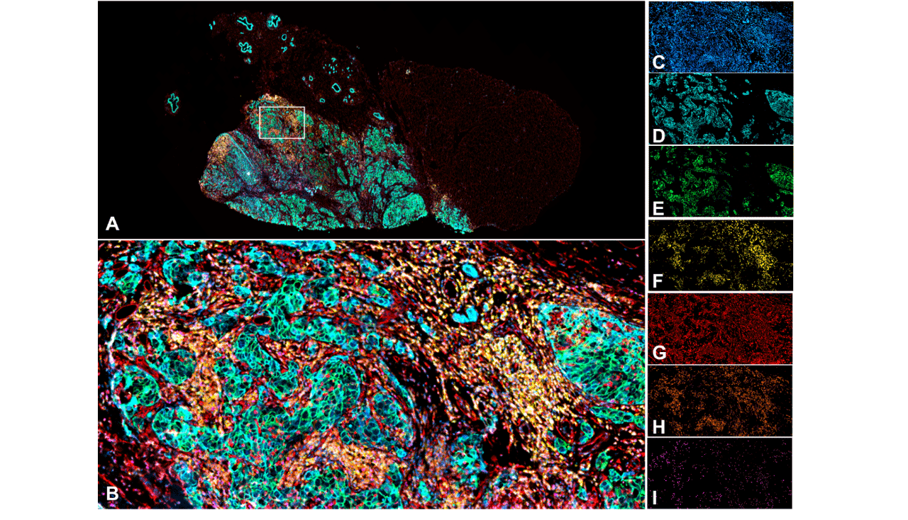
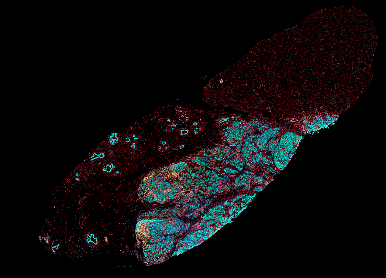
Multiplex seven color panel on human breast cancer FFPE section imaged with ZEISS Axioscan digital slide scanner. A) Overview image, B) Inset, C) Hoechst (blue, nuclei), D) Atto425 (cyan, cancer cells), E) AF488 (green, E-cadherin, epithelial marker), F) AF555 (yellow, CD3, T lymphocytes), G) AF594 (red, Vimentin, mesenchymal marker), H) AF647 (orange, CD8, cytotoxic T lymphocytes), I) CF754 (purple, Foxp3, Treg cells)
Multiplex seven color panel on human breast cancer FFPE section imaged with ZEISS Axioscan digital slide scanner. A) Overview image, B) Inset, C) Hoechst (blue, nuclei), D) Atto425 (cyan, cancer cells), E) AF488 (green, E-cadherin, epithelial marker), F) AF555 (yellow, CD3, T lymphocytes), G) AF594 (red, Vimentin, mesenchymal marker), H) AF647 (orange, CD8, cytotoxic T lymphocytes), I) CF754 (purple, Foxp3, Treg cells)
Published Protocol for Multiplex Immunofluorescence Combined with Spatial Image Analysis
Dr. Bouzin and colleagues have published their protocol for multiplex immunofluorescence combined with spatial image analysis for the staining of two to six antigens along with a nuclear fluorescent dye. The total number of fluorophores possible will depend on the slide scanner available in the laboratory.
In the article, N. Huyghe et al., 2023, they describe several mIF panels for different cancer types. After staining and digital scanning, they describe image analysis and bioinformatics scripts for spatial biology.
Their freely available workflow adds a spatial dimension to the classical density analysis already routinely performed for several markers. They propose that this protocol could equip scientists to better study the complex interactions between cancer cells and the tumor microenvironment.
While this protocol is enabling many new projects in our facility, looking forward, I see two challenges we will address. First, to combine multiplex immunofluorescence workflows on a single tissue section to subsequent multiplex chromogenic immunostaining or to mRNA detection. Also, to expand to 3D imaging.
Disclaimer
ZEISS explicitly excludes the possibility of making a diagnosis or recommending treatment for possibly affected patients on the basis of the information generated with ZEISS Axioscan digital slide scanner.


