State-of-the-art microscopy equipment gives students unique experiences - from training to faculty research to R&D
At the Keck Center for Science and Engineering at Chapman University, USA, undergraduate students are given a competitive edge - access to train on some of the most cutting-edge microscopy tools in the world. The High-Resolution Imaging Facility was conceived and implemented when Dr. Andrew Lyon was serving as Dean of the Schmid College of Science and Technology at Chapman. As a physical chemist with research focusing on nanostructures and colloidal particles in the development of new materials, Dr. Lyon understands personally how mastery of multi-modal imaging can be a powerful tool for developing new scientists. This equipment not only gives students a step up in their education, but also in the opportunities they receive in faculty research labs and possible partnerships within the R&D community.
I know how transformative it can be when an undergraduate student acquires the highly transferrable skills that come along with robust training in microscopy. It was clear as we expanded our research mission at Chapman, that the need for microscopy would increase rapidly. For these reasons, and so many more, we decided to push forward with a bold plan to assemble a world-class imaging suite.
Dr. Molla Islam (left) working with a student at the ZEISS Sigma field emission scanning electron microscope.
Dr. Molla Islam (left) working with a student at the ZEISS Sigma field emission scanning electron microscope.
State-of-the-Art Microscopy Equipment at the High-Resolution Imaging Facility
The facility is managed by Dr. Molla Islam, a former research scientist and now full-time Director. Dr. Islam oversees everything from training new users to offering advice on improving sample preparation and experimental outcomes. The facility supports both advanced scientific coursework as well as independent student research projects and faculty research labs.
The high-resolution imaging suite at Chapman gave me a ton of hands-on experience with various microscopy techniques. Easy access to high-quality microscopy tools as an undergrad helped me thoroughly engage in the work we were doing in the Lyon Group and enabled me to hit the ground running with the work I am doing now as a Ph.D. student. It was incredibly beneficial to have these microscopes at my disposal, which most students don’t experience until graduate school.
Students Use Advanced Microscopy in Faculty Research Labs to Help Drive Scientific Discovery
From photochemistry to evolutionary biology to biomaterials and more
Photochemistry
Evolution
Biomaterials
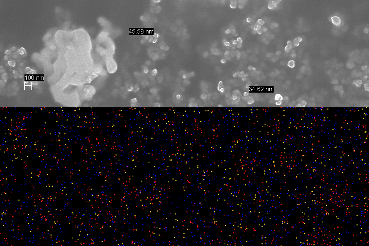
SEM and EDS images of Au-Ru core-shell plasmonic nanoparticles acquired with ZEISS Sigma 300 to obtain precise elemental distribution across nanoparticle samples.
SEM and EDS images of Au-Ru core-shell plasmonic nanoparticles acquired with ZEISS Sigma 300 to obtain precise elemental distribution across nanoparticle samples.
Studying Catalysts to Combat Global Warming
Using field emission SEM and EDS for photochemistry research
Dr. Jerry LaRue is an Assistant Professor of Chemistry who studies catalysts. One real world problem students in his lab work on is how to reduce greenhouse gas emissions which are contributing to global warming. With the right catalyst, the combustion reactions used by the world's petroleum industries could become more energy efficient.
Plasmonic metal nanoparticles are potential catalysts since they can efficiently generate large amounts of electrons critical to excited state chemistry upon interaction with visible light. Scanning electron microscopy (SEM) with EDS has been vastly practical and beneficial throughout the course of his photochemistry research. His students study the synthesized single metallic and bimetallic plasmonic nanoparticles to understand their surface structure.
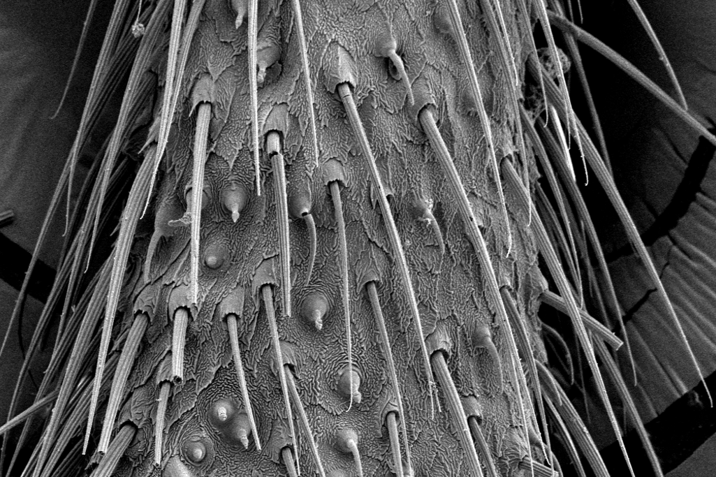
Chemical sensing hairs on a firefly antenna visualized by scanning electron microscopy.
Chemical sensing hairs on a firefly antenna visualized by scanning electron microscopy.
Investigating the Evolution of Flexible Structures that Interact with Their Environment
Scanning electron microscopy reveals the tiny hairs on firefly antennae
From researching odor detection in domesticated dogs to the study of odor capture by crustaceans to understanding how valveless tubular hearts drive flow, students in Dr. Lindsay Waldrop's lab investigate many diverse phenomena.
In this example, scanning electron microscopy is used to show how the antennae of fireflies may be evolving in response to sexual selection. Most fireflies flash in the evening to attract mates, but some have reverted back to using chemical signals, or pheromones, to attract mates similar to other beetle groups. Since chemical sensors are on the beetles’ antennae, students in the Waldrop lab are imaging antennae to study the size and distribution of chemical sensing hairs on a variety of species of fireflies.
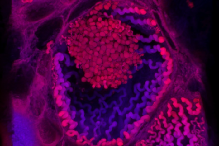
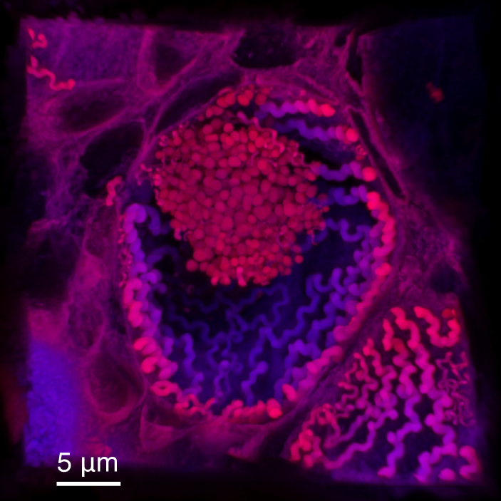
An epidermal thread cell from the Pacific hagfish (Eptatretus stoutii) which contain a proteinaceous thread that is loosely packed along the plasma membrane, measuring between 0.1 – 1 μm in diameter and ~2 mm in length. Imaged with a ZEISS confocal with Airyscan detector from a hematoxylin-eosin (H&E)-stained cross-sectional slide.
Striving to make high performance, eco-friendly materials
3D confocal microscopy of hagfish slime
At the Comparative Biomaterials Lab, Dr. Douglas Fudge and his students study a wide range of materials made by animals, including nano-scale filaments within cells, slimes secreted to ward off predators, and large structures like the keratinous plates of baleen whales. They are committed to applying what we learn from the study of biomaterials to real-life challenges, such as how we can make high performance materials for industry that are more eco-friendly in their manufacture and disposal.
Dr. Fudge explains that animals make outstanding materials for a wide variety of functions without the benefit of petroleum and without fouling their environment. He believes that humans could do the same if they listen to the lessons biology has to teach.
Shown here is an example of a hagfish epidermal thread cell imaged using confocal microscopy. Dr. Fudge and his team are studying hagfish to better understand the remarkable and emergent properties of hagfish slime, one of the most unusual complex fluids found in nature.
Poised to Partner with the Start-Up Community
Chapman University has two campuses located in Orange and Irvine, California – home to numerous tech Fortune 1,000 companies and fast-growing start-ups. Many of these companies want to move quickly and either do not have the funding to buy high end imaging equipment or have experienced microscopists on staff. The facility is ready to offer imaging resources to biotech startups who lack the necessary imaging tools and/or staff to advance their R&D mission.
Additionally, bringing companies such as these onto campus allows students to get a glimpse into working with start-ups as well as open up possible opportunities for work collaborations and internships.
I am impressed by what our students have accomplished using these tools thus far, but I am even more excited about what is going to come next. As the research programs in the Schmid College of Science and Technology become even more vibrant, and as our new Fowler School of Engineering continues to expand, it is clear that the microscopy facility will become an even more vital component in enabling our research and education missions to have national and global impact.


 working with a student at the ZEISS Sigma field emission scanning electron microscope.")
 working with a student at the ZEISS Sigma field emission scanning electron microscope.")
 working with a student at the ZEISS Sigma field emission scanning electron microscope.")
 working with a student at the ZEISS Sigma field emission scanning electron microscope.")