

Microbiology & Microbial Research
Visualize the microbial world with advanced microscopy solutions.
Turning microbial complexity into actionable insights
Visualizing pathogens with clarity, specificity and confidence
Most pathogens are less than two micrometers in size, making high magnification and oil immersion objectives essential for accurate morphological examination, whether distinguishing cocci, bacilli, or spirochetes. Fluorescence methods such as immunofluorescence and fluorescence in situ hybridization (FISH) provide even greater specificity, giving microbiologists critical insight into pathogen identity.
From protozoa and Plasmodia in blood smears to cysts, worm eggs, and mycobacteria in tissue sections, crystal-clear visualization is vital for reliable diagnosis. Brightfield, phase contrast, darkfield, and fluorescence microscopy each offer unique advantages, while electron microscopy extends resolution to the nanoscale, revealing microbial surfaces, biofilm architecture, and host–pathogen interfaces. Combined with digital documentation and advanced analysis tools like ZEISS arivis for segmentation and quantification, these technologies ensure reproducible, data-driven results, helping labs work with clarity, efficiency, and confidence.
Visualizing tuberculosis with the Primostar 3 iLED
Auramine staining for tuberculosis diagnosis with fluorescence microscopyRecommended Products for Microbiology


ZEISS lab essentials


 in the upper three cell layers.")
 in the upper three cell layers.")


Microbiology FAQs
-
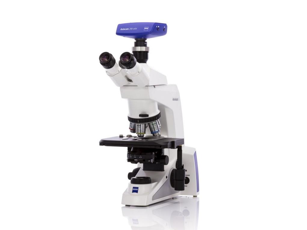
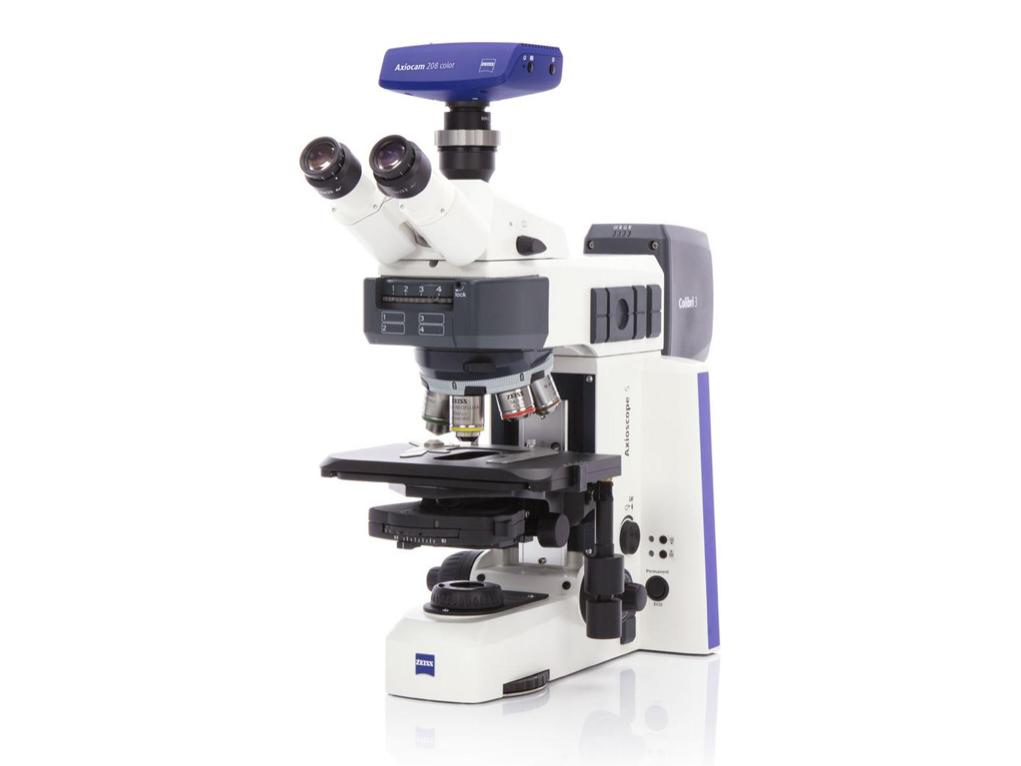
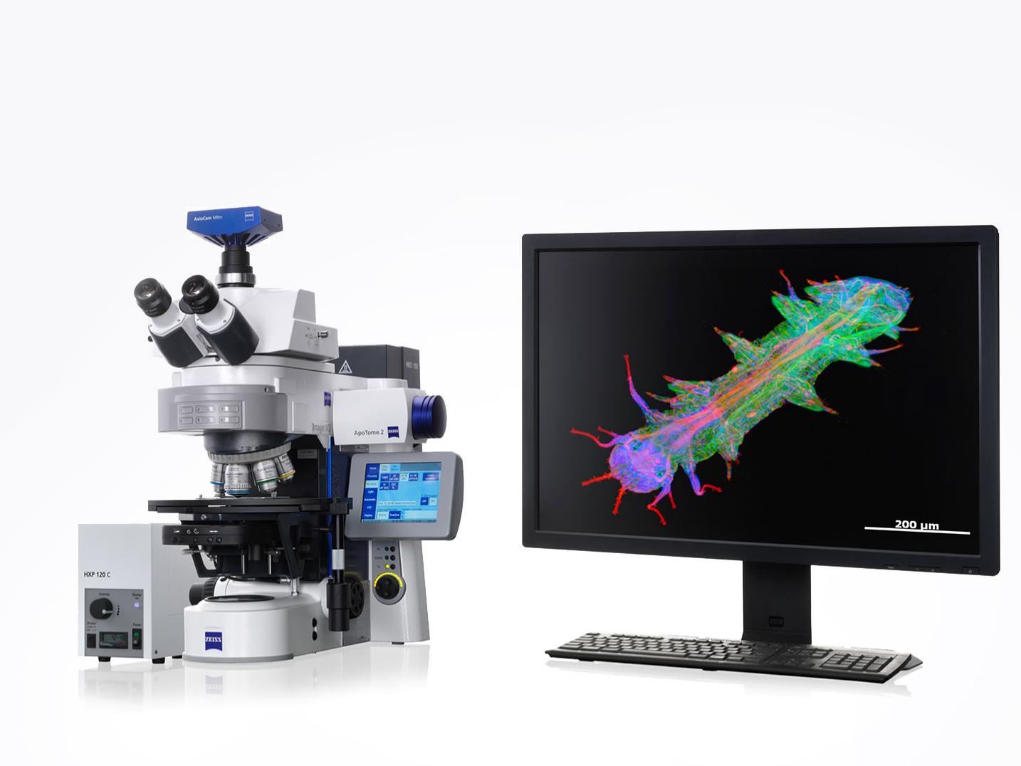
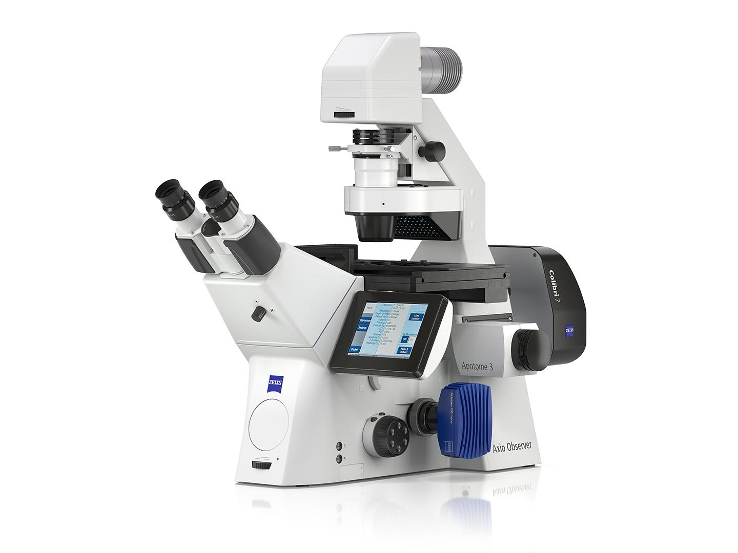
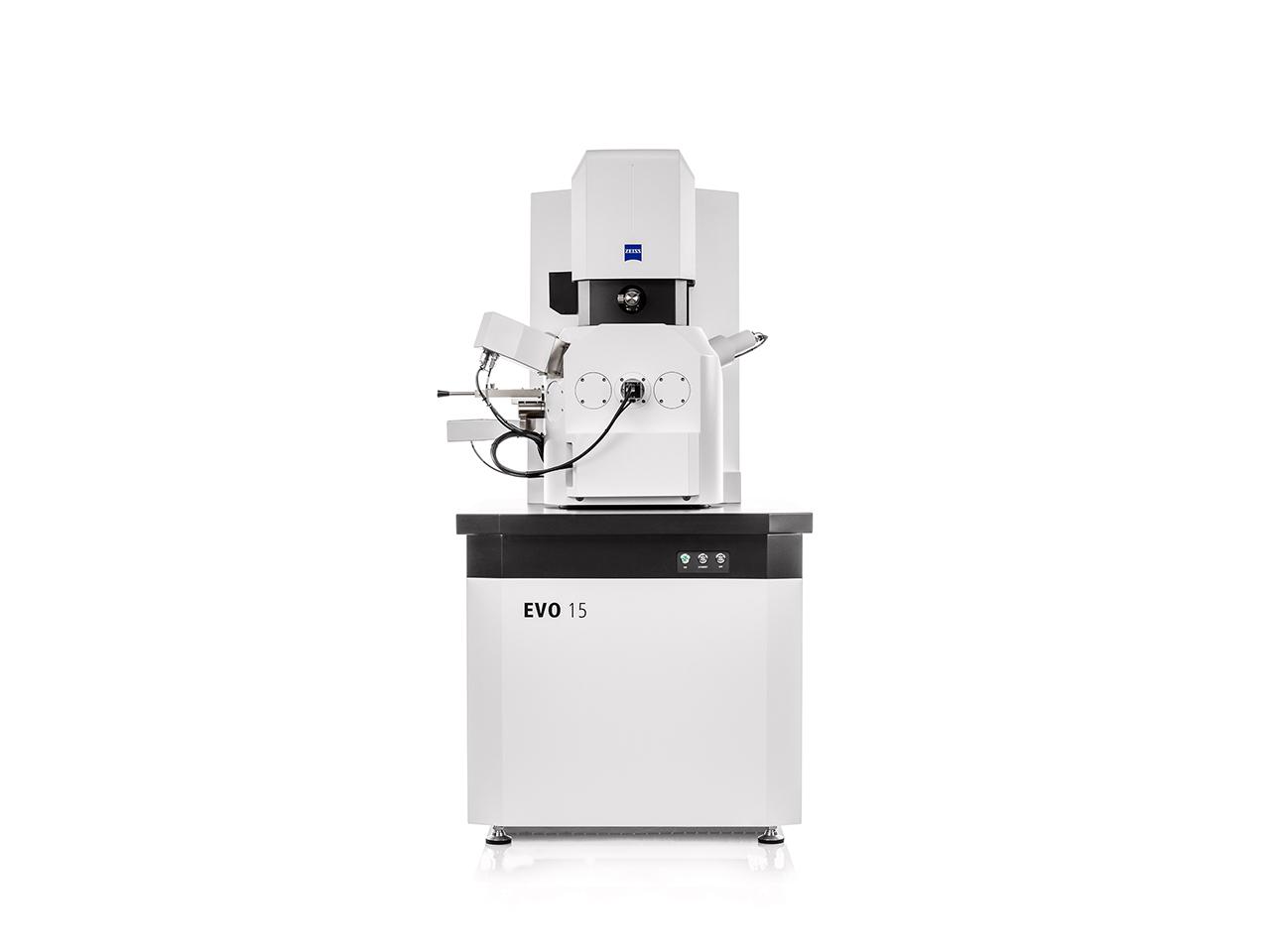
ZEISS offers a full portfolio tailored to microbiology workflows. For routine diagnostics, Primostar 3 and Primostar 3 iLED provide reliable brightfield and fluorescence imaging for Gram stains and acid-fast screening. For daily lab work, Axiolab 5 combines ergonomic design with one-button digital documentation. Advanced platforms like Axioscope 5 and Axio Imager 2 support fluorescence, FISH, and complex sample analysis, while the Axio Observer enables live-cell studies of microbial dynamics and host–pathogen interactions. When ultrastructural detail is required, ZEISS EVO SEM and Crossbeam FIB-SEM reveal biofilm architecture and microbial morphology at nanometer resolution.
-
Pathogens are typically less than two micrometers in size, making them difficult to resolve without advanced optics. ZEISS microscopes provide high-resolution objectives, oil immersion capabilities, and proven contrast techniques (brightfield, phase contrast, darkfield, fluorescence) to ensure confident identification. This level of clarity is essential for distinguishing microbial morphologies, detecting parasites in blood or stool samples, and documenting results for diagnostics, research, or quality control.
-
Accurate documentation is essential for quality assurance, teaching, and clinical decision-making. ZEISS Axiocam cameras, Labscope software, and ZEN software workflows make it easy to capture true-to-color images, automatically include scale bars, and share results instantly. For high-throughput needs, Axioscan 7 digitizes entire slides, while arivis software provides advanced segmentation and analysis pipelines. These tools enable reproducibility, remote collaboration, and compliance with laboratory standards.
-
ZEISS microscopes can contribute to antimicrobial resistance research by enabling detailed imaging and analysis of microbial interactions, drug effects, and resistance mechanisms. Techniques such as live-cell imaging and high-throughput screening can be employed to study the efficacy of antimicrobial agents.
-
Routine microbiology often involves high sample volumes and repetitive tasks like Gram staining and acid-fast screening. ZEISS solutions such as Axiolab 5 and Axioscope 5 feature “smart microscopy” with one-button digital documentation, automated exposure settings, and ergonomic designs. These features reduce fatigue, minimize user error, and speed up daily workflows without compromising accuracy.
-
Biofilms are highly structured microbial communities that require imaging at multiple scales to fully understand their formation, architecture, and resistance mechanisms. ZEISS offers a broad range of techniques to address these challenges:
- Light Microscopy (Axiolab 5, Axioscope 5, Axio Imager 2): Brightfield, phase contrast, darkfield, and fluorescence imaging reveal overall biofilm morphology, cell density, and viability.
- Confocal & Super-Resolution (Axio Observer, Lattice SIM, Elyra): Optical sectioning and high-resolution fluorescence uncover 3D structure, microbial interactions, and protein localization within biofilms.
- Lightsheet Microscopy (Lightsheet 7, Lattice Lightsheet): Non-destructive, volumetric imaging of biofilm growth and dynamics over time in 3D.
- Electron Microscopy (EVO SEM, Sigma SEM, GeminiSEM, Crossbeam FIB-SEM): Ultrastructural views of microbial surfaces, extracellular polymeric substances (EPS), and host–biofilm interfaces at nanometer resolution.
- X-ray Microscopy (VersaXRM): 3D visualization of biofilms in larger host tissues or industrial materials, without sectioning.
- Digital Analysis (ZEISS arivis software suite): Segmentation, quantification, and 3D visualization transform complex imaging datasets into reproducible, quantitative insights.
Together, these techniques allow researchers to study biofilms from the macro scale of community growth down to the nano scale of microbial ultrastructure, helping to unravel their role in infection, antimicrobial resistance, and environmental processes.