Multi-scale battery characterization workflows
ZEISS supports multi-scale battery characterization workflows with imaging, analysis, and correlative microscopy solutions for structural and degradation analysis.
Skip to main content

ZEISS supports battery research with microscopy workflows for studying material behavior, degradation, safety, charging performance, and long-term reliability across scales.
Battery research depends on understanding how materials and structures behave across scales. From whole cell architecture to nanoscale interfaces within electrodes, features such as pore connectivity, particle morphology, cracking, and dendrite formation all influence battery performance, safety, and longevity. At the same time, battery materials are often air-sensitive and challenging to prepare, while destructive workflows can alter critical structures before imaging.
ZEISS microscopy solutions help researchers overcome these challenges through connected workflows that streamline sample handling, reduce manual intervention, and enable simultaneous structural and chemical analysis while preserving sample integrity.
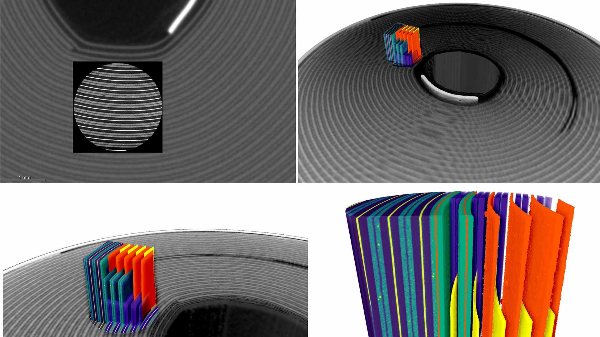
Analyze intact batteries non-destructively, then zoom into regions of interest for high-resolution characterization.
Correlate data across imaging and analytical techniques to build a more complete understanding of battery materials.
Study degradation pathways, structural evolution, and failure mechanisms that influence battery safety, performance, and longevity.
ZEISS supports multi-scale battery characterization workflows with imaging, analysis, and correlative microscopy solutions for structural and degradation analysis.
FE-SEMs allow for imaging of battery raw material powders at the micro- and nanometer scale, helping researchers to determine grain size and shape, detect impurities, enhance the process and quality control.


NMC cathode particle. Imaged with scanning electron microscopy.
NMC cathode particle. Imaged with scanning electron microscopy.
Microscopy plays a key role in studying powders and particles used to produce battery electrodes. By observing the structure, morphology, and chemistry of these raw materials, scientists can better understand their electrochemical behavior at a fundamental level, and gain insight to how they will perform once integrated into a functioning cell.


Porous polymer separator. Imaged with scanning electron microscopy.
Porous polymer separator. Imaged with scanning electron microscopy.
Other materials within battery cells also play critical roles, such as providing structural support and enabling electronic and ionic conduction (current collectors, binders, separators, electrolytes). Microscopic analysis can reveal structural impacts of these materials on transport processes, as well as phenomena occurring at interfaces.
ZEISS VersaXRM, GeminiSEM, and Crossbeam support high-resolution imaging and analytical workflows for studying electrode materials, interfaces, porosity, particle morphology, and microstructural evolution linked to battery performance.


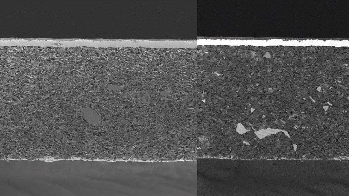
Battery materials often exhibit subtle structural and compositional differences, making it hard for a single imaging approach to distinguish them. ZEISS electron microscopy workflows provide complementary imaging modes for analyzing interfaces, particle distributions, coatings, porosity, and material contrast within complex battery structures.
Imaging the same lithium-ion battery cross-section with different detector signals reveals striking variations in material contrast and microstructural detail. By comparing these imaging modes, researchers gain useful insights into interfaces, coatings, particle distributions, and other structural features that shape battery performance and degradation.


Cycled cathode layers delaminated from current collector. Imaged with 3D X-ray microscopy.
Cycled cathode layers delaminated from current collector. Imaged with 3D X-ray microscopy.
Battery electrodes consist of complex three-dimensional networks of active material, binder, and electrolyte (porosity). By using 3D microscopy techniques, these micro- and nano-scale networks can be imaged directly to enable measurement of critical morphological characteristics or even produce discrete volumetric modeling domains for input to numerical simulations of battery electrochemistry.
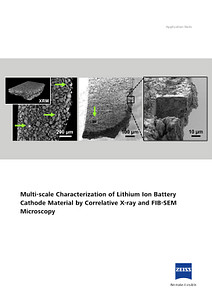
ZEISS correlative workflows can be applied to samples such as assembled cells and combine X-ray, electron, ion, and light microscopy techniques to support transitions from overview imaging to targeted high-resolution structural and chemical characterizations.
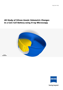
With ZEISS in situ X-ray microscopy workflows, researchers can unlock insights into structural changes, deformation, crack propagation, and degradation mechanisms as batteries operate under real or simulated conditions.
Finally, keep your battery research efficient with ZEISS service, training, and technical support solutions, providing long-term research success.
ZEISS provides service, training, and technical support to help laboratories maintain efficient battery imaging and analysis workflows. From installation and onboarding to system maintenance and technical assistance, ZEISS helps researchers maximize instrument performance and support long-term workflow reliability.
In situ characterization allows researchers to observe structural changes, degradation, and failure mechanisms while a battery operates under real or simulated conditions.
Non-destructive imaging preserves sensitive structures and allows researchers to investigate intact batteries before targeted downstream analysis.
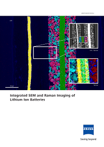
Battery workflows often combine X-ray microscopy, SEM, FIB-SEM, Raman microscopy, EDS, EBSD, and SIMS to connect structural and chemical information across scales.
Electron microscopy provides high-resolution imaging and analysis of battery materials, interfaces, defects, and microstructures linked to performance and degradationproperty.
Microscopy supports investigations into degradation, cracking, dendrite formation, porosity, contamination, structural evolution, and failure analysis.
Correlative workflows connect data across instruments and scales, helping researchers move from overview imaging into targeted high-resolution characterization.