

ZEISS Versa X-ray Microscopes
Discover More with 3D X-ray Imaging at Submicron ResolutionExperience the power of ZEISS Versa X-ray microscopes (XRM), the proven choice for researchers and scientists worldwide. VersaXRM feature intuitive user interfaces, ensuring that every user can maximize their productivity and achieve exceptional results. Prioritizing real resolution in practical settings, VersaXRM provide you with the capability to observe even the smallest details with unparalleled clarity. Renowned for their stability and precision, the ZEISS commitment to quality is evident in every aspect of VersaXRM, including with the purpose-built ZXR-1 X-ray source on the advanced VersaXRM 730. Trust that your investment will withstand the test of time, meeting your needs for years to come.


VersaXRM 730
ZEISS VersaXRM® 730 offers submicron imaging with 450-500 nm resolution across 30-160 kV with the exclusive 40x-Prime objective. Its ZEN navx® automated user guidance and control system and AI-driven DeepRecon Pro streamline workflows, optimize image quality, and speed throughput. Designed for accessibility, it supports a broad user spectrum, enabling advanced research capabilities with ease for your whole team. ZXR-1, the innovative, ZEISS purpose-built
X-ray source standard on VersaXRM 730, further fulfills the VersaXRM “Protect Your Investment” promise, offering a platform for continued innovation.
Mouse models are valuable tools in genetic research since they closely resemble humans in terms of physiology and genetics. Non-destructive XRM is an ideal imaging technology for such a sample. This iodine-contrasted E15.5 mouse embryo was imaged using FAST Mode on VersaXRM 730 with a total scanning time of 6 minutes. Sample courtesy of Chih-Wei Logan Hsu, Baylor College of Medicine.
VersaXRM 730
Perfect tomographies.¹ Every user. Every sample. Every time.Explore the limitless potential of the ZEISS VersaXRM 730, featuring the exclusive 40x-Prime objective, the award-winning ZEN navx, and the purpose-built ZXR-1 X-ray source. This system redefines submicron imaging with unparalleled resolution performance, unlocking groundbreaking capabilities for your research. ZEN navx streamlines workflows with intelligent system insights, ensuring efficient and effortless results. Benefit from AI-based reconstruction for superior image quality and increased throughput from DeepRecon Pro, while FAST Mode enables sub-one-minute tomographies. Experience a new era of understanding your samples with the ZEISS VersaXRM 730.


Head of objectives with the 40X-P
Revolutionize Your Research with Unmatched Resolution Performance
ZEISS 40x-Prime ObjectiveExclusive to ZEISS VersaXRM 730, the 40x-Prime objective lens enables you to achieve unparalleled resolution performance of 450-500 nm across the full range of source voltage, from 30 kV to 160 kV. This unique feature unlocks entirely new application capabilities for researchers, pushing industry standards of submicron imaging resolution. And, with more X-ray photons available, achieve even faster time to results for varied samples with uncompromised resolution. Known for their ability to achieve Resolution at a Distance (RaaD™), ZEISS VersaXRM have always enabled high resolution imaging of a wide array of sample types and sizes over a long range of length scales. Now, experience submicron imaging like never before with the higher energy capabilities and exclusive 40x-Prime objective of VersaXRM 730.


AI-based Image Perfection
ZEISS DeepRecon Pro from the Advanced Reconstruction ToolboxZEISS DeepRecon Pro has become such a powerful tool for XRM reconstruction that the ART high performance workstation and DeepRecon Pro with a two-year license are included with your VersaXRM 730.
DeepRecon Pro is an innovative AI-based technology bringing superior throughput and image quality benefits across a wide range of applications. DeepRecon Pro is applicable to both unique samples as well as semi-repetitive and repetitive workflows. Customers can now self-train new machine learning network models on-site with an extremely easy-to-use interface. The one-click workflow of DeepRecon Pro eliminates the need for a machine learning expert and can be seamlessly operated by even a novice user.


Non-destructive three-dimensional grain map of an Armco iron sample with illustrations of the various grain analysis that can be performed on a typical LabDCT Pro dataset.


Unlocking Crystallographic Information
LabDCT Pro for diffraction contrast tomography (DCT)LabDCT Pro for diffraction contrast tomography (DCT), an option available exclusively on ZEISS VersaXRM 730, enables nondestructive mapping of grain orientation and microstructure in 3D. Direct visualization of 3D crystallographic grain orientation opens up a new dimension in the characterization of polycrystalline materials like metal alloys, geomaterials, ceramics, or pharmaceuticals.
- LabDCT Pro supports specimens with crystal structures from cubic symmetry to systems with lower symmetry such as monoclinic materials.
- Acquire high resolution crystallographic information using the dedicated 4X DCT objective. For even larger samples, use large area mapping and increase your throughput with the Flat Panel Extension (FPX).
- Obtain comprehensive 3D microstructure analysis from larger representative volumes and wide-ranging sample geometries.
- Investigate microstructural evolution with 4D imaging experiments.
- Combine 3D crystallographic information with 3D microstructural features.
- Combine modalities to understand structure-property relationships.


Gain an Edge in Contrast
Dual Scan Contrast Visualizer (DSCoVer), exclusive to VersaXRM 730, extends the detail captured in a single energy absorption image by combining information from tomographies taken at two different X-ray energies. DSCoVer takes advantage of how X-rays interact with matter based on effective atomic number and density. This provides you with a unique capability for distinguishing, for example, mineralogical differences within rocks as well as among difficult-to-discern materials such as silicon and aluminum.


Achieve New Degrees of Freedom
The flagship VersaXRM 730 offers additional unique features and imaging capabilities.
- Improve scan speed and accuracy of large, flat, or irregular samples with advanced acquisition techniques such as High Aspect Ratio Tomography (HART).
- Flexibly image larger samples with Wide Field Mode (WFM) to stitch projections horizontally to form an extended lateral field of view for either higher voxel density for a given field of view or a wide lateral field of view to provide larger 3D volume for large samples.
- Automated Filter Changer (AFC) enables seamless filter changing without manual intervention, and your selection can be programmed and recorded for each recipe.
-
1
The ability to consistently generate “perfect tomographies” by all users for all samples is made possible through the use of ZEN navx with its sample-oriented parameter and user guidance, built-in workflows, and system/sample intelligence and protection, combined with the use of DeepRecon Pro AI-based 3D reconstruction.


VersaXRM 615
Unlock new degrees of versatility for your scientific and industrial research with the ZEISS VersaXRM® 615 platform. This cost-effective solution for a high-end X-ray microscope delivers advanced resolution and contrast, pushing non-destructive imaging further for accelerated research. With innovative X-ray source and optics technology, enjoy rapid tomography scans that maintain quality. Seamless workflows and AI-based DeepRecon Pro deep learning, facilitate the discovery of high resolution interest areas without sample alteration.
Smart watch battery. ZEISS VersaXRM 615 scans the intact battery to identify areas of interest and zoom-in for high resolution imaging.
Discover New Levels of Versatility with ZEISS VersaXRM 615
Extending the Limits of Your ExplorationUnlock new degrees of versatility for your scientific discovery and industrial research with ZEISS VersaXRM 615. This cost-effective, high-end X-ray microscope is an ideal choice for the modern analytical laboratory. With its advanced resolution and contrast capabilities, VersaXRM 615 pushes the boundaries of non-destructive imaging, offering greater flexibility and accelerating your research.
X-ray source and in optics technology provide higher X-ray flux, enabling faster tomography scans without sacrificing resolution and contrast. Innovative acquisition workflows empower you to locate high-resolution regions of interest without cutting your sample. Seamlessly move from exploration to discovery with VersaXRM 615.


Xradia 515 Versa
Experience exceptional versatility in your scientific and industrial research with ZEISS Xradia 515 Versa, a trusted X-ray microscope for modern labs. Its hallmark Resolution at a Distance (RaaD) capability ensures class-leading resolution over longer working distances, fostering groundbreaking insights for a wide variety of sample types. Paired with powerful contrast and 4D/in situ capabilities for a variety of research needs, this flexible platform ensures a fast time to results.
Copper sulfide ore imaged with Versa, using Mineralogic 3D for classification of mineralogy


ZEISS Xradia 515 Versa Features
- Scout-and-Scan control system
- 500 nm spatial resolution and minimal achievable voxel size of 40 nm with the optional 40x objective
- Two-stage magnification process based on synchrotron caliber optics
- SmartShield for system / sample protection
- System stability for highest performance
- Optional Flat Panel Extension (FPX) for large field of view imaging
- Optional Advanced Reconstruction Toolbox for enhanced performance
- Optional accessories: Autoloader programmable robot for handling up to 14 samples, In Situ Interface Kit for in situ / 4D imaging
Perfect Tomographies. Every user. Every sample. Every time.
A quick view of the capabilities of ZEISS VersaXRM 730.The Technology Behind ZEISS Versa X-ray Microscopes
-


The Versatile Advantage of RaaD
The Versatile Advantage of RaaD
The Versatile Advantage of RaaD
The two-stage magnification technique offered by ZEISS Xradia Versa uniquely achieves Resolution at a Distance, or RaaD, which enables you to effectively study the widest range of sample sizes, including those within in situ chambers.
Images are initially magnified via geometric projection as with conventional microCT. The projected image is cast onto a scintillator, converting X-rays to a visible light image which is then optically magnified by microscope optics before acquisition by a CCD detector.
Reducing dependence on geometric magnification enables ZEISS Xradia Versa solutions to maintain submicron spatial resolution down to 500 nm at large working distances.
-


Powering Accessibility with the Principles of Human-centered Design
ZEN navx Guidance and Control System
ZEISS researchers studied XRM users to develop a solid understanding of the issues and challenges you face, including your biases, how you compensate, and your workarounds. With that data in hand, our expert team developed systematic built-in guidance, automated workflows, and intelligent system insights to enable even novice users to achieve results more easily and efficiently. If you are an expert user, ZEN navx offers significant efficiency benefits, "unlocking" the system to allow you to explore the full versatility of ZEISS Versa XRM. ZEN navx also provides infographic-like visualization that helps you to understand the trade-offs of your choices, e.g., between resolution, field of view, and throughput.
ZEN navx includes an embedded 3D viewer to integrate Volume Scout capability for end-to-end 3D navigation. This streamlines access to your sample to pinpoint and identify specific regions of interest to target for higher resolution imaging.
Additional capabilities include the ZEN navx File Transfer Utility, or FTU, which puts your microscope data exactly where you need it, when you need it, without having to manually transfer from system to workstation, or to save on a hard-drive to carry from place to place. SmartShield works within ZEN navx to protect your system and your sample from damage.

Winner of the Red Dot 2024 Best of the Best Interface award, ZEN navx intuitive navigation follows the evolution of the XRM user base, revolutionizing X-ray navigation and control with seamless and integrated workflows. It also complements the planning and execution of advanced correlative workflows with other ZEISS platforms in the ZEN environment.
ZEN navx is available for advanced VersaXRM platforms: VersaXRM 730 and VersaXRM 615.
-

ZXR-1: Your Source for Innovation
The ZXR-1 X-ray source is a unique innovation purpose-built for the capabilities of the advanced VersaXRM 730. Designed to push the boundaries of X-ray technology, the 25W ZEISS-built ZXR-1 offers unparalleled reliability, cost efficiency, and cutting-edge features. With the largest installed base of XRM, ZEISS is privy to the needs, and can anticipate the demands, of our user base.
Innovation Showcase
The ZXR-1 source represents a significant advancement in X-ray technology. It provides a platform for advanced capabilities that enhance imaging precision and quality, making it an ideal choice for a wide range of requirements. Experience the future of X-ray technology with ZXR-1, where cutting-edge design meets unparalleled performance. Our commitment to innovation is driven by customer insights, ensuring that the ZXR-1 meets the highest standards of excellence.Reliability Assurance
ZXR-1 is engineered to deliver exceptional reliability while maintaining performance standards with minimal downtime. With more operating hours, ZXR-1 sets a new standard in longevity and dependability. This means fewer interruptions and more productive hours for your operations.Lower Cost of Ownership
Investing in VersaXRM with ZXR-1 source means significant savings over time. The extended operating life for ZXR-1 reduces the frequency and cost of source replacements.Contact us to discuss your imaging requirements so we can provide you with more information about the benefits of ZXR-1 for your specific needs.
-
One-Minute Tomographies with Fast Acquisition Scanning Technology
FAST Mode, enabled by Your Flat Panel Extension (FPX)
FAST Mode on ZEISS VersaXRM enables rapid 3D image acquisition for all samples through continuous motion scanning. Used with the optional flat panel detector (FPX), this mode allows non-stop sample rotation during X-ray image capture at various angles, eliminating the overhead delay of traditional step-and-shoot acquisition. Thus, dramatically faster scan times can be attained when exposure times are below 0.5 seconds, typical for the large, sensitive FPX detector. Expect acquisition times generally from <1 to 5 minutes, and even below 20 seconds for less stringent image quality needs.
FAST Mode enables true, nearly real-time 3D navigation for all samples thanks to full integration with the Volume Scout workflow in ZEN navx. FAST Mode acquisition integrates seamlessly with Volume Scout to allow for nearly immediate feedback and true 3D navigation to the correct region of interest in your complex samples.
-

Tensile testing of laser welded steel under increasing load.
Tensile testing of laser welded steel under increasing load.
Push the Limits of Scientific Advancement
ZEISS Versa X-ray microscopes provide the industry's premier 3D imaging solution for the widest variety of in situ rigs, from high-pressure flow cells to tension, compression, and thermal stages. Moving beyond the three dimensions of space, leverage the non-destructive nature of X-ray investigation to extend your studies into the dimension of time with 4D experiments.
These studies require samples to be further away from the X-ray source to accommodate various types of in situ rigs. On traditional microCT systems, this significantly limits the resolution achievable for your samples. ZEISS Versa XRM are uniquely equipped with dual-stage magnification architecture with RaaD technology that enable the highest resolution for in situ imaging.
ZEISS Versa XRM platforms can accommodate a variety of in situ rigs, including user-customized designs. You can add the optional in situ Interface Kit to your ZEISS Xradia XRM, which includes a mechanical integration kit, a robust cabling guide, and other facilities (feed-throughs) along with recipe-based software that simplifies your control from within the Versa Scout-and-Scan or ZEN navx user interfaces. When your needs require pushing the resolution limits of your in situ experiments, convert your ZEISS Xradia microCT or XRM to VersaXRM 730 X-ray microscope and leverage RaaD technology for the maximum performance tomographic imaging of samples within in situ chambers or rigs.
-
Begin Your Multi-Scale, Multi-Modal, Multi-Dimensional Microscopy with Non-destructive 3D Imaging
Because of the non-destructive nature of X-rays and the versatile array of sample types and sizes they are able to image, correlative microscopy often begins with, or is enabled by, ZEISS Versa XRM.
Using the Scout-and-Zoom or Volume Scout capabilities of Versa, you are able to clearly define your region of interest (ROI) before sacrificing your sample to premature cutting or other sample prep. Rapidly scout at low resolution with a large field of view, and then zoom to the ROI at higher resolution, whether using the range of Versa objectives (up to 40x-P), nanoscale ZEISS Ultra XRM, or ZEISS light, electron, or FIB-SEM microscopes. This prevents premature sample destruction and allows for maximum workflow efficiency while combining full sample context with key sample information.
Additionally, the ability to perform interior tomography, or to clearly see inside your sample in 3D, further reduces the risk of losing sight of your ROI. Achieve greater efficiency by pinpointing a specific “address” to which to navigate for accurate and efficient next steps for interrogating your sample.
Finally, examine your sample under varying conditions and over time with in situ and 4D studies before performing further analysis — chemical, surface, etc.— with other ZEISS modalities.
Leverage the widest array of microscopy solutions available — exclusively from ZEISS — to perform multi-modal, multi-length-scale, multi-dimensional analyses, by starting with non-destructive 3D X-ray microscopy.

Full correlative sample workflow for the project. Initial XRM scans highlight key areas for higher resolution imaging and target locations for thin section orientation within the volume. Subsequent 2D analysis includes electron and light microscopy, leading to correlation with in situ microanalytical data.
-
Investment Protection
Continuous improvement and upgradeability
As your imaging needs evolve, so should your instrument. The ZEISS Versa XRM family is built on the established ZEISS Versa 3D X-ray microscope platform that is upgradeable, expandable, and reliable, paving the way for future enhancements and protecting your investment. Select the system that is right for you today and expand as your needs require.
To make certain your system offers the latest capabilities and remains serviceable, you can field-convert your platform to the latest X-ray technology: your ZEISS Context microCT can become a CrystalCT® or higher performance Versa X-ray microscope. Your CrystalCT can become VersaXRM 730 with LabDCT. And every mid-tier Versa platform can be upgraded to the most advanced VersaXRM from ZEISS.
In addition to instrument conversions at your facility, new modules are continuously developed that will enhance your instrument to provide advanced capabilities such as in situ sample environments, unique imaging modalities, and productivity enhancing modules. Also, periodic major software releases include important new features that are made available to existing instruments, thereby enhancing and extending the capabilities of your research.

What is the source technology?
Both VersaXRM 615 and VersaXRM 730 offer as standard the 25W, fast-activation ZXR-1 X-ray source, purpose-built for stability for high performance VersaXRM platforms. ZXR-1 is the field-upgradeable ZEISS designed-and-built source for future VersaXRM innovation, offers greater reliability and lower cost of ownership
What is the resolution of VersaXRM?
500 nm spatial resolution with the 40x objective for VersaXRM 615
450 nm spatial with the 40x-P objective on VersaXRM 730. Additionally, VersaXRM 730 offers resolution performance of 500 nm across the full voltage range of 30-160 kV.What is the highest resolution detector?
VersaXRM 615 offers the optional 40x objective, and VersaXRM 730 offers the optional 40x-P objective. The 40x-P objective enables high resolution imaging for larger, denser, high Z samples such as metals and composites and for interior tomographies, while maintaining quality.
What is the user interface?
Both VersaXRM 615 and 730 offer the user-centered ZEN navx, developed by ZEISS X-ray Microscopy by studying user biases, habits, and workarounds. It offers three built-in workflows to enable users at every skill level to be immediately successful. ZEN navx also enables both sample and system protection with its SmartShield options. It also includes an embedded viewer that enables users to integrate interior tomography, or Volume Scout, capabilities without destroying or prematurely harvesting your sample.
How long does it take to do a tomography?
Using a mode of data collection called Fast Acquisition Scanning Technology, or FAST, tomographies can approach the sub-one-minute range. Using FAST Mode, the sample rotates continuously while X-ray transmission images are acquired at different angles. Integrated into ZEN navx, it allows for nearly immediate feedback and 3D navigation to the correct region of interest, even for complex samples.
Do VersaXRM have AI-based reconstruction capabilities?
Every VersaXRM 615 and 730 now comes standard with DeepRecon Pro, an AI-based deep learning method. DeepRecon Pro enables faster throughput or greater image clarity, dependent on user requirements. Also included with your VersaXRM is a high-performance workstation, enabling all reconstruction methods in the Advanced Reconstruction Toolbox created for Versa platforms.
How do I get an upgrade of my current Versa?
The ZEISS “Protect Your Investment” promise that has been a standard practice enables you to upgrade your system to the latest technology. Contact your ZEISS sales representative today for details.


Discover the Advantages in Your Research Area
ZEISS VersaXRM - Microscopic Solutions for all Applications-

Flux grown layered KBiS2 semiconductor crystal. 3D volume rendering shows the complex 3D microstructure consisting of rod and needle structure. Sample courtesy of Prof. Daniel Shoemaker, UIUC
Flux grown layered KBiS2 semiconductor crystal. 3D volume rendering shows the complex 3D microstructure consisting of rod and needle structure. Sample courtesy of Prof. Daniel Shoemaker, UIUC
Materials Research
- Experience the unique benefits of ZEISS VersaXRM, including non-destructive views of deeply buried microstructures, compositional contrast for studying challenging materials, and the ability to maintain RaaD for in situ imaging.
- Enjoy fast and intuitive 3D navigation technology for macro-scale inspection and easily identify regions of interest for high-resolution imaging.
- Benefit from faster throughput, improved image quality, and resolution performance, allowing for better data, increased sample statistics, more users, and enhanced instrument utilization.


Segmented active ingredient particles in an antihistamine tablet. Following imaging on a ZEISS Versa XRM, the data was reconstructed using ZEISS DeepRecon Pro to improve contrast between similar low density materials for better segmentation. Widest width of tablet is 5 mm.


3D rendering of a bund le of Rayon polymer fibers that have been imaged in propagation phase contrast mode. The 3D rendering shows a high-resolution 3D dataset processed using ZEISS PhaseEvolve to enhance the microvoids in individual fibers. Color represents void volume.


Tomography and segmentation of multiple phases in a high density concrete nuclear reactor vessel. 3D view shows segmentation of pores (red) and high-density minerals titanomagnetite and ilmenite (yellow) in chips of dolerite within the concrete. Concrete core 15 mm diameter. Sample courtesy of Giacomo Torelli, University of Sheffield, UK

Carbon fiber reinforced polymer composite.
-
3D dataset of a mouse brain that was imaged with the 40×-P objective of ZEISS Versa XRM and reconstructed using ZEISS DeepRecon Pro. Sample courtesy of Dr. Kevin Boergens, the University of Illinois at Chicago, US
Life Sciences
- Capture whole samples at multiple length scales with ZEISS VersaXRM, utilizing RaaD and FAST Mode to easily navigate and capture high-resolution regions of interest.
- Overcome limitations in imaging large sample volumes by leveraging ZEISS DeepScout, generating high-resolution overviews that were previously unreachable.
- Benefit from high-contrast images acquired with VersaXRM, enabling precise identification of structures of interest for fail-proof segmentation and localization for higher resolution acquisition using electron microscopy.


Dragonfly, imaged in its native structure without any sample preparation and sectioning.


Greyscale image is a single slice from a 3D dataset of a mouse brain that was imaged with the 40×-P objective of ZEISS Versa XRM and reconstructed using ZEISS DeepRecon Pro. Sample courtesy of Dr. Kevin Boergens, the University of Illinois at Chicago, US.

The XRM micrograph of a blossom reveals its components in a new 3D view. Sepals (yellow) and petals (purple) can be distinguished.
Embedded plant root in soil: the root can be recognized as a dominant structure within the soil which consists of grains of different sizes and shapes. Voxel size: 5.5 µm.
-
Porosity and permeability measurements in key sedimentary rocks for carbon capture and storage can be made with detailed characterization of porous media in 3D. High resolution data allow for detailed analysis of connected (green) and isolated (red) porosity.
Porosity and permeability measurements in key sedimentary rocks for carbon capture and storage can be made with detailed characterization of porous media in 3D. High resolution data allow for detailed analysis of connected (green) and isolated (red) porosity.
Geological Research
- Experience the fast and precise nanoscale tomography imaging capabilities of ZEISS VersaXRM for geological samples, enabling detailed examination of samples from Earth and beyond.
- Benefit from the accurate 3D nanoscale support for in situ studies, fluid flow analysis, mineral reactivity studies, mineral phase segmentation, and diffraction contrast tomography with ZEISS LabDCT Pro.
- Enjoy high throughput multiscale imaging and characterization of rock and fossil samples, leading to improved efficiencies and more time for data interpretation.
- Achieve higher quality data for enhanced image analysis and AI applications, and combine the power of ZEISS Versa XRM with automated segmentation software for CT automated quantitative mineralogy.

Granulite facies metagabbro sample from the Lewisian complex that has been analyzed using Mineralogic 3D software for quantitative analysis of mineralogy, grain size, shape, and distributions, as well as mineral relationships, inclusion assemblages and more all prior to destructive sample preparation.


Quantitative XRM give a unique opportunity to identify key minerals in the battery raw materials supply chain. Spodumene and plagioclase feldspar can be clearly differentiated, and segmentation provides associated heavy mineral relationships.


Cu-Ni ore: 4-minute FAST Mode scan with DeepRecon Pro AI-assisted automated mineralogy with Mineralogic 3D provides particle analysis and mineral identification direct from XRM data for process mineralogy, liberation, and locking.


Segmentation of heavy minerals (orange) in a silicate-rich Vesta meteorite sample
-
Comprehensive characterization of an AM aluminum gear wheel reveals inclusions, pores, and deviation of dimensions relative to the CAD model. Sample courtesy of Timo Bernthaler, University of Aalen, Germany.
Additive Manufacturing
- Utilize Scout-and-Zoom technology to swiftly access inner structures without any sample manipulation, saving time and effort.
- Enhance the speed of inspection throughout the additive manufacturing process chain, ensuring high-quality results.
- Benefit from class-leading submicron resolution to thoroughly analyze process parameters and material characteristics with precision.
.")
Surface roughness evaluation of an AM printed duct (Ti-6Al-4V); high resolution scan acquired at ~1.7 mm voxel over a ~3.4 mm area.

Imaging of different A205 AM powder qualities at 3.9 µm voxel resolution.


Display lattice metal additive manufacturing manifold. Sample courtesy from Penn United Technologies Inc.

Additive manufactured lattice structure.


Inner structure of an AM manufactured aluminum gear wheel; 3 µm voxel resolution imaging is used to see unmelted particles, high-Z inclusions, and small voids.
-


3D view of a radio frequency package acquired at 1.2 µm voxel resolution with FAST Mode acquisition for 10 min scan.
3D view of a radio frequency package acquired at 1.2 µm voxel resolution with FAST Mode acquisition for 10 min scan.
Electronics and Semiconductor Packaging
- Harness groundbreaking RaaD capability and AI-enabled fast scans to non-destructively image IC packages and internal defects.
- Simplify and optimize your experience with the intuitive ZEN navx user interface, which improves operational efficiency through built-in onscreen guidance, sample intelligence, and streamlined workflows.
- Achieve faster time-to-results with faster throughput at a large field of view (FOV), enabling quicker identification of failures, root causes, and facilitating more sample runs for failure analysis, packaging development, and competitive analysis applications.


Visualization of C4 bumps, TSVs, and Cu-pillar micro bumps in a 2.5D package, enabling high-resolution views from within the intact package, 1 µm/voxel.


2D slice view of solder fatigue cracks in a thermal cycled smartphone control board at 2.5 μm voxel resolution.


Non-destructive visualization and characterization of solder fatigue cracks in a thermal cycled smartphone control board at 2.5 μm voxel resolution.


3D visualization at 1 μm voxel resolution of the defective Cu pillar solder pins in a fingerprint sensor device.
-
X-ray microscopy scan of a medical device, a dry powder inhaler. A cross-sectional virtual slice of a scan obtained using FPX is shown on the left and a clipped 3D rendering on the right. Different grayscale values on the left side c orrespond to different density materials.
Industrial Inspection and Quality Control
- Empower fast access to internal features of a part without the need for destruction or disassembly, thanks to Volume Scout technology embedded in ZEN navx.
- Achieve high-quality inspection of manufactured parts and assembled devices with faster throughput, while preserving their integrity.
- Conduct detailed analysis of microstructures in parts and evaluate their material characteristics with the class-leading submicron resolution.

Asthma inhaler, showing details of a drug particulates blockage at the exit of the actuator. 2D slice in the center was obtained from a full 3D scan of the device using FPX.


3D printed plastic lattice imaged in 17 seconds in FAST Mode on FPX.


Twisted iron honeycomb prepared by hydrogel infusion additive manufacturing (HIAM).
Number of DCT projections: 16652
Number of grains: > 100,000
Sample courtesy: Dr. Sammy Shaker, CalTech
X-ray microscopy scan of a small carburetor with a semi-transparent visualization of a 3D rendering showing its components, segmented in false colors, including porosity details segmented in red.
-
3D renderings and 2D slice view of rechargeable lithium ion 2025 coin cell battery.
Lithium Ion Batteries
- Achieve high-resolution imaging of intact pouch and cylindrical cells for longitudinal studies of aging effects across hundreds of charge cycles with Resolution at a Distance.
- Benefit from the unmatched fidelity of the only tool capable of looking into an intact battery.
- Identify the region of interest for high-resolution investigations using Scout-and-Zoom.
- Enjoy dramatically reduced high-resolution scan times with VersaXRM.
- Conduct high-resolution interior tomographies across larger samples with ZEISS DeepScout.
")
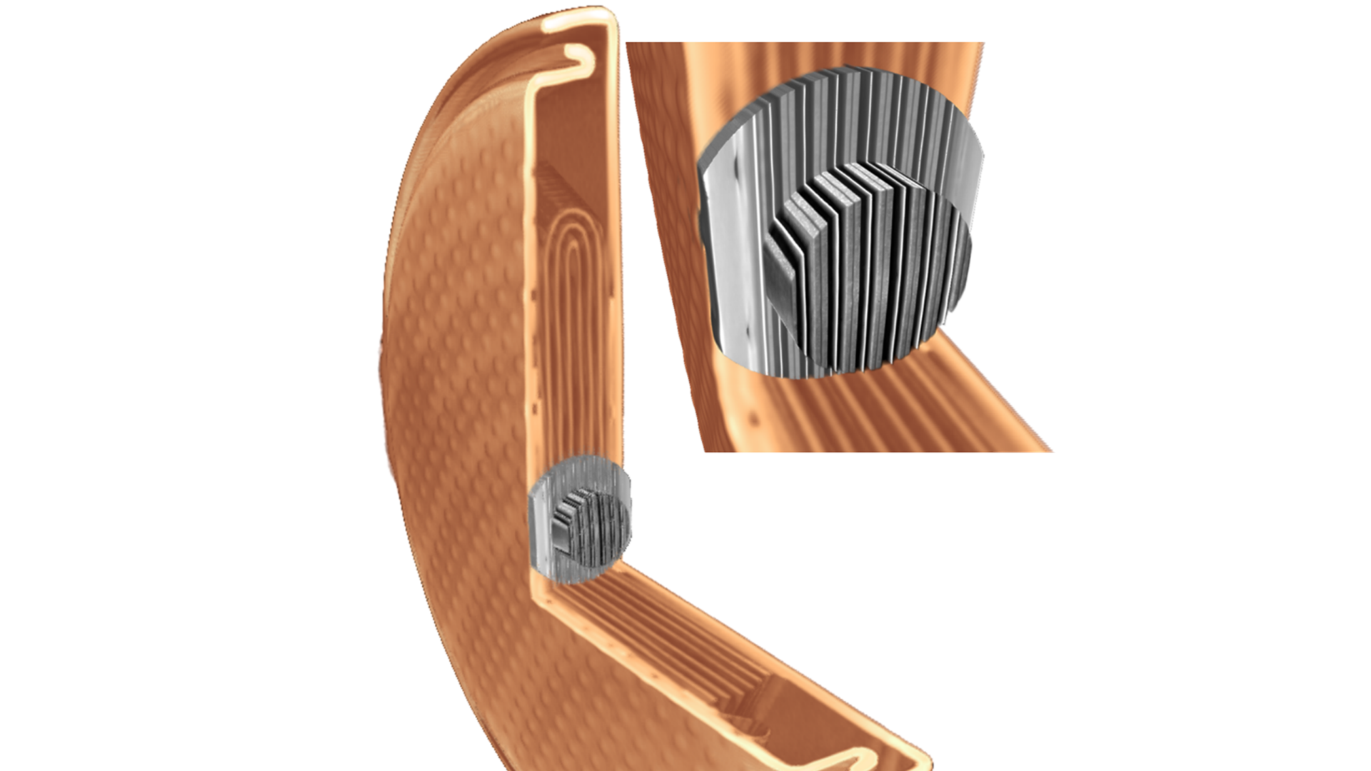
Intact cylinder cell (160 kV) – welding burrs, metallic inclusions, folds and kinks in conductive layers.
")
Small pouch cell (80 kV) – in situ microstructure, aging effect at cathode grain level, separator layer.

Small pouch cell: 0.4x overview scan; 4x Resolution at a Distance; 20x RaaD.


3D volume of materials within black mass, a powder generated from the crushing & shredding of recycled batteries. Cathode particles (blue) and residual foils (turquoise) individually segmented using Mineralogic 3D for quantification and analyses.


.")


")
")


Accessories
Upgrade your microscope with additional accessories to enhance its capabilities is available on all Versa X-ray microscopes")
 is available on all Versa X-ray microscopes")
 is available on all Versa X-ray microscopes")
 is available on all Versa X-ray microscopes")
Flat Panel Extension (FPX)
Large Sample, High Throughput ScanningFPX enhances imaging flexibility and creates workflow efficiencies for industrial and academic research. Scout-and-Zoom is a unique capability of ZEISS Versa X-ray microscopes that leverages FPX to perform a low resolution, large field of view, scout-scan to identify interior regions for higher resolution zoom-scans on a variety of different sample types. The Volume Scout workflow streamlines this process within ZEN navx. On ZEISS VersaXRM 730 and VersaXRM 615 platforms, FPX enables FAST Mode, allowing for sub-one-minute tomographies for efficient 3D navigation and rapid sample inspection. Combine with Volume Scout for end-to-end 3D navigation.


Autoloader
Maximize Your Instrument’s UtilizationMaximize use and minimize user intervention with the optional ZEISS Autoloader. Reduce the frequency of user interaction and increase productivity by enabling multiple jobs to run. Load up to 14 sample stations, which can support up to 70 samples, queue, and allow to run all day, or off-shift.


In Situ Interface Kit
Push the Limits of Scientific DiscoveryZEISS Versa platforms can accommodate a variety of in situ rigs, from high-pressure flow cells to tension, compression, and thermal stages, to user-customized designs. Moving beyond the three dimensions of space, leverage the non-destructive nature of X-ray investigation to extend your studies into the dimension of time with 4D experiments.


ZEISS arivis Pro
ZEISS arivis Pro empowers you to automate image analysis and visualization pipelines. Leverage traditional methods or AI models effortlessly to create pipelines for any image size, dimension, or modality without the need to code.


ZEN AI Toolkit including Intellesis
Machine learning can exponentially increase the throughput of image analysis and reduce the risk of human error. This toolkit contains solutions for image denoising, image segmentation, and object classification.


Lithium ion battery
Lithium ion battery
Dragonfly 3D World ZEISS edition
An advanced analysis and visualization software solution for your 3D data acquired by a variety of technologies including X-ray, FIB-SEM, SEM and helium ion microscopy. Available exclusively through ZEISS, 3D World ZEISS edition an intuitive, complete, and customizable toolkit for visualization and analysis of large 3D grayscale data. 3D World allows for navigation, annotation, creator files, including video production, of your 3D data. Perform image processing, segmentation, and object analysis to quantify your results.


