ZEISS Crossbeam 350 FIB-SEM microscope designed for flexible imaging and sample preparation in multi-user research laboratories.

ZEISS Crossbeam 350 FIB-SEM

Flexible FIB-SEM for multi-user lab applications

ZEISS Crossbeam 350 combines ZEISS Ion-sculptor FIB milling with ZEISS Gemini 1 imaging and Variable Pressure operation to support a wide range of applications, including non-conductive, magnetic, and not high-vacuum compatible samples. Designed for shared facilities, it delivers dependable high-resolution imaging analytics, sample preparation, and flexible configurations for research and industrial workflows.

  • Variable Pressure SEM for non-conductive and challenging materials
  • Fast, precise Ion-sculptor FIB milling, using low-kV finishing to minimize sample damage
  • Flexible chamber and accessory options for analysis and nanofabrication
  • Stable alignment and easy operation make it ideal for shared facilities
ZEISS Crossbeam 350 FIB-SEM microscope used for flexible materials analysis and sample preparation workflows.

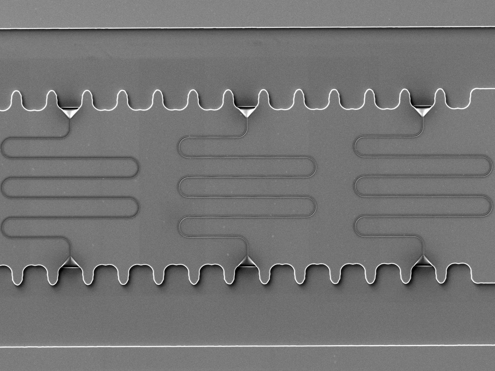
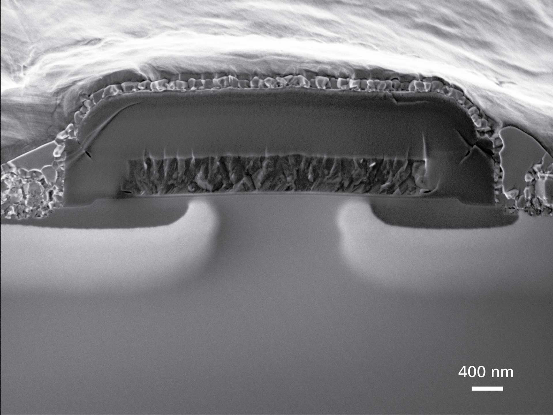
What’s different about Crossbeam 350

Crossbeam 350 is tailored for routine workflows, including low vacuum operation, standard FIB processing, and TEM lamella preparation. Guided and semi-automated workflows reduce manual effort while improving reproducibility across users and sites. Optional airlock NavCam imaging accelerates navigation and ROI targetin g large samples and wafers, enabling faster transitions from sample to result. Additionally, expanded gas injection support with up to five channels adds flexibility for deposition and etching, allowing workflows to evolve as applications grow in complexity.

Illustration of focused ion beam milling used to prepare thin TEM lamellae for analysis with the ZEISS Crossbeam 350 FIB-SEM.

Key specifications

  • Electron optics and ion beam technology: Gemini 1 FE-SEM + Ion-sculptor FIB
  • VP pressure range: Supports non-conductive and not high vacuum compatible materials
  • Resolution: surface sensitive, high resolution at low-kV SEM for routine nanoscale analysis
  • Handles non-conductive, magnetic, outgassing, and not high-vacuum compatible samples, as well as metals, polymers, ceramics, biomaterials, cells, tissues, and semiconductor specimens.
SEM image of a TEM lamella trench prepared by focused ion beam milling for lamella thinning and sample preparation.

Integrated TEM lamella preparation & imaging workflows

ZEISS Crossbeam 350 streamlines FIB-SEM sample preparation, failure analysis, and exploratory 3D tomography. Researchers can perform chunking, thinning, and inspection with live SEM monitoring, followed by low-energy FIB polishing to minimize ion damage. Guided ZEN workflows support manual and semi-automated TEM lamella preparation, batch execution across multiple sites, and recipe-driven reproducibility — making Crossbeam 350 an accessible entry point for 3D analysis.

SEM image of a milled sample cross-section displayed in ZEISS software for FIB-SEM analysis and microscopy workflow automation.

Advanced software workflows for automation, analysis, and scalable microscopy data processing

ZEISS delivers an integrated software ecosystem that accelerates microscopy workflows from acquisition to insight. ZEN core for EM orchestrates automated recipes, batch execution, and integrated SEM/FIB imaging. Complementing core control, the ZEISS arivis suite extends capabilities within scalable, AI-driven image analysis.

arivis Pro enables flexible multi-dimensional visualization and segmentation pipelines; arivis Hub accelerates and batches high-volume processing; and arivis Cloud provides cloud-based AI model training to automate and scale advanced analysis without coding. Together, these tools enhance nanofabrication, large-area imaging, and correlative workflows with reproducible, top-quality results.

Related applications

SEM micrograph of nanofluidic channels fabricated by focused ion beam milling for micro- and nanofluidics research.

Materials Research

  • Perform nanostructure analysis with low-kV imaging enabled surface sensitivity
  • Execute site specific cross section and 3D tomography runs
  • Prepare TEM samples with low-energy milling
  • Adapt workflows to a range of samples such as ceramics, polymers, metals, and composites
  • Characterize contact layers, interconnects, and advanced nodes with VP imaging
  • Reduce sample charging without coating using Variable Pressure
SEM image of a cleaved SiC MOSFET device showing dopant contrast between N+ diffusion and P-type body regions, imaged at low voltage with ZEISS Crossbeam 350 FIB-SEM.

Electronics & Semiconductors

  • Analyze larger devices: Ideal for MEMS, analog, mature nodes, and power devices
  • Prepare TEM lamellae across multiple devices with consistent thickness and minimal damage
3D FIB-SEM reconstruction of the nematode C. elegans showing internal anatomy at nanometer resolution.

Life Sciences & Biological Research

  • Reveal ultrastructure with low-kV, surface-sensitive SEM imaging
  • Access targeted regions of cells or tissue using precise FIB milling
  • Perform 3D tomography to reconstruct organelles and biological interfaces
  • Prepare high-quality TEM lamellae with low-energy thinning for minimal damage

Customer testimonial

There are many different physical interactions that are happening within these cells and when we’re doing 3D electron microscopy, we can see all of these things. This allows us to take an integrated viewpoint on muscle cell structure.

Dr. Brian Glancy Earl Stadtman Investigator, Muscle Energetics Laboratory, National Heart, Lung, and Blood Institute at the National Institutes of Health, USA

Services & support

Expert guidance for daily sample preparation and analysis

ZEISS supports Crossbeam 350 users well beyond installation. Application specialists help teams get up and running quickly, refine workflows, and build confidence with FIB-SEM sample preparation and 3D analysis. Flexible service agreements, remote diagnostics, and training options make it easy to maintain performance as needed to evolve without overcommitting resources early on.

Contact ZEISS Microscopy

Contact

Form is loading...

/ 4
Next Step:
  • Step 1
  • Step 2
  • Step 3
Contact us
Required Information
Optional Information

If you want to have more information on data processing at ZEISS please refer to our data privacy notice.

FAQ

  • ZEISS Crossbeam 350 is best suited for labs that need reliable FIB-SEM performance across a wide range of samples, with flexibility for exploratory research, routine lamella preparation, and shared-facility use. It uses Gemini 1 electron optics with Ion-sculptor FIB milling and Variable Pressure SEM to support a wide range of materials, including non-conductive, magnetic, and non-high-vacuum compatible samples. Its guided workflows and flexible configurations make it ideal for shared facilities and multi-user labs.

  • Crossbeam 350 uses ZEN core for EM to provide guided workflows and recipe-based operations, helping reduce user-to-user variability and simplifying training in shared environments.

  • Yes. Crossbeam 350 supports controlled imaging and milling workflows for beam-sensitive samples, though it is not optimized for the most demanding low-damage or ultra-high-throughput applications.

  • You should choose Crossbeam 350 or Crossbeam 750 when you need a flexible, reliable FIB-SEM system for a wide range of applications, shared-facility use, or exploratory research — without the higher throughput and performance requirements that drive selection of Crossbeam 550.