Array Tomography
Non-destructive Volume Imaging Using Standard, Scanning Electron MicroscopySchematic Representation of a Typical Workflow


1
A resin-embedded sample is cut into an array of serial sections, each with a section thickness of typically 30 – 70 nm, and attached to a sample carrier in the order they were cut.


2
Each serial section is imaged with the scanning electron microscope (SEM).


3
The acquired EM images are processed and digitally aligned into a 3D data set. Cell compartments can be identified and segmented.


4
The segmented 3D data set can be visualized, investigated, and statistically analyzed.
Application Example
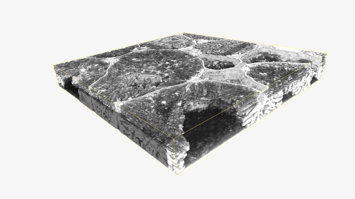
3D reconstruction of serial sections from root nodules with the distribution of plasmodesmataNew Discoveries from the Ultrastructure of Life Virtual Seminar Series
In a series of six webinars, explore the technological underpinnings of Volume EM imaging and its growing number of application areas in neurobiology, cancer research, developmental biology, plant science, and more.
Learn about vEM-specific sample preparation and technologies (array tomography, serial block-face SEM, and FIB-SEM), advanced image processing, data analysis, and result visualization capabilities of workflow-oriented software solutions.


The symbiotic relationship between plants and bacteria
Courtesy of D. Sherrier, J. Caplan, and S. Modla, University of Delaware, USA.
The Symbiotic Relationship between Plants and Bacteria
Understanding the Impact of Bacteria in Root Nodules on the Health and Condition of PlantsThe root network of a plant provides access to all of the water and nutrients that are crucial components for all plant growth. Exploring the whole root network as well as understanding the influence of external microbes is important for optimizing plant health and yield. Investigating the symbiotic relationship between plants and bacteria in root nodules requires knowledge of root nodule and bacteria distribution and a combination of both fluorescence and high-resolution structural assessment is vital to understand this in detail.
Correlative array tomography enables the overlay of both fluorescence and structural data to enable visualization of root nodule and bacteria distribution.